
Mouse WISP-1 (Wnt-induced secreted protein-1; also named CNN4) is a 40 kDa, secreted, heparin-binding glycoprotein that is a member of the CCN (or CTGF/Cyr61/Nov) cysteine-rich protein family (1- 6). It is synthesized as a 367 amino acid (aa) precursor that contains a series of structural homology modules. Following a 22 aa signal sequence, there is a 68 aa IGFBP-like domain (aa 53-120), a 66 aa von Willebrand factor type C (VWC) module (aa 121-186), a 41 aa TSP type I domain (aa 220-260) and a 75 aa, C-terminal cysteine knot motif (aa 273-347) (1, 6). The VWC module is associated with protein-protein interaction, the TSP domain binds sulfated glycoconjugates, and the cysteine knot mediates dimerization and receptor binding (4). It is likely that WISP-1 normally circulates as an 80 kDa homodimer (2). Multiple splice forms are reported for human WISP-1, but none for rodent (7-9). Mature mouse WISP-1 is 98%, 84%, and 85% aa identical to rat, canine, and human WISP-1, respectively. WISP-1 is expressed by fibroblasts and osteoblasts, and may contribute to fracture healing by promoting bone cell formation (1, 10, 11).
Mouse WISP-1/CCN4 Antibody (214203)
R&D Systems | Catalog # MAB1680

Scientific images may be resized, cropped, or adjusted for brightness or contrast for presentation purposes. These updates are limited to presentation and do not affect the underlying scientific interpretation of the data. Where available, additional source data may be provided upon request.
Key Product Details
Species Reactivity
Validated:
Cited:
Applications
Validated:
Cited:
Label
Antibody Source
Product Specifications
Immunogen
Leu21-Asn367
Accession # O54775
Specificity
Clonality
Host
Isotype
Scientific Data Images for Mouse WISP-1/CCN4 Antibody (214203)
Detection of WISP-1/CCN4 by Flow Cytometry
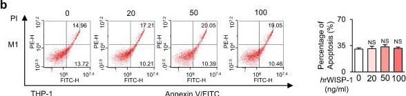
Recombinant WISP-1 reduces M2 macrophage survival, induces apoptosis, and promotes reprogramming toward an M1-like phenotype. (a) Cell viability assay of THP-1-derived M1 (M1) and M2 macrophages (M2) treated with 20-100 ng/ml mouse (rWISP-1) or human WISP-1 (hrWISP-1) for 3 days. (b) Left: Flow cytometry analysis after Annexin V-FICT/PI dual staining was employed to evaluate the apoptosis of THP-1- derived M1 and M2 macrophages after treatment with hrWISP- (20-100 ng/ml) for 3 days. Right: Apoptotic cells were quantified as the sum of the percentages of early and late stages of apoptosis. (c) qRT-PCR analysis of relative mRNA levels of M1 (Nos2, MhcII, and Il12p40) and M2 (Tgf beta 1, Il10, and Il4) markers in M2 macrophages treated with 20-100 ng/ml hrWISP-1 for 3 days. (d) ELISA of TNF-alpha, IL-1 beta, IL-4, and IL-13 in the culture supernatant of M2 macrophages treated with 20-100 ng/ml hrWISP-1 for 3 days. (e) Flow cytometry analysis of the population of CD80+ and CD206+ cells among M2 macrophages after treatment with hrWISP-1 (20-100 ng/ml) for 2 or 3 days. Mean fluorescence intensity (MFI) values (right). (f) Heatmap showing differentially expressed macrophage polarization-related genes in THP-1-derived M2 macrophages (left). Red: high expression; blue: low expression. Relative expression of selected genes from PCR array profiling of macrophage polarization markers (right). Log2 fold-change values (hrWISP-1 vs. Vehicle). THP-1-derived M2 macrophages were treated with hrWISP-1 (50 ng/ml) for 3 days. NS: not significant; *P < 0.05, **P < 0.01, ***P < 0.001, two-tailed Student's t-test. The data are from one experiment representative of three independent experiments with similar results (b, e, and f left) or from three independent experiments (mean ± standard error: a, c, d; b, e and f right). Image collected and cropped by CiteAb from the following open publication (https://www.ijbs.com/v22p0995.htm), licensed under a CC-BY license. Not internally tested by R&D Systems.Applications for Mouse WISP-1/CCN4 Antibody (214203)
Western Blot
Sample: Recombinant Mouse WISP‑1/CCN4 (Catalog # 1680-WS)
under non-reducing conditions only
Formulation, Preparation, and Storage
Purification
Reconstitution
Reconstitute at 0.5 mg/mL in sterile PBS. For liquid material, refer to CoA for concentration.
Formulation
Shipping
Stability & Storage
- 12 months from date of receipt, -20 to -70 °C as supplied.
- 1 month, 2 to 8 °C under sterile conditions after reconstitution.
- 6 months, -20 to -70 °C under sterile conditions after reconstitution.
Calculators
Background: WISP-1/CCN4
References
- Pennica, D. et al. (1998) Proc. Natl. Acad. Sci. USA 95:14717.
- Tanada, S. et al. (2001) Oncogene 20:5525.
- Brigstock, D.R. et al. (2003) J. Clin. Pathol. Mol. Pathol. 56:127.
- Desnoyers, L. (2004) Curr. Pharm. Des. 10:3913.
- Brigstock, D.R. (2003) J. Endocrinol. 178:169.
- Hashimoto, Y. et al. (1998) J. Exp. Med. 187:289.
- SwissProt Accession # O54775.
- SwissProt Accession # Q99PP0.
- SwissProt Accession # O95388
- French, D.M. et al. (2004) Am. J. Pathol. 165:855.
- Parisi, M.S. et al. (2006) Bone 38:671.
Long Name
Alternate Names
Gene Symbol
UniProt
Additional WISP-1/CCN4 Products
Product Documents for Mouse WISP-1/CCN4 Antibody (214203)
Certificate of Analysis
To download a Certificate of Analysis, please enter a lot or batch number in the search box below.
Note: Certificate of Analysis not available for kit components.
Product Specific Notices for Mouse WISP-1/CCN4 Antibody (214203)
For research use only
Related Research Areas
Citations for Mouse WISP-1/CCN4 Antibody (214203)
 Powered by Bioz
Customer Reviews for Mouse WISP-1/CCN4 Antibody (214203)
There are currently no reviews for this product. Be the first to review Mouse WISP-1/CCN4 Antibody (214203) and earn rewards!
Have you used Mouse WISP-1/CCN4 Antibody (214203)?
Submit a review and receive an Amazon gift card!
$25/€18/£15/$25CAN/¥2500 for a review with an image
$10/€7/£6/$10CAN/¥1110 for a review without an image
Submit a review
Protocols
Find general support by application which include: protocols, troubleshooting, illustrated assays, videos and webinars.
- Cellular Response to Hypoxia Protocols
- R&D Systems Quality Control Western Blot Protocol
- Troubleshooting Guide: Western Blot Figures
- Western Blot Conditions
- Western Blot Protocol
- Western Blot Protocol for Cell Lysates
- Western Blot Troubleshooting
- Western Blot Troubleshooting Guide
- View all Protocols, Troubleshooting, Illustrated assays and Webinars