MxA/Mx1 Antibody - BSA Free
Novus Biologicals | Catalog # NBP1-32905
![Immunocytochemistry/ Immunofluorescence: MxA/Mx1 Antibody [NBP1-32905]](https://resources.rndsystems.com/images/products/MxA-Mx1-Antibody-Immunocytochemistry-Immunofluorescence-NBP1-32905-img0015.jpg "Immunocytochemistry/ Immunofluorescence: MxA/Mx1 Antibody [NBP1-32905]")
Loading...
Key Product Details
Species Reactivity
Validated:
Human, Bovine, Monkey, Rabbit
Predicted:
Chimpanzee (98%), Rhesus Macaque (92%). Backed by our 100% Guarantee.
Applications
Immunohistochemistry, Immunohistochemistry-Paraffin, Immunohistochemistry-Frozen, Western Blot, Immunocytochemistry/ Immunofluorescence
Label
Unconjugated
Antibody Source
Polyclonal Rabbit IgG
Format
BSA Free
Loading...
Product Specifications
Immunogen
Recombinant protein encompassing a sequence within the center region of human MxA/Mx1. The exact sequence is proprietary.
Localization
Cytoplasm
Clonality
Polyclonal
Host
Rabbit
Isotype
IgG
Theoretical MW
76 kDa.
Disclaimer note: The observed molecular weight of the protein may vary from the listed predicted molecular weight due to post translational modifications, post translation cleavages, relative charges, and other experimental factors.
Disclaimer note: The observed molecular weight of the protein may vary from the listed predicted molecular weight due to post translational modifications, post translation cleavages, relative charges, and other experimental factors.
Scientific Data Images for MxA/Mx1 Antibody - BSA Free
Immunocytochemistry/ Immunofluorescence: MxA/Mx1 Antibody [NBP1-32905]
Immunocytochemistry/Immunofluorescence: MxA/Mx1 Antibody [NBP1-32905] - Detects MX1 protein at cytoplasm by immunofluorescent analysis. Sample: HeLa cells were fixed in 4% paraformaldehyde at RT for 15 min. Green: MX1 protein stained by MX1 antibody, Internal diluted at 1:200. Red: alpha Tubulin, a cytoskeleton marker, stained by alpha Tubulin antibody diluted at 1:1000. Blue: Hoechst 33342 staining.![Immunohistochemistry-Paraffin: MxA/Mx1 Antibody [NBP1-32905]](https://resources.rndsystems.com/images/products/MxA-Mx1-Antibody-Immunohistochemistry-Paraffin-NBP1-32905-img0021.jpg "Immunohistochemistry-Paraffin: MxA/Mx1 Antibody [NBP1-32905]")
Immunohistochemistry-Paraffin: MxA/Mx1 Antibody [NBP1-32905]
Immunohistochemistry-Paraffin: MxA/Mx1 Antibody [NBP1-32905] - Human lymph node. Antigen retrieval in citrate buffer, 95C for 30 min. Image submitted by a verified customer review.![Western Blot: MxA/Mx1 Antibody [NBP1-32905]](https://resources.rndsystems.com/images/products/MxA-Mx1-Antibody-Western-Blot-NBP1-32905-img0018.jpg "Western Blot: MxA/Mx1 Antibody [NBP1-32905]")
Western Blot: MxA/Mx1 Antibody [NBP1-32905]
Western Blot: MxA/Mx1 Antibody [NBP1-32905] - A. 30 ug 293T whole cell lysate/extract B. 30 ug whole cell lysate/extract of human MX1-transfected 293T cells. 7.5 % SDS-PAGE MX1 antibody [N2C2], Internal dilution: 1:10000![Immunohistochemistry-Paraffin: MxA/Mx1 Antibody [NBP1-32905]](https://resources.rndsystems.com/images/products/MxA-Mx1-Antibody-Immunohistochemistry-Paraffin-NBP1-32905-img0007.jpg "Immunohistochemistry-Paraffin: MxA/Mx1 Antibody [NBP1-32905]")
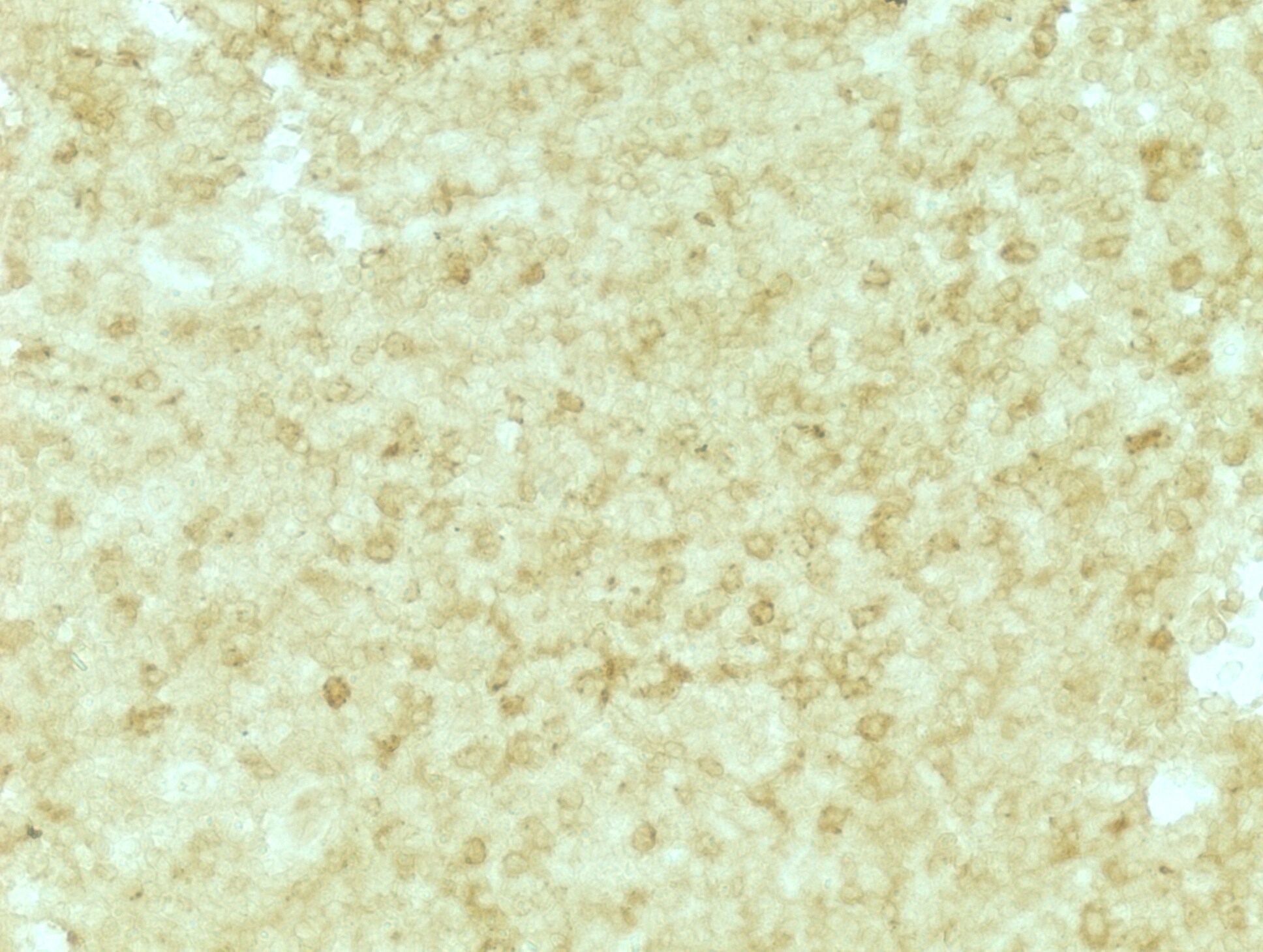
Immunohistochemistry-Paraffin: MxA/Mx1 Antibody [NBP1-32905]
Immunohistochemistry-Paraffin: MxA/Mx1 Antibody [NBP1-32905] - MDA-MB-468 xenograft, using MX1antibody at 1:500 dilution. Antigen Retrieval: Citrate buffer, pH 6.0, 15 min.![Immunohistochemistry-Paraffin: MxA/Mx1 Antibody [NBP1-32905]](https://resources.rndsystems.com/images/products/MxA-Mx1-Antibody-Immunohistochemistry-Paraffin-NBP1-32905-img0016.jpg "Immunohistochemistry-Paraffin: MxA/Mx1 Antibody [NBP1-32905]")
Immunohistochemistry-Paraffin: MxA/Mx1 Antibody [NBP1-32905]
Immunohistochemistry-Paraffin: MxA/Mx1 Antibody [NBP1-32905] - Human colon carcinoma. MX1 antibody [N2C2], Internal diluted at 1:500. Antigen Retrieval: Citrate buffer, pH 6.0, 15 min.
Western Blot: MxA/Mx1 Antibody [NBP1-32905] -
Various whole cell extracts (30 ug) were separated by 7.5% SDS-PAGE, and the membrane was blotted with MX1 antibody [N2C2], Internal (NBP1-32905) diluted at 1:1000. The HRP-conjugated anti-rabbit IgG antibody was used to detect the primary antibody. Corresponding RNA expression data for the same cell lines are based on Human Protein Atlas program.
Flow Cytometry: MxA/Mx1 Antibody - BSA Free [NBP1-32905] -
The correlation between miR-141 level and the expression profile of MxA and STAT3 in infected A549 cells. (A) Quantification of steady-state miR-141 in infected A549 cells with MOI of 0.5 and transfected with either pre-miR-141 or miR-141 inhibitor compared with noninfected cells (control) using qRT-PCR. (B) Relative gene expression of MxA and STAT3 in infected A549 cells transfected with either specific inhibitor against miR-141 or pre- miR-141 compared with control-transfected cells using qRT-PCR. Error bars indicate the STD of three independent experiments. Student two-tailed t-test used for statistical analysis, (*) indicates P-values ≤ 0.05, and (**) indicates P ≤ 0.01. (C) Flow cytometric assay quantifies the kinetic proteins expression profile of MxA (in blue dots) and STAT3 (in red dots) in infected and transfected A549 cells compared with control cells. (D) Western blot analysis reveals the protein expression level of MxA and STAT3 in infected and transfected cells compared to control cells, beta -actin expression profile severed as an internal control Image collected and cropped by CiteAb from the following open publication (https://pubmed.ncbi.nlm.nih.gov/37596622), licensed under a CC-BY license. Not internally tested by Novus Biologicals.
Flow Cytometry: MxA/Mx1 Antibody - BSA Free [NBP1-32905] -
The correlation between miR-141 level and the expression profile of MxA and STAT3 in infected A549 cells. (A) Quantification of steady-state miR-141 in infected A549 cells with MOI of 0.5 and transfected with either pre-miR-141 or miR-141 inhibitor compared with noninfected cells (control) using qRT-PCR. (B) Relative gene expression of MxA and STAT3 in infected A549 cells transfected with either specific inhibitor against miR-141 or pre- miR-141 compared with control-transfected cells using qRT-PCR. Error bars indicate the STD of three independent experiments. Student two-tailed t-test used for statistical analysis, (*) indicates P-values ≤ 0.05, and (**) indicates P ≤ 0.01. (C) Flow cytometric assay quantifies the kinetic proteins expression profile of MxA (in blue dots) and STAT3 (in red dots) in infected and transfected A549 cells compared with control cells. (D) Western blot analysis reveals the protein expression level of MxA and STAT3 in infected and transfected cells compared to control cells, beta -actin expression profile severed as an internal control Image collected and cropped by CiteAb from the following open publication (https://pubmed.ncbi.nlm.nih.gov/37596622), licensed under a CC-BY license. Not internally tested by Novus Biologicals.Applications for MxA/Mx1 Antibody - BSA Free
Application
Recommended Usage
Immunocytochemistry/ Immunofluorescence
1:100-1:1000
Immunohistochemistry
1:100-1:1000
Immunohistochemistry-Frozen
Assay dependent
Immunohistochemistry-Paraffin
1:100-1:1000
Western Blot
1:500-1:3000
Reviewed Applications
Read 1 review rated 4 using NBP1-32905 in the following applications:
Formulation, Preparation, and Storage
Purification
Antigen Affinity-purified
Formulation
PBS, 20% Glycerol
Format
BSA Free
Preservative
0.025% Proclin 300
Concentration
Concentrations vary lot to lot. See vial label for concentration. If unlisted please contact technical services.
Shipping
The product is shipped with polar packs. Upon receipt, store it immediately at the temperature recommended below.
Stability & Storage
Aliquot and store at -20C or -80C. Avoid freeze-thaw cycles.
Background: MxA/Mx1
Long Name
Interferon-regulated Resistance GTP-binding protein MxA
Alternate Names
IFI-78K, IFI78, Mx1
Gene Symbol
MX1
Additional MxA/Mx1 Products
Product Documents for MxA/Mx1 Antibody - BSA Free
Certificate of Analysis
To download a Certificate of Analysis, please enter a lot or batch number in the search box below.
Product Specific Notices for MxA/Mx1 Antibody - BSA Free
This product is for research use only and is not approved for use in humans or in clinical diagnosis. Primary Antibodies are guaranteed for 1 year from date of receipt.
⚠ WARNING: This product can expose you to chemicals including mercury, which is known to the State of California to cause reproductive toxicity with developmental effects. For more information go to www.P65Warnings.ca.gov.Related Research Areas
Citations for MxA/Mx1 Antibody - BSA Free
 Powered by Bioz
Customer Reviews for MxA/Mx1 Antibody - BSA Free (1)
4 out of 5
1 Customer Rating
Have you used MxA/Mx1 Antibody - BSA Free?
Submit a review and receive an Amazon gift card!
$25/€18/£15/$25CAN/¥2500 Yen for a review with an image
$10/€7/£6/$10CAN/¥1110 Yen for a review without an image
Submit a review
Customer Images
Showing
1
-
1 of
1 review
Showing All
Filter By:
-
Application: Immunohistochemistry-ParaffinSample Tested: Human lymph nodeSpecies: HumanVerified Customer | Posted 09/25/2018Heat mediated antigen retrieval performed in citrate buffer for 30 mins at 95C

There are no reviews that match your criteria.
Protocols
Find general support by application which include: protocols, troubleshooting, illustrated assays, videos and webinars.
- Antigen Retrieval Protocol (PIER)
- Antigen Retrieval for Frozen Sections Protocol
- Appropriate Fixation of IHC/ICC Samples
- Cellular Response to Hypoxia Protocols
- Chromogenic IHC Staining of Formalin-Fixed Paraffin-Embedded (FFPE) Tissue Protocol
- Chromogenic Immunohistochemistry Staining of Frozen Tissue
- ClariTSA™ Fluorophore Kits
- Detection & Visualization of Antibody Binding
- Fluorescent IHC Staining of Frozen Tissue Protocol
- Graphic Protocol for Heat-induced Epitope Retrieval
- Graphic Protocol for the Preparation and Fluorescent IHC Staining of Frozen Tissue Sections
- Graphic Protocol for the Preparation and Fluorescent IHC Staining of Paraffin-embedded Tissue Sections
- Graphic Protocol for the Preparation of Gelatin-coated Slides for Histological Tissue Sections
- ICC Cell Smear Protocol for Suspension Cells
- ICC Immunocytochemistry Protocol Videos
- ICC for Adherent Cells
- IHC Sample Preparation (Frozen sections vs Paraffin)
- Immunocytochemistry (ICC) Protocol
- Immunocytochemistry Troubleshooting
- Immunofluorescence of Organoids Embedded in Cultrex Basement Membrane Extract
- Immunofluorescent IHC Staining of Formalin-Fixed Paraffin-Embedded (FFPE) Tissue Protocol
- Immunohistochemistry (IHC) and Immunocytochemistry (ICC) Protocols
- Immunohistochemistry Frozen Troubleshooting
- Immunohistochemistry Paraffin Troubleshooting
- Preparing Samples for IHC/ICC Experiments
- Preventing Non-Specific Staining (Non-Specific Binding)
- Primary Antibody Selection & Optimization
- Protocol for Heat-Induced Epitope Retrieval (HIER)
- Protocol for Making a 4% Formaldehyde Solution in PBS
- Protocol for VisUCyte™ HRP Polymer Detection Reagent
- Protocol for the Fluorescent ICC Staining of Cell Smears - Graphic
- Protocol for the Fluorescent ICC Staining of Cultured Cells on Coverslips - Graphic
- Protocol for the Preparation & Fixation of Cells on Coverslips
- Protocol for the Preparation and Chromogenic IHC Staining of Frozen Tissue Sections
- Protocol for the Preparation and Chromogenic IHC Staining of Frozen Tissue Sections - Graphic
- Protocol for the Preparation and Chromogenic IHC Staining of Paraffin-embedded Tissue Sections
- Protocol for the Preparation and Chromogenic IHC Staining of Paraffin-embedded Tissue Sections - Graphic
- Protocol for the Preparation and Fluorescent ICC Staining of Cells on Coverslips
- Protocol for the Preparation and Fluorescent ICC Staining of Non-adherent Cells
- Protocol for the Preparation and Fluorescent ICC Staining of Stem Cells on Coverslips
- Protocol for the Preparation and Fluorescent IHC Staining of Frozen Tissue Sections
- Protocol for the Preparation and Fluorescent IHC Staining of Paraffin-embedded Tissue Sections
- Protocol for the Preparation of Gelatin-coated Slides for Histological Tissue Sections
- Protocol for the Preparation of a Cell Smear for Non-adherent Cell ICC - Graphic
- R&D Systems Quality Control Western Blot Protocol
- TUNEL and Active Caspase-3 Detection by IHC/ICC Protocol
- The Importance of IHC/ICC Controls
- Troubleshooting Guide: Immunohistochemistry
- Troubleshooting Guide: Western Blot Figures
- Western Blot Conditions
- Western Blot Protocol
- Western Blot Protocol for Cell Lysates
- Western Blot Troubleshooting
- Western Blot Troubleshooting Guide
- View all Protocols, Troubleshooting, Illustrated assays and Webinars
Loading...