MYH6 Antibody (3-48) - Azide and BSA Free
Novus Biologicals | Catalog # NB300-284
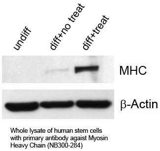
![Western Blot: MYH6 Antibody (3-48)Azide and BSA Free [NB300-284]](https://resources.rndsystems.com/images/products/MYH6-Antibody-3-48-Western-Blot-NB300-284-img0004.jpg "Western Blot: MYH6 Antibody (3-48)Azide and BSA Free [NB300-284]")
Key Product Details
Species Reactivity
Validated:
Cited:
Predicted:
Applications
Validated:
Cited:
Label
Antibody Source
Format
Product Specifications
Immunogen
Reactivity Notes
Specificity
Clonality
Host
Isotype
Scientific Data Images for MYH6 Antibody (3-48) - Azide and BSA Free
Western Blot: MYH6 Antibody (3-48)Azide and BSA Free [NB300-284]
Western Blot: MYH6 Antibody (3-48) [NB300-284] - analysis of total cardiac myosin expression in tissue lysates of rat heart (lanes 1-5), liver (lanes 6-7), and lung (lanes 8-9). Increasing amounts of total protein were loaded as follow: lanes 1, 6 and 8: 0 ug; lane 2: 50 ug; lanes 3, 7 and 9: 250 ug; lane 5: 500 ug. (anti-cMHC was used at 1:500; secondary antibody: Goat anti-mouse IgG Fc-HRP; 1:5000)![Immunohistochemistry-Paraffin: MYH6 Antibody (3-48) - Azide and BSA Free [NB300-284]](https://resources.rndsystems.com/images/products/MYH6-Antibody-3-48-Immunohistochemistry-Paraffin-NB300-284-img0005.jpg "Immunohistochemistry-Paraffin: MYH6 Antibody (3-48) - Azide and BSA Free [NB300-284]")
Immunohistochemistry-Paraffin: MYH6 Antibody (3-48) - Azide and BSA Free [NB300-284]
Immunohistochemistry-Paraffin: MYH6 Antibody (3-48) [NB300-284] - Formaldehyde-fixed paraffin-embedded (FFPE) Tissue Slides. Human heart ventricle following immunostaining with cMHC 3-48 and irrelevant NeuAc monoclonal antibody at low microscopical magnification.![Western Blot: MYH6 Antibody (3-48)Azide and BSA Free [NB300-284]](https://resources.rndsystems.com/images/products/MYH6-Antibody-3-48-Western-Blot-NB300-284-img0006.jpg "Western Blot: MYH6 Antibody (3-48)Azide and BSA Free [NB300-284]")
Western Blot: MYH6 Antibody (3-48)Azide and BSA Free [NB300-284]
MYH6-Antibody-3-48-Western-Blot-NB300-284-img0006.jpg - Azide and BSA Free [NB300-284] -")
Western Blot: MYH6 Antibody (3-48) - Azide and BSA Free [NB300-284] -
Western blot immunodetection of cardiac MyHC isoforms from left ventricle (LV) homogenates and extracts of adult euthyroid (EU), hypothyroid (HY), and hyperthyroid (TH) Lewis rats. The membranes were stained by NB300-284 antibody recognizing both MyHC alpha and MyHC beta isoforms (a) or by BA.G5 antibody, which was solely specific for cardiac MyHC alpha isoform (b). - Azide and BSA Free [NB300-284] -")
Western Blot: MYH6 Antibody (3-48) - Azide and BSA Free [NB300-284] -
Western Blot: MYH6 Antibody (3-48) - Azide and BSA Free [NB300-284] - Western blot immunodetection of cardiac MyHC isoforms from left ventricle (LV) homogenates & extracts of adult euthyroid (EU), hypothyroid (HY), & hyperthyroid (TH) Lewis rats. The membranes were stained by NB300-284 antibody recognizing both MyHC alpha & MyHC beta isoforms (a) or by BA.G5 antibody, which was solely specific for cardiac MyHC alpha isoform (b). Image collected & cropped by CiteAb from the following publication (https://pubmed.ncbi.nlm.nih.gov/22187528), licensed under a CC-BY license. Not internally tested by Novus Biologicals.Applications for MYH6 Antibody (3-48) - Azide and BSA Free
Immunohistochemistry
Immunohistochemistry-Paraffin
Western Blot
Reviewed Applications
Read 2 reviews rated 4.5 using NB300-284 in the following applications:
Formulation, Preparation, and Storage
Purification
Formulation
Format
Preservative
Concentration
Shipping
Stability & Storage
Background: MYH6
Long Name
Alternate Names
Gene Symbol
UniProt
Additional MYH6 Products
Product Documents for MYH6 Antibody (3-48) - Azide and BSA Free
Certificate of Analysis
To download a Certificate of Analysis, please enter a lot or batch number in the search box below.
Product Specific Notices for MYH6 Antibody (3-48) - Azide and BSA Free
This product is for research use only and is not approved for use in humans or in clinical diagnosis. Primary Antibodies are guaranteed for 1 year from date of receipt.
Related Research Areas
Citations for MYH6 Antibody (3-48) - Azide and BSA Free
 Powered by Bioz
Customer Reviews for MYH6 Antibody (3-48) - Azide and BSA Free (2)
Have you used MYH6 Antibody (3-48) - Azide and BSA Free?
Submit a review and receive an Amazon gift card!
$25/€18/£15/$25CAN/¥2500 Yen for a review with an image
$10/€7/£6/$10CAN/¥1110 Yen for a review without an image
Submit a review
Customer Images
-
Application: Western BlotSample Tested:Species: HumanVerified Customer | Posted 11/25/2014

-
Application: Western BlotSample Tested: RatSpecies: RatVerified Customer | Posted 12/30/2011
There are no reviews that match your criteria.
Protocols
View specific protocols for MYH6 Antibody (3-48) - Azide and BSA Free (NB300-284):
Materials
1) 1 Phosphate buffered saline (pH 7.6): NaCl 137mmol/L, KCl 2.7mmol/L, Na2HPO4 4.3mmol/L, KH2PO4 1.4 mmol/L
2) Citrate buffer, 0.01 M, pH6.0, Sodium Citrate 3g, Citric acid 0.4g
3) 3% Hydrogen peroxide
4) Primary antibody
5) Blocking serum (normal serum)
6) Biotinylated secondary antibody
7) DAB staining kit
Methods
1. Dewax and hydration of slides using xylene and EtOH:
Dry slides for 20 min in a 60 C oven
Add Xylene, 2 x 10 min
100%, 95%, 80%, and 70% EtOH, 5 min each EtOH concentration
Rinse in PBS, 5'
2 Antigen retrieval method (only for paraffin slides)
1a. High-pressure antigen retrieval procedure (recommended method)
Place slides in a glass slide holder (ensure that the slide holder is completely filled with slides, slides without sections if necessary, to ensure even heating. The entire slide holder is immersed in 1000 ml of Citrate buffer (0.01M, pH6.0) within a pressure cooker
Once steam is produced, and ONLY when steam is visible, from the pressure cooker (usually 15-20 min), the required high-pressure will have been reached, and slides will be incubated for 2 min.
Turn off heat, and allow buffer and slides to cool to room temperature
Slides are then rinsed in PBS for 5 minutes
2. Add 3% hydrogen peroxide solution, 10'at RT, then PBS, 3X5'
3. Normal blocking serum, 20'at RT
4. Incubate with Primary Ab, 4C overnight or 1.5 hours at 37C
5. Rinse with PBS, 3 X 5' each rinse
6. Add Biotin-conjugated second antibody, 10'at RT
7. Rinse with PBS, 3 X 5' each rinse
8. Add Streptavidin-Peroxidase, 10'at RT
9. Rinse with PBS, 3 X 5' each rinse
10. Staining with DAB solution, 2-5'under microscope
11. Stop the reaction by washing in tap water
12. Counterstain in Haematoxylin for 3-5 minutes
13. 75%, 80%, 95% and 100% ethanol, 5x2', xylene 2 x 10'
Find general support by application which include: protocols, troubleshooting, illustrated assays, videos and webinars.
- Antigen Retrieval Protocol (PIER)
- Antigen Retrieval for Frozen Sections Protocol
- Appropriate Fixation of IHC/ICC Samples
- Cellular Response to Hypoxia Protocols
- Chromogenic IHC Staining of Formalin-Fixed Paraffin-Embedded (FFPE) Tissue Protocol
- Chromogenic Immunohistochemistry Staining of Frozen Tissue
- ClariTSA™ Fluorophore Kits
- Detection & Visualization of Antibody Binding
- Fluorescent IHC Staining of Frozen Tissue Protocol
- Graphic Protocol for Heat-induced Epitope Retrieval
- Graphic Protocol for the Preparation and Fluorescent IHC Staining of Frozen Tissue Sections
- Graphic Protocol for the Preparation and Fluorescent IHC Staining of Paraffin-embedded Tissue Sections
- Graphic Protocol for the Preparation of Gelatin-coated Slides for Histological Tissue Sections
- ICC Cell Smear Protocol for Suspension Cells
- ICC Immunocytochemistry Protocol Videos
- ICC for Adherent Cells
- IHC Sample Preparation (Frozen sections vs Paraffin)
- Immunocytochemistry (ICC) Protocol
- Immunocytochemistry Troubleshooting
- Immunofluorescence of Organoids Embedded in Cultrex Basement Membrane Extract
- Immunofluorescent IHC Staining of Formalin-Fixed Paraffin-Embedded (FFPE) Tissue Protocol
- Immunohistochemistry (IHC) and Immunocytochemistry (ICC) Protocols
- Immunohistochemistry Frozen Troubleshooting
- Immunohistochemistry Paraffin Troubleshooting
- Preparing Samples for IHC/ICC Experiments
- Preventing Non-Specific Staining (Non-Specific Binding)
- Primary Antibody Selection & Optimization
- Protocol for Heat-Induced Epitope Retrieval (HIER)
- Protocol for Making a 4% Formaldehyde Solution in PBS
- Protocol for VisUCyte™ HRP Polymer Detection Reagent
- Protocol for the Fluorescent ICC Staining of Cell Smears - Graphic
- Protocol for the Fluorescent ICC Staining of Cultured Cells on Coverslips - Graphic
- Protocol for the Preparation & Fixation of Cells on Coverslips
- Protocol for the Preparation and Chromogenic IHC Staining of Frozen Tissue Sections
- Protocol for the Preparation and Chromogenic IHC Staining of Frozen Tissue Sections - Graphic
- Protocol for the Preparation and Chromogenic IHC Staining of Paraffin-embedded Tissue Sections
- Protocol for the Preparation and Chromogenic IHC Staining of Paraffin-embedded Tissue Sections - Graphic
- Protocol for the Preparation and Fluorescent ICC Staining of Cells on Coverslips
- Protocol for the Preparation and Fluorescent ICC Staining of Non-adherent Cells
- Protocol for the Preparation and Fluorescent ICC Staining of Stem Cells on Coverslips
- Protocol for the Preparation and Fluorescent IHC Staining of Frozen Tissue Sections
- Protocol for the Preparation and Fluorescent IHC Staining of Paraffin-embedded Tissue Sections
- Protocol for the Preparation of Gelatin-coated Slides for Histological Tissue Sections
- Protocol for the Preparation of a Cell Smear for Non-adherent Cell ICC - Graphic
- R&D Systems Quality Control Western Blot Protocol
- TUNEL and Active Caspase-3 Detection by IHC/ICC Protocol
- The Importance of IHC/ICC Controls
- Troubleshooting Guide: Immunohistochemistry
- Troubleshooting Guide: Western Blot Figures
- Western Blot Conditions
- Western Blot Protocol
- Western Blot Protocol for Cell Lysates
- Western Blot Troubleshooting
- Western Blot Troubleshooting Guide
- View all Protocols, Troubleshooting, Illustrated assays and Webinars
FAQs for MYH6 Antibody (3-48) - Azide and BSA Free
-
Q:
We are looking for a Myosin heavy chain antibody. The sample is from mouse, and I would like to perform ICC. I found NB300-284. From its specific reference (https://www.sciencedirect.com/science/article/abs/pii/S0171933511002123), there are ICC images. However, I cannot confirm if those ICC images are produced by NB300-284. Is it possible for you to confirm for us?
A: The catalog # NB300-284 refers to our myosin heavy chain antibody (3-48) and this antibody is generated from clone 3-48. If you see page number 151 of the publication (Eur J Cell Biol. 2012 Feb;91(2):150-5. Epub 2011 Dec 10), you can see a mention on Novus's myosin heavy chain antibody with clone 3-48 that was used for ICC-IF and WB applications.