MYL9 Antibody - BSA Free
Novus Biologicals | Catalog # NB100-82061
![Immunocytochemistry/ Immunofluorescence: MYL9 Antibody [NB100-82061]](https://resources.rndsystems.com/images/products/Myosin-Light-Chain-2-Antibody-Immunocytochemistry-Immunofluorescence-NB100-82061-img0001.jpg "Immunocytochemistry/ Immunofluorescence: MYL9 Antibody [NB100-82061]")
Loading...
Key Product Details
Species Reactivity
Human, Mouse, Rat
Applications
Western Blot, Immunocytochemistry/ Immunofluorescence
Label
Unconjugated
Antibody Source
Polyclonal Rabbit IgG
Format
BSA Free
Loading...
Product Specifications
Immunogen
The antiserum was produced against synthesized non-phosphopeptide derived from human MYL9 around amino acids 17~21 (A-T-S-N-V).
Specificity
Detects endogenous levels of total MYL9 protein.
Clonality
Polyclonal
Host
Rabbit
Isotype
IgG
Theoretical MW
18 kDa.
Disclaimer note: The observed molecular weight of the protein may vary from the listed predicted molecular weight due to post translational modifications, post translation cleavages, relative charges, and other experimental factors.
Disclaimer note: The observed molecular weight of the protein may vary from the listed predicted molecular weight due to post translational modifications, post translation cleavages, relative charges, and other experimental factors.
Scientific Data Images for MYL9 Antibody - BSA Free
Immunocytochemistry/ Immunofluorescence: MYL9 Antibody [NB100-82061]
Immunocytochemistry/Immunofluorescence: MYL9 Antibody [NB100-82061] - Staining of methanol-fixed Hela cells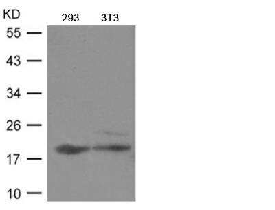
MYL9 Antibody [NB100-82061] - Western blot analysis of extracts from 293 and 3T3 cells
Applications for MYL9 Antibody - BSA Free
Application
Recommended Usage
Immunocytochemistry/ Immunofluorescence
1:100-1:200
Western Blot
1:500-1:1000
Formulation, Preparation, and Storage
Purification
Immunogen affinity purified
Formulation
PBS (without Mg2+ and Ca2+), pH 7.4, 150 mM NaCl, 50% glycerol
Format
BSA Free
Preservative
0.02% Sodium Azide
Concentration
1.0 mg/ml
Shipping
The product is shipped with polar packs. Upon receipt, store it immediately at the temperature recommended below.
Stability & Storage
Store at 4C short term. Aliquot and store at -20C long term. Avoid freeze-thaw cycles.
Background: MYL9
Alternate Names
LC2020 kDa myosin light chain, MLC2Myosin RLC, MRLC1MLC-2C, myosin regulatory light chain 1, Myosin regulatory light chain 2, smooth muscle isoform, Myosin regulatory light chain 9, Myosin regulatory light chain MRLC1, myosin regulatory light polypeptide 9, myosin, light chain 9, regulatory, myosin, light polypeptide 9, regulatory, MYRL2MGC3505
Entrez Gene IDs
363925 (Rat)
Gene Symbol
MYL9
UniProt
Additional MYL9 Products
Product Documents for MYL9 Antibody - BSA Free
Certificate of Analysis
To download a Certificate of Analysis, please enter a lot or batch number in the search box below.
Product Specific Notices for MYL9 Antibody - BSA Free
This product is for research use only and is not approved for use in humans or in clinical diagnosis. Primary Antibodies are guaranteed for 1 year from date of receipt.
Customer Reviews for MYL9 Antibody - BSA Free
There are currently no reviews for this product. Be the first to review MYL9 Antibody - BSA Free and earn rewards!
Have you used MYL9 Antibody - BSA Free?
Submit a review and receive an Amazon gift card!
$25/€18/£15/$25CAN/¥2500 Yen for a review with an image
$10/€7/£6/$10CAN/¥1110 Yen for a review without an image
Submit a review
Protocols
Find general support by application which include: protocols, troubleshooting, illustrated assays, videos and webinars.
- Appropriate Fixation of IHC/ICC Samples
- Cellular Response to Hypoxia Protocols
- ClariTSA™ Fluorophore Kits
- Detection & Visualization of Antibody Binding
- ICC Cell Smear Protocol for Suspension Cells
- ICC Immunocytochemistry Protocol Videos
- ICC for Adherent Cells
- Immunocytochemistry (ICC) Protocol
- Immunocytochemistry Troubleshooting
- Immunofluorescence of Organoids Embedded in Cultrex Basement Membrane Extract
- Immunohistochemistry (IHC) and Immunocytochemistry (ICC) Protocols
- Preparing Samples for IHC/ICC Experiments
- Preventing Non-Specific Staining (Non-Specific Binding)
- Primary Antibody Selection & Optimization
- Protocol for VisUCyte™ HRP Polymer Detection Reagent
- Protocol for the Fluorescent ICC Staining of Cell Smears - Graphic
- Protocol for the Fluorescent ICC Staining of Cultured Cells on Coverslips - Graphic
- Protocol for the Preparation and Fluorescent ICC Staining of Cells on Coverslips
- Protocol for the Preparation and Fluorescent ICC Staining of Non-adherent Cells
- Protocol for the Preparation and Fluorescent ICC Staining of Stem Cells on Coverslips
- Protocol for the Preparation of a Cell Smear for Non-adherent Cell ICC - Graphic
- R&D Systems Quality Control Western Blot Protocol
- TUNEL and Active Caspase-3 Detection by IHC/ICC Protocol
- The Importance of IHC/ICC Controls
- Troubleshooting Guide: Western Blot Figures
- Western Blot Conditions
- Western Blot Protocol
- Western Blot Protocol for Cell Lysates
- Western Blot Troubleshooting
- Western Blot Troubleshooting Guide
- View all Protocols, Troubleshooting, Illustrated assays and Webinars
Loading...