NFATC1/NFAT2 Antibody - BSA Free
Novus Biologicals | Catalog # NBP2-57739
![Immunocytochemistry/ Immunofluorescence: NFATC1/NFAT2 Antibody [NBP2-57739]](https://resources.rndsystems.com/images/products/NFATC1-NFAT2-Antibody-Immunocytochemistry-Immunofluorescence-NBP2-57739-img0001.jpg "Immunocytochemistry/ Immunofluorescence: NFATC1/NFAT2 Antibody [NBP2-57739]")
Loading...
Key Product Details
Species Reactivity
Validated:
Human
Cited:
Mouse
Applications
Validated:
Immunocytochemistry/ Immunofluorescence, Chromatin Immunoprecipitation-exo-Seq
Cited:
Western Blot
Label
Unconjugated
Antibody Source
Polyclonal Rabbit IgG
Format
BSA Free
Loading...
Product Specifications
Immunogen
This antibody was developed against a recombinant protein corresponding to the following amino acid sequence: GYGAALDGGPAGYFLSSGHTRPDGAPALESPRIEITSCLGLYHNNNQFFHDVEVEDVLPSSKRSPSTATLSLPSLEAYRD
Reactivity Notes
Rat 84%. Use in Mouse reported in secitific publication PMID: 32729236
Clonality
Polyclonal
Host
Rabbit
Isotype
IgG
Scientific Data Images for NFATC1/NFAT2 Antibody - BSA Free
Immunocytochemistry/ Immunofluorescence: NFATC1/NFAT2 Antibody [NBP2-57739]
Immunocytochemistry/Immunofluorescence: NFATC1/NFAT2 Antibody [NBP2-57739] - Staining of human cell line MCF7 shows localization to nucleoplasm & nuclear bodies.![NFATC1/NFAT2 Antibody - BSA Free Chromatin Immunoprecipitation-exo-Seq: NFATC1/NFAT2 Antibody - BSA Free [NBP2-57739]](https://resources.rndsystems.com/images/products/nbp2-57739_rabbit-polyclonal-nfatc1-nfat2-antibody-25620259103138.jpg "Chromatin Immunoprecipitation-exo-Seq: NFATC1/NFAT2 Antibody - BSA Free [NBP2-57739]")
Chromatin Immunoprecipitation-exo-Seq: NFATC1/NFAT2 Antibody - BSA Free [NBP2-57739]
ChIP-Exo-Seq composite graph for Anti-NFATC1 (NBP2-57739) tested in K562 cells. Strand-specific reads (blue: forward, red: reverse) and IgG controls (black: forward, grey: reverse) are plotted against the distance from a composite set of reference binding sites. The antibody exhibits robust target enrichment compared to a non-specific IgG control and precisely reveals its structural organization around the binding site. Data generated by Prof. B. F. Pugh´s Lab at Cornell University.
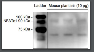
Western Blot: NFATC1/NFAT2 Antibody - BSA Free [NBP2-57739] -
Effects of low‐dose lithium supplementation on soleus and extensor digitorum longus (EDL) NFAT activation, PGC‐1 alpha and MHC isoform expression. (a) Representative Western blot images of phosphorylated and total nuclear factor activated T cells (p‐NFATc1 and NFATc1) and peroxisome proliferator‐activated receptor gamma coactivator 1‐alpha (PGC1‐ alpha ), all of which are downstream markers of calcineurin signaling. For pNFATc1, NFATc1, and PGC‐1 alpha, 7.5 μg of total protein was loaded into each well. (b) Quantification of p‐NFATc1 and NFATC1 were first normalized to ponceau prior to calculating the p‐NFATc1/NFATc1 ratio. (c) Quantification of PGC1‐ alpha content in soleus and EDL muscles in control‐fed and LiCl groups normalized to ponceau. (d) Representative Western blot images of MHC I and IIa in soleus muscles from control and lithium‐fed mice. For both isoforms, 5 μg of total protein was loaded into each well. Quantification of MHC I (e) and MHC IIa (f) normalized to ponceau. For (b,c) and (e,f), all data are expressed relative to control. *p <.05, significantly different from control, using a Student's t tests (n = 6–9 per group). All data are expressed as means +/- SD Image collected and cropped by CiteAb from the following open publication (https://pubmed.ncbi.nlm.nih.gov/32729236), licensed under a CC-BY license. Not internally tested by Novus Biologicals.Applications for NFATC1/NFAT2 Antibody - BSA Free
Application
Recommended Usage
Chromatin Immunoprecipitation-exo-Seq
1-10ug per reaction
Immunocytochemistry/ Immunofluorescence
0.25-2 ug/ml
Application Notes
ICC/IF, Fixation/Permeabilization: PFA/Triton X-100
Reviewed Applications
Read 1 review rated 3 using NBP2-57739 in the following applications:
Formulation, Preparation, and Storage
Purification
Affinity purified
Formulation
PBS (pH 7.2) and 40% Glycerol
Format
BSA Free
Preservative
0.02% Sodium Azide
Concentration
Concentrations vary lot to lot. See vial label for concentration. If unlisted please contact technical services.
Shipping
The product is shipped with polar packs. Upon receipt, store it immediately at the temperature recommended below.
Stability & Storage
Store at 4C short term. Aliquot and store at -20C long term. Avoid freeze-thaw cycles.
Background: NFATC1
Long Name
Nuclear Factor of Activated T Cells C1
Alternate Names
NFAT2
Gene Symbol
NFATC1
Additional NFATC1 Products
Product Documents for NFATC1/NFAT2 Antibody - BSA Free
Certificate of Analysis
To download a Certificate of Analysis, please enter a lot or batch number in the search box below.
Product Specific Notices for NFATC1/NFAT2 Antibody - BSA Free
This product is for research use only and is not approved for use in humans or in clinical diagnosis. Primary Antibodies are guaranteed for 1 year from date of receipt.
Citations for NFATC1/NFAT2 Antibody - BSA Free
 Powered by Bioz
Customer Reviews for NFATC1/NFAT2 Antibody - BSA Free (1)
3 out of 5
1 Customer Rating
Have you used NFATC1/NFAT2 Antibody - BSA Free?
Submit a review and receive an Amazon gift card!
$25/€18/£15/$25CAN/¥2500 Yen for a review with an image
$10/€7/£6/$10CAN/¥1110 Yen for a review without an image
Submit a review
Customer Images
Showing
1
-
1 of
1 review
Showing All
Filter By:
-
Application: Western BlotSample Tested: Mouse skeletal muscleSpecies: MouseVerified Customer | Posted 06/15/20171:2500 primary dilution overnight in milk 1:2000 secondary dilution 1 hour in milk

There are no reviews that match your criteria.
Protocols
Find general support by application which include: protocols, troubleshooting, illustrated assays, videos and webinars.
- Appropriate Fixation of IHC/ICC Samples
- Cellular Response to Hypoxia Protocols
- ClariTSA™ Fluorophore Kits
- Detection & Visualization of Antibody Binding
- ICC Cell Smear Protocol for Suspension Cells
- ICC Immunocytochemistry Protocol Videos
- ICC for Adherent Cells
- Immunocytochemistry (ICC) Protocol
- Immunocytochemistry Troubleshooting
- Immunofluorescence of Organoids Embedded in Cultrex Basement Membrane Extract
- Immunohistochemistry (IHC) and Immunocytochemistry (ICC) Protocols
- Preparing Samples for IHC/ICC Experiments
- Preventing Non-Specific Staining (Non-Specific Binding)
- Primary Antibody Selection & Optimization
- Protocol for VisUCyte™ HRP Polymer Detection Reagent
- Protocol for the Fluorescent ICC Staining of Cell Smears - Graphic
- Protocol for the Fluorescent ICC Staining of Cultured Cells on Coverslips - Graphic
- Protocol for the Preparation and Fluorescent ICC Staining of Cells on Coverslips
- Protocol for the Preparation and Fluorescent ICC Staining of Non-adherent Cells
- Protocol for the Preparation and Fluorescent ICC Staining of Stem Cells on Coverslips
- Protocol for the Preparation of a Cell Smear for Non-adherent Cell ICC - Graphic
- TUNEL and Active Caspase-3 Detection by IHC/ICC Protocol
- The Importance of IHC/ICC Controls
- View all Protocols, Troubleshooting, Illustrated assays and Webinars

