NFkB p105/p50 Antibody (2J10D7) - BSA Free
Novus Biologicals | Catalog # NB100-56583
Key Product Details
Species Reactivity
Validated:
Human, Rat, Rabbit
Cited:
Human, Rat, Rabbit
Applications
Validated:
Knockout Validated, Immunohistochemistry, Immunohistochemistry-Paraffin, Western Blot, Flow Cytometry, Simple Western
Cited:
IF/IHC
Label
Unconjugated
Antibody Source
Monoclonal Mouse IgG1 kappa Clone # 2J10D7
Format
BSA Free
Loading...
Product Specifications
Immunogen
A portion of amino acids 150-200 of human NF-kB (p50) was used as the immunogen.
Reactivity Notes
Immunogen displays the following percentage of sequence identity for non-tested species: Mouse (86%), Rat (86%). Rat reactivity reported in scientific literature (PMID:31850807).
Clonality
Monoclonal
Host
Mouse
Isotype
IgG1 kappa
Scientific Data Images for NFkB p105/p50 Antibody (2J10D7) - BSA Free
![Western Blot: NFkB p105/p50 Antibody (2J10D7)BSA Free [NB100-56583]](https://resources.rndsystems.com/images/products/NFkB-p105-p50-Antibody-2J10D7-Western-Blot-NB100-56583-img0004.jpg "Western Blot: NFkB p105/p50 Antibody (2J10D7)BSA Free [NB100-56583]")
Western Blot: NFkB p105/p50 Antibody (2J10D7)BSA Free [NB100-56583]
Western Blot: NFkB p105/p50 Antibody (2J10D7) [NB100-56583] - Analysis of p50 in HeLa lysate in the A) absence and B) presence of immunizing peptide using p50 antibody at 5 ug/ml.![Immunohistochemistry-Paraffin: NFkB p105/p50 Antibody (2J10D7) - BSA Free [NB100-56583]](https://resources.rndsystems.com/images/products/NFkB-p105-p50-Antibody-2J10D7-Immunohistochemistry-Paraffin-NB100-56583-img0009.jpg "Immunohistochemistry-Paraffin: NFkB p105/p50 Antibody (2J10D7) - BSA Free [NB100-56583]")
Immunohistochemistry-Paraffin: NFkB p105/p50 Antibody (2J10D7) - BSA Free [NB100-56583]
Immunohistochemistry-Paraffin: NFkB p105/p50 Antibody (2J10D7) [NB100-56583] - IHC-P of rabbit aorta using NB100-56583. Secondary antibody: anti-mouse histofine ( Nisherei Bioscience Inc. ref: 414131F). Development: DAB (Dako ref: K346811-2) and counterstained with Hematoxylin. Submitted via verified customer review![Flow Cytometry: NFkB p105/p50 Antibody (2J10D7) - BSA Free [NB100-56583]](https://resources.rndsystems.com/images/products/NFkB-p105-p50-Antibody-2J10D7-Flow-Cytometry-NB100-56583-img0008.jpg "Flow Cytometry: NFkB p105/p50 Antibody (2J10D7) - BSA Free [NB100-56583]")
Flow Cytometry: NFkB p105/p50 Antibody (2J10D7) - BSA Free [NB100-56583]
Flow Cytometry: NFkB p105/p50 Antibody (2J10D7) [NB100-56583] - An intracellular stain was performed on U937 cells with NFkB p105/p50 [2J10D7] Antibody NB100-56583C (blue) and a matched isotype control (orange). Cells were fixed with 4% PFA and then permeabilized with 0.1% saponin. Cells were incubated in an antibody dilution of 2.5 ug/mL for 30 minutes at room temperature. Both antibodies were conjugated to DyLight 650.![Immunohistochemistry-Paraffin: NFkB p105/p50 Antibody (2J10D7) - BSA Free [NB100-56583]](https://resources.rndsystems.com/images/products/NFkB-p105-p50-Antibody-2J10D7-Immunohistochemistry-Paraffin-NB100-56583-img0003.jpg "Immunohistochemistry-Paraffin: NFkB p105/p50 Antibody (2J10D7) - BSA Free [NB100-56583]")
Immunohistochemistry-Paraffin: NFkB p105/p50 Antibody (2J10D7) - BSA Free [NB100-56583]
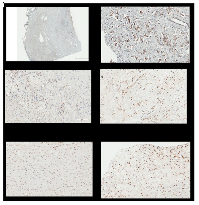
Immunohistochemistry-Paraffin: NFkB p105/p50 Antibody (2J10D7) [NB100-56583] - analysis of human breast tumor tissue using an isotype control antibody (top) and p50 antibody (bottom) at 5 ug/ml.![Flow Cytometry: NFkB p105/p50 Antibody (2J10D7) - BSA Free [NB100-56583]](https://resources.rndsystems.com/images/products/NFkB-p105-p50-Antibody-2J10D7-Flow-Cytometry-NB100-56583-img0005.jpg "Flow Cytometry: NFkB p105/p50 Antibody (2J10D7) - BSA Free [NB100-56583]")
Flow Cytometry: NFkB p105/p50 Antibody (2J10D7) - BSA Free [NB100-56583]
Flow Cytometry: NFkB p105/p50 Antibody (2J10D7) [NB100-56583] - An intracellular stain was performed on Jurkat cells with NFkB p105/p50 Antibody (2J10D7) NB100-56583C (blue) and a matched isotype control (orange). Cells were fixed with 4% PFA and then permeablized with 0.1% saponin. Cells were incubated in an antibody dilution of 2.5 ug/mL for 30 minutes. Both antibodies were conjugated to Dylight 650.![Flow Cytometry: NFkB p105/p50 Antibody (2J10D7) - BSA Free [NB100-56583]](https://resources.rndsystems.com/images/products/NFkB-p105-p50-Antibody-2J10D7-Flow-Cytometry-NB100-56583-img0006.jpg "Flow Cytometry: NFkB p105/p50 Antibody (2J10D7) - BSA Free [NB100-56583]")
Flow Cytometry: NFkB p105/p50 Antibody (2J10D7) - BSA Free [NB100-56583]
Flow Cytometry: NFkB p105/p50 Antibody (2J10D7) [NB100-56583] - An intracellular stain was performed on HeLa cells with NFkB p105/p50 Antibody (2J10D7) NB100-56583C (blue) and a matched isotype control (orange). Cells were fixed with 4% PFA and then permeablized with 0.1% saponin. Cells were incubated in an antibody dilution of 2.5 ug/mL for 30 minutes. Both antibodies were conjugated to Dylight 650.![Flow Cytometry: NFkB p105/p50 Antibody (2J10D7) - BSA Free [NB100-56583]](https://resources.rndsystems.com/images/products/NFkB-p105-p50-Antibody-2J10D7-Flow-Cytometry-NB100-56583-img0007.jpg "Flow Cytometry: NFkB p105/p50 Antibody (2J10D7) - BSA Free [NB100-56583]")
Flow Cytometry: NFkB p105/p50 Antibody (2J10D7) - BSA Free [NB100-56583]
Flow Cytometry: NFkB p105/p50 Antibody (2J10D7) [NB100-56583] - An intracellular stain was performed on U2-OS cells with NFkB p105/p50 [2J10D7] Antibody NB100-56583C (blue) and a matched isotype control (orange). Cells were fixed with 4% PFA and then permeabilized with 0.1% saponin. Cells were incubated in an antibody dilution of 2.5 ug/mL for 30 minutes at room temperature. Both antibodies were conjugated to DyLight 650.![NFkB p105/p50 Antibody (2J10D7) - BSA Free Immunohistochemistry-Paraffin: NFkB p105/p50 Antibody (2J10D7) - BSA Free [NB100-56583]](https://resources.rndsystems.com/images/products/antibody/nb100-56583_mouse-monoclonal-nfkb-p105-p50-antibody-2j10d7-imgenex-img-5812a-2210202585646.jpg "Immunohistochemistry-Paraffin: NFkB p105/p50 Antibody (2J10D7) - BSA Free [NB100-56583]")
Immunohistochemistry-Paraffin: NFkB p105/p50 Antibody (2J10D7) - BSA Free [NB100-56583]
Immunohistochemistry-Paraffin: NFkB p105/p50 Antibody (2J10D7) [NB100-56583] - Analysis of a FFPE tissue section of human lymph node using 1:200 dilution of NFkB p105/p50 (2J10D7) antibody. The staining was developed using HRP labeled anti-mouse secondary antibody and DAB reagent, and nuclei of cells were counter-stained with hematoxylin.Applications for NFkB p105/p50 Antibody (2J10D7) - BSA Free
Application
Recommended Usage
Immunohistochemistry
1:100 - 1:200
Immunohistochemistry-Paraffin
1:100 - 1:200
Simple Western
1:10 - 1:250
Western Blot
2-5 ug/ml
Application Notes
See Simple Western Antibody Database for Simple Western validation: Tested in HeLa Lysate, separated by Size, antibody dilution of 1:10, 1:50, 1:250, apparent MW was 56, 126 kDa
Reviewed Applications
Read 1 review rated 5 using NB100-56583 in the following applications:
Flow Cytometry Panel Builder
Bio-Techne Knows Flow Cytometry
Save time and reduce costly mistakes by quickly finding compatible reagents using the Panel Builder Tool.
Advanced Features
- Spectra Viewer - Custom analysis of spectra from multiple fluorochromes
- Spillover Popups - Visualize the spectra of individual fluorochromes
- Antigen Density Selector - Match fluorochrome brightness with antigen density

Formulation, Preparation, and Storage
Purification
Protein G purified
Formulation
PBS
Format
BSA Free
Preservative
0.05% Sodium Azide
Concentration
1.0 mg/ml
Shipping
The product is shipped with polar packs. Upon receipt, store it immediately at the temperature recommended below.
Stability & Storage
Store at 4C short term. Aliquot and store at -20C long term. Avoid freeze-thaw cycles.
Background: NFkB p105/p50
Alternate Names
DKFZp686C01211, DNA binding factor KBF1, DNA-binding factor KBF1, EBP-1, KBF1, NF-kappaB, NF-kappa-B, NF-kappabeta, NFKB-p105, NFKB-p50, nuclear factor kappa-B DNA binding subunit, nuclear factor NF-kappa-B p105 subunit, nuclear factor NF-kappa-B p50 subunit, nuclear factor of kappa light polypeptide gene enhancer in B-cells 1MGC54151, p105, p50
Entrez Gene IDs
4790 (Human)
Gene Symbol
NFKB1
UniProt
Additional NFkB p105/p50 Products
Product Documents for NFkB p105/p50 Antibody (2J10D7) - BSA Free
Certificate of Analysis
To download a Certificate of Analysis, please enter a lot or batch number in the search box below.
Product Specific Notices for NFkB p105/p50 Antibody (2J10D7) - BSA Free
This product is for research use only and is not approved for use in humans or in clinical diagnosis. Primary Antibodies are guaranteed for 1 year from date of receipt.
Citations for NFkB p105/p50 Antibody (2J10D7) - BSA Free
 Powered by Bioz
Customer Reviews for NFkB p105/p50 Antibody (2J10D7) - BSA Free (1)
5 out of 5
1 Customer Rating
Have you used NFkB p105/p50 Antibody (2J10D7) - BSA Free?
Submit a review and receive an Amazon gift card!
$25/€18/£15/$25CAN/¥2500 Yen for a review with an image
$10/€7/£6/$10CAN/¥1110 Yen for a review without an image
Submit a review
Customer Images
Showing
1
-
1 of
1 review
Showing All
Filter By:
-
Application: Immunohistochemistry-ParaffinSample Tested: aorta arterySpecies: RabbitVerified Customer | Posted 10/02/2017Antigen Retrieval: pressure cooker with solution Trilogy TM (Cell Marque ref: 920P-07). Antibody dilution: 0.07ug/mL (diluted 1/4000). Secondary antibody: anti-mouse histofine ( Nisherei Bioscience Inc. ref: 414131F). Development: DAB (Dako ref: K346811-2) and counterstained with Hematoxylin.

There are no reviews that match your criteria.
Protocols
View specific protocols for NFkB p105/p50 Antibody (2J10D7) - BSA Free (NB100-56583):
Immunohistochemistry-Paraffin Embedded Sections
Antigen Unmasking:
Bring slides to a boil in 10 mM sodium citrate buffer (pH 6.0) then maintain at a sub-boiling temperature for 10 minutes. Cool slides on bench-top for 30 minutes (keep slides in the sodium citrate buffer at all times).
Staining:
1. Wash sections in deionized water three times for 5 minutes each.
2. Wash sections in PBS for 5 minutes.
3. Block each section with 100-400 ul blocking solution (1% BSA in PBS) for 1 hour at room temperature.
4. Remove blocking solution and add 100-400 ul diluted primary antibody. Incubate overnight at 4 C.
5. Remove antibody solution and wash sections in wash buffer three times for 5 minutes each.
6. Add 100-400 ul HRP polymer conjugated secondary antibody. Incubate 30 minutes at room temperature.
7. Wash sections three times in wash buffer for 5 minutes each.
8. Add 100-400 ul DAB substrate to each section and monitor staining closely.
9. As soon as the sections develop, immerse slides in deionized water.
10. Counterstain sections in hematoxylin.
11. Wash sections in deionized water two times for 5 minutes each.
12. Dehydrate sections.
13. Mount coverslips.
Western Blot Protocol
1. Perform SDS-PAGE on samples to be analyzed, loading 10-25 ug of total protein per lane.
2. Transfer proteins to PVDF membrane according to the instructions provided by the manufacturer of the membrane and transfer apparatus.
3. Stain the membrane with Ponceau S (or similar product) to assess transfer success, and mark molecular weight standards where appropriate.
4. Rinse the blot TBS -0.05% Tween 20 (TBST).
5. Block the membrane in 5% Non-fat milk in TBST (blocking buffer) for at least 1 hour.
6. Wash the membrane in TBST three times for 10 minutes each.
7. Dilute primary antibody in blocking buffer and incubate overnight at 4C with gentle rocking.
8. Wash the membrane in TBST three times for 10 minutes each.
9. Incubate the membrane in diluted HRP conjugated secondary antibody in blocking buffer (as per manufacturer's instructions) for 1 hour at room temperature.
10. Wash the blot in TBST three times for 10 minutes each (this step can be repeated as required to reduce background).
11. Apply the detection reagent of choice in accordance with the manufacturer's instructions.
Find general support by application which include: protocols, troubleshooting, illustrated assays, videos and webinars.
- 7-Amino Actinomycin D (7-AAD) Cell Viability Flow Cytometry Protocol
- Antigen Retrieval Protocol (PIER)
- Antigen Retrieval for Frozen Sections Protocol
- Appropriate Fixation of IHC/ICC Samples
- Cellular Response to Hypoxia Protocols
- Chromogenic IHC Staining of Formalin-Fixed Paraffin-Embedded (FFPE) Tissue Protocol
- Chromogenic Immunohistochemistry Staining of Frozen Tissue
- ClariTSA™ Fluorophore Kits
- Detection & Visualization of Antibody Binding
- Extracellular Membrane Flow Cytometry Protocol
- Flow Cytometry Protocol for Cell Surface Markers
- Flow Cytometry Protocol for Staining Membrane Associated Proteins
- Flow Cytometry Staining Protocols
- Flow Cytometry Troubleshooting Guide
- Fluorescent IHC Staining of Frozen Tissue Protocol
- Graphic Protocol for Heat-induced Epitope Retrieval
- Graphic Protocol for the Preparation and Fluorescent IHC Staining of Frozen Tissue Sections
- Graphic Protocol for the Preparation and Fluorescent IHC Staining of Paraffin-embedded Tissue Sections
- Graphic Protocol for the Preparation of Gelatin-coated Slides for Histological Tissue Sections
- IHC Sample Preparation (Frozen sections vs Paraffin)
- Immunofluorescent IHC Staining of Formalin-Fixed Paraffin-Embedded (FFPE) Tissue Protocol
- Immunohistochemistry (IHC) and Immunocytochemistry (ICC) Protocols
- Immunohistochemistry Frozen Troubleshooting
- Immunohistochemistry Paraffin Troubleshooting
- Intracellular Flow Cytometry Protocol Using Alcohol (Methanol)
- Intracellular Flow Cytometry Protocol Using Detergents
- Intracellular Nuclear Staining Flow Cytometry Protocol Using Detergents
- Intracellular Staining Flow Cytometry Protocol Using Alcohol Permeabilization
- Intracellular Staining Flow Cytometry Protocol Using Detergents to Permeabilize Cells
- Preparing Samples for IHC/ICC Experiments
- Preventing Non-Specific Staining (Non-Specific Binding)
- Primary Antibody Selection & Optimization
- Propidium Iodide Cell Viability Flow Cytometry Protocol
- Protocol for Heat-Induced Epitope Retrieval (HIER)
- Protocol for Liperfluo
- Protocol for Making a 4% Formaldehyde Solution in PBS
- Protocol for VisUCyte™ HRP Polymer Detection Reagent
- Protocol for the Characterization of Human Th22 Cells
- Protocol for the Characterization of Human Th9 Cells
- Protocol for the Preparation & Fixation of Cells on Coverslips
- Protocol for the Preparation and Chromogenic IHC Staining of Frozen Tissue Sections
- Protocol for the Preparation and Chromogenic IHC Staining of Frozen Tissue Sections - Graphic
- Protocol for the Preparation and Chromogenic IHC Staining of Paraffin-embedded Tissue Sections
- Protocol for the Preparation and Chromogenic IHC Staining of Paraffin-embedded Tissue Sections - Graphic
- Protocol for the Preparation and Fluorescent IHC Staining of Frozen Tissue Sections
- Protocol for the Preparation and Fluorescent IHC Staining of Paraffin-embedded Tissue Sections
- Protocol for the Preparation of Gelatin-coated Slides for Histological Tissue Sections
- Protocol: Annexin V and PI Staining by Flow Cytometry
- Protocol: Annexin V and PI Staining for Apoptosis by Flow Cytometry
- R&D Systems Quality Control Western Blot Protocol
- TUNEL and Active Caspase-3 Detection by IHC/ICC Protocol
- The Importance of IHC/ICC Controls
- Troubleshooting Guide: Fluorokine Flow Cytometry Kits
- Troubleshooting Guide: Immunohistochemistry
- Troubleshooting Guide: Western Blot Figures
- Western Blot Conditions
- Western Blot Protocol
- Western Blot Protocol for Cell Lysates
- Western Blot Troubleshooting
- Western Blot Troubleshooting Guide
- View all Protocols, Troubleshooting, Illustrated assays and Webinars
Loading...