NK1R Antibody - BSA Free
Novus Biologicals | Catalog # NB300-119
![Western Blot: NK1R AntibodyBSA Free [NB300-119]](https://resources.rndsystems.com/images/products/NK1R-Antibody-Western-Blot-NB300-119-img0009.jpg "Western Blot: NK1R AntibodyBSA Free [NB300-119]")
Key Product Details
Species Reactivity
Validated:
Cited:
Predicted:
Applications
Validated:
Cited:
Label
Antibody Source
Format
Product Specifications
Immunogen
Reactivity Notes
Localization
Clonality
Host
Isotype
Theoretical MW
Disclaimer note: The observed molecular weight of the protein may vary from the listed predicted molecular weight due to post translational modifications, post translation cleavages, relative charges, and other experimental factors.
Scientific Data Images for NK1R Antibody - BSA Free
Western Blot: NK1R AntibodyBSA Free [NB300-119]
Western Blot: NK1R Antibody [NB300-119] - Neurokinin 1 Receptor Antibody [NB300-119] - Analysis of Neurokinin 1 Receptor in 1: human brain lysate, 2: rat brain lysate and 3: monkey brain lysate.![Immunocytochemistry/ Immunofluorescence: NK1R Antibody - BSA Free [NB300-119]](https://resources.rndsystems.com/images/products/NK1R-Antibody-Immunocytochemistry-Immunofluorescence-NB300-119-img0004.jpg "Immunocytochemistry/ Immunofluorescence: NK1R Antibody - BSA Free [NB300-119]")
Immunocytochemistry/ Immunofluorescence: NK1R Antibody - BSA Free [NB300-119]
Immunocytochemistry/Immunofluorescence: NK1R Antibody [NB300-119] - Neurokinin 1 Receptor Antibody [NB300-119] - Immunostain of NK1R in rat enteric glial cells. Image from verified customer review.![Immunohistochemistry: NK1R Antibody - BSA Free [NB300-119]](https://resources.rndsystems.com/images/products/NK1R-Antibody-Immunohistochemistry-NB300-119-img0006.jpg "Immunohistochemistry: NK1R Antibody - BSA Free [NB300-119]")
Immunohistochemistry: NK1R Antibody - BSA Free [NB300-119]
Immunohistochemistry: NK1R Antibody [NB300-119] - Neurokinin 1 Receptor Antibody [NB300-119] - Staining of human brain, basal nucleus of Meynert, neurons.![Western Blot: NK1R AntibodyBSA Free [NB300-119]](https://resources.rndsystems.com/images/products/NK1R-Antibody-Western-Blot-NB300-119-img0010.jpg "Western Blot: NK1R AntibodyBSA Free [NB300-119]")
Western Blot: NK1R AntibodyBSA Free [NB300-119]
Western Blot: NK1R Antibody [NB300-119] - Total protein from human, mouse and rat brain was separated on a 12% gel by SDS-PAGE, transferred to PVDF membrane and blocked in 5% non-fat milk in TBST. The membrane was probed with 2.0 ug/mL anti-NK1R in 1% non-fat milk in TBST and detected with an anti-rabbit HRP secondary antibody using chemiluminescence.![Western Blot: NK1R AntibodyBSA Free [NB300-119]](https://resources.rndsystems.com/images/products/NK1R-Antibody-Western-Blot-NB300-119-img0011.jpg "Western Blot: NK1R AntibodyBSA Free [NB300-119]")
Western Blot: NK1R AntibodyBSA Free [NB300-119]
NK1R-Antibody-Western-Blot-NB300-119-img0011.jpg
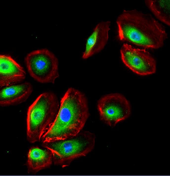
Immunocytochemistry/Immunofluorescence: NK1R Antibody [NB300-119] -
Immunocytochemistry/Immunofluorescence: NK1R Antibody [NB300-119] - Overnight incubation at 4C with NK1R antibody diluted 1:100 in PBS/BSA 1%. Red: Phalloidin, Blue: DAPI, Green: NK1R antibody. Image from verified customer review.
Western Blot: NK1R Antibody - BSA Free [NB300-119] -
Western Blot: NK1R Antibody - BSA Free [NB300-119] - TACR1 & its downstream targets SRC & p-SRC are expressed in neuroblastoma cell lines(A) Endogenous TACR1, SRC & p-SRC expression in whole-cell extracts of neuroblastoma cell lines was visualized by western blotting. GAPDH was used as a loading control. (B) Quantitative RT-PCR of TACR1 mRNA in a panel of neuroblastoma cell lines (n = 3, error bars indicate standard deviation). (C–E) Quantification of SRC (C), p-SRC (D) & TACR1 (E) protein expression using densitometry analysis of western immunoblots (n = 3, error bars indicate standard deviation). Image collected & cropped by CiteAb from the following publication (https://www.oncotarget.com/lookup/doi/10.18632/oncotarget.13440), licensed under a CC-BY license. Not internally tested by Novus Biologicals.
Western Blot: NK1R Antibody - BSA Free [NB300-119] -
Western Blot: NK1R Antibody - BSA Free [NB300-119] - Striatal beta 3 subunit protein reduced conditional KO mice. (A) Fluorescent images demonstrating colocalization between endogenous tdTomato fluorescence & DARPP-32 expression. Approximately half of the DARPP-32+ cells expressed endogenous tdTomato, indicative of Cre expression. Scale bar is 25 μm. (B) Representative western blot analysis of the GABAA receptor beta 3 subunit from individual 30-day-old mice revealed reduced amounts of beta 3 subunit protein in striatum (B) from Cre positive ( beta 3f/fDrd2) animals compared to Cre negative ( beta 3f/+ & beta 3f/f) control animals. The amount of beta 3 protein in cortex (C) did not differ between genotypes. Blots were reprobed for beta -actin. (D) Summary graph of western blot analysis demonstrating a significant reduction in beta 3 protein in striatum, but not cortex. Data are expressed as percent change in band intensity relative to Cre negative controls following normalization to actin. Image collected & cropped by CiteAb from the following publication (https://pubmed.ncbi.nlm.nih.gov/21847370), licensed under a CC-BY license. Not internally tested by Novus Biologicals.
Immunohistochemistry: NK1R Antibody - BSA Free [NB300-119] -
A An image of slides showing the hematoxyline eosin (HE)-stained and immunohistochemical analysis of neurokinin receptor 1 (NK1R)- and beta 2-adrenergic receptor (ADRB2)-stained cells in the pre-pandemic (upper row) and in-pandemic endometria (lower row) with corresponding positive controls (basal nucleus, Meynert neurons of human brain tissue for NK1R and melanoma for ADRB2) and negative controls. The immunoreactivities of NK1R B and ADRB2 C are shown as measured by the immunoreactive score (IRS) in pre-pandemic and in-pandemic endometria. The procedure of IRS measurement is described in methods. Mann–Whitney U test indicated that while no significance difference in the endometrial expression of NK1R was observed between the groups, ADRB2 expression in the endometria was significantly higher in the in-pandemic group than that in pre-pandemic group (p = 0.015) (C). The boxes represent the interquartile ranges and horizontal lines in the boxes represent median values. Scale bar = 50 μm for each slide Image collected and cropped by CiteAb from the following open publication (https://pubmed.ncbi.nlm.nih.gov/37142998), licensed under a CC-BY license. Not internally tested by Novus Biologicals.Applications for NK1R Antibody - BSA Free
Flow Cytometry
Immunocytochemistry/ Immunofluorescence
Immunohistochemistry
Immunohistochemistry-Frozen
Immunohistochemistry-Paraffin
Western Blot
Reviewed Applications
Read 3 reviews rated 4.3 using NB300-119 in the following applications:
Flow Cytometry Panel Builder
Bio-Techne Knows Flow Cytometry
Save time and reduce costly mistakes by quickly finding compatible reagents using the Panel Builder Tool.
Advanced Features
- Spectra Viewer - Custom analysis of spectra from multiple fluorochromes
- Spillover Popups - Visualize the spectra of individual fluorochromes
- Antigen Density Selector - Match fluorochrome brightness with antigen density

Formulation, Preparation, and Storage
Purification
Formulation
Format
Preservative
Concentration
Shipping
Stability & Storage
Background: NK1R
Long Name
Alternate Names
Gene Symbol
Additional NK1R Products
Product Documents for NK1R Antibody - BSA Free
Certificate of Analysis
To download a Certificate of Analysis, please enter a lot or batch number in the search box below.
Product Specific Notices for NK1R Antibody - BSA Free
This product is for research use only and is not approved for use in humans or in clinical diagnosis. Primary Antibodies are guaranteed for 1 year from date of receipt.
Citations for NK1R Antibody - BSA Free
 Powered by Bioz
Customer Reviews for NK1R Antibody - BSA Free (3)
Have you used NK1R Antibody - BSA Free?
Submit a review and receive an Amazon gift card!
$25/€18/£15/$25CAN/¥2500 Yen for a review with an image
$10/€7/£6/$10CAN/¥1110 Yen for a review without an image
Submit a review
Customer Images
-
Application: ImmunocytochemistrySample Tested: Cultured Human KeratinocytesSpecies: HumanVerified Customer | Posted 06/28/2023Overnight incubation at 4ºC with NK1R antibody diluted 1:100 in PBS/BSA 1% Red: Phalloidin Blue: DAPI Green: NK1R antibody

-
Application: Western BlotSample Tested: MouseSpecies: MouseVerified Customer | Posted 12/27/2011
-
Application: Immunohistochemistry-ParaffinSample Tested: Rat enteric glial cellsSpecies: RatVerified Customer | Posted 04/26/2010
There are no reviews that match your criteria.
Protocols
View specific protocols for NK1R Antibody - BSA Free (NB300-119):
Culture cells to appropriate density in 35 mm culture dishes or 6-well plates.
1. Remove culture medium and wash the cells briefly in PBS. Add 10% formalin to the dish and fix at room temperature for 10 minutes.
2. Remove the formalin and wash the cells in PBS.
3. Permeablize the cells with 0.1% Triton X100 or other suitable detergent for 10 min.
4. Remove the permeablization buffer and wash three times for 10 minutes each in PBS. Be sure to not let the specimen dry out.
5. To block nonspecific antibody binding, incubate in 10% normal goat serum from 1 hour to overnight at room temperature.
6. Add primary antibody at appropriate dilution and incubate overnight at 4C.
7. Remove primary antibody and replace with PBS. Wash three times for 10 minutes each.
8. Add secondary antibody at appropriate dilution. Incubate for 1 hour at room temperature.
9. Remove secondary antibody and replace with PBS. Wash three times for 10 minutes each.
10. Counter stain DNA with DAPi if required.
Antigen Unmasking:
Bring slides to a boil in 10 mM sodium citrate buffer (pH 6.0) then maintain at a sub-boiling temperature for 10 minutes. Cool slides on bench-top for 30 minutes (keep slides in the sodium citrate buffer at all times).
Staining:
1. Wash sections in deionized water three times for 5 minutes each.
2. Wash sections in PBS for 5 minutes.
3. Block each section with 100-400 ul blocking solution (1% BSA in PBS) for 1 hour at room temperature.
4. Remove blocking solution and add 100-400 ul diluted primary antibody. Incubate overnight at 4 C.
5. Remove antibody solution and wash sections in wash buffer three times for 5 minutes each.
6. Add 100-400 ul HRP polymer conjugated secondary antibody. Incubate 30 minutes at room temperature.
7. Wash sections three times in wash buffer for 5 minutes each.
8. Add 100-400 ul DAB substrate to each section and monitor staining closely.
9. As soon as the sections develop, immerse slides in deionized water.
10. Counterstain sections in hematoxylin.
11. Wash sections in deionized water two times for 5 minutes each.
12. Dehydrate sections.
13. Mount coverslips.
1. Perform SDS-PAGE on samples to be analyzed, loading 10-25 ug of total protein per lane.
2. Transfer proteins to PVDF membrane according to the instructions provided by the manufacturer of the membrane and transfer apparatus.
3. Stain the membrane with Ponceau S (or similar product) to assess transfer success, and mark molecular weight standards where appropriate.
4. Rinse the blot TBS -0.05% Tween 20 (TBST).
5. Block the membrane in 5% Non-fat milk in TBST (blocking buffer) for at least 1 hour.
6. Wash the membrane in TBST three times for 10 minutes each.
7. Dilute primary antibody in blocking buffer and incubate overnight at 4C with gentle rocking.
8. Wash the membrane in TBST three times for 10 minutes each.
9. Incubate the membrane in diluted HRP conjugated secondary antibody in blocking buffer (as per manufacturer's instructions) for 1 hour at room temperature.
10. Wash the blot in TBST three times for 10 minutes each (this step can be repeated as required to reduce background).
11. Apply the detection reagent of choice in accordance with the manufacturer's instructions.
Find general support by application which include: protocols, troubleshooting, illustrated assays, videos and webinars.
- 7-Amino Actinomycin D (7-AAD) Cell Viability Flow Cytometry Protocol
- Antigen Retrieval Protocol (PIER)
- Antigen Retrieval for Frozen Sections Protocol
- Appropriate Fixation of IHC/ICC Samples
- Cellular Response to Hypoxia Protocols
- Chromogenic IHC Staining of Formalin-Fixed Paraffin-Embedded (FFPE) Tissue Protocol
- Chromogenic Immunohistochemistry Staining of Frozen Tissue
- ClariTSA™ Fluorophore Kits
- Detection & Visualization of Antibody Binding
- Extracellular Membrane Flow Cytometry Protocol
- Flow Cytometry Protocol for Cell Surface Markers
- Flow Cytometry Protocol for Staining Membrane Associated Proteins
- Flow Cytometry Staining Protocols
- Flow Cytometry Troubleshooting Guide
- Fluorescent IHC Staining of Frozen Tissue Protocol
- Graphic Protocol for Heat-induced Epitope Retrieval
- Graphic Protocol for the Preparation and Fluorescent IHC Staining of Frozen Tissue Sections
- Graphic Protocol for the Preparation and Fluorescent IHC Staining of Paraffin-embedded Tissue Sections
- Graphic Protocol for the Preparation of Gelatin-coated Slides for Histological Tissue Sections
- ICC Cell Smear Protocol for Suspension Cells
- ICC Immunocytochemistry Protocol Videos
- ICC for Adherent Cells
- IHC Sample Preparation (Frozen sections vs Paraffin)
- Immunocytochemistry (ICC) Protocol
- Immunocytochemistry Troubleshooting
- Immunofluorescence of Organoids Embedded in Cultrex Basement Membrane Extract
- Immunofluorescent IHC Staining of Formalin-Fixed Paraffin-Embedded (FFPE) Tissue Protocol
- Immunohistochemistry (IHC) and Immunocytochemistry (ICC) Protocols
- Immunohistochemistry Frozen Troubleshooting
- Immunohistochemistry Paraffin Troubleshooting
- Intracellular Flow Cytometry Protocol Using Alcohol (Methanol)
- Intracellular Flow Cytometry Protocol Using Detergents
- Intracellular Nuclear Staining Flow Cytometry Protocol Using Detergents
- Intracellular Staining Flow Cytometry Protocol Using Alcohol Permeabilization
- Intracellular Staining Flow Cytometry Protocol Using Detergents to Permeabilize Cells
- Preparing Samples for IHC/ICC Experiments
- Preventing Non-Specific Staining (Non-Specific Binding)
- Primary Antibody Selection & Optimization
- Propidium Iodide Cell Viability Flow Cytometry Protocol
- Protocol for Heat-Induced Epitope Retrieval (HIER)
- Protocol for Liperfluo
- Protocol for Making a 4% Formaldehyde Solution in PBS
- Protocol for VisUCyte™ HRP Polymer Detection Reagent
- Protocol for the Characterization of Human Th22 Cells
- Protocol for the Characterization of Human Th9 Cells
- Protocol for the Fluorescent ICC Staining of Cell Smears - Graphic
- Protocol for the Fluorescent ICC Staining of Cultured Cells on Coverslips - Graphic
- Protocol for the Preparation & Fixation of Cells on Coverslips
- Protocol for the Preparation and Chromogenic IHC Staining of Frozen Tissue Sections
- Protocol for the Preparation and Chromogenic IHC Staining of Frozen Tissue Sections - Graphic
- Protocol for the Preparation and Chromogenic IHC Staining of Paraffin-embedded Tissue Sections
- Protocol for the Preparation and Chromogenic IHC Staining of Paraffin-embedded Tissue Sections - Graphic
- Protocol for the Preparation and Fluorescent ICC Staining of Cells on Coverslips
- Protocol for the Preparation and Fluorescent ICC Staining of Non-adherent Cells
- Protocol for the Preparation and Fluorescent ICC Staining of Stem Cells on Coverslips
- Protocol for the Preparation and Fluorescent IHC Staining of Frozen Tissue Sections
- Protocol for the Preparation and Fluorescent IHC Staining of Paraffin-embedded Tissue Sections
- Protocol for the Preparation of Gelatin-coated Slides for Histological Tissue Sections
- Protocol for the Preparation of a Cell Smear for Non-adherent Cell ICC - Graphic
- Protocol: Annexin V and PI Staining by Flow Cytometry
- Protocol: Annexin V and PI Staining for Apoptosis by Flow Cytometry
- R&D Systems Quality Control Western Blot Protocol
- TUNEL and Active Caspase-3 Detection by IHC/ICC Protocol
- The Importance of IHC/ICC Controls
- Troubleshooting Guide: Fluorokine Flow Cytometry Kits
- Troubleshooting Guide: Immunohistochemistry
- Troubleshooting Guide: Western Blot Figures
- Western Blot Conditions
- Western Blot Protocol
- Western Blot Protocol for Cell Lysates
- Western Blot Troubleshooting
- Western Blot Troubleshooting Guide
- View all Protocols, Troubleshooting, Illustrated assays and Webinars
FAQs for NK1R Antibody - BSA Free
-
Q: I have been trying this antibody for Western blot incortex and spinal cord from rat without results. Is there any reference where they have done WB with this antibody?
A:
At this time we do not have any references found where this has been described in WB, and of the references we have they are all for use in staining applications. We do have a product review however from a customer who observed positive results in WB as well as our own QC testing data that show positive staining in brain samples. This link (Product Specific Protocol) is also the recommended protocol for this one that may be helpful to you. If you have any difficulties with this one we would be happy to have the lab take a look at your results and compare them to our QC tests.
-
Q: One of our customers is willing to purchase this antibody, before placing the order she would like to know if it can be used for formalin-fix, paraffin-embedded sections in immunohistochemistry?
A: Yes, this product has been validated for use in IHC paraffin embedded sections.
-
Q: Our customer would like to know the MW that this antibody can detect in western blot.
A: The MW of this protein is 46KDa.
-
Q: What amino acid residues are recognized by the anti- Neurokinin 1 Receptor antibody NB300-119?
A: The exact immunogen sequence is deemed proprietary by the lab, and we have not epitope mapped this particular antibody. Therefore we do not know exactly where NB300-119 binds to its target. However, the immunogen was derived from the N-terminus of the human Neurokinin 1 Receptor protein. If there is a specific region that you are interested in targeting, or excluding, please send your desired sequence information to our Technical Support team at technical@novusbio.com, for comparison to the immunogen sequence used to synthesize the antibody.
-
Q: I have been trying this antibody for Western blot incortex and spinal cord from rat without results. Is there any reference where they have done WB with this antibody?
A:
At this time we do not have any references found where this has been described in WB, and of the references we have they are all for use in staining applications. We do have a product review however from a customer who observed positive results in WB as well as our own QC testing data that show positive staining in brain samples. This link (Product Specific Protocol) is also the recommended protocol for this one that may be helpful to you. If you have any difficulties with this one we would be happy to have the lab take a look at your results and compare them to our QC tests.
-
Q: One of our customers is willing to purchase this antibody, before placing the order she would like to know if it can be used for formalin-fix, paraffin-embedded sections in immunohistochemistry?
A: Yes, this product has been validated for use in IHC paraffin embedded sections.
-
Q: Our customer would like to know the MW that this antibody can detect in western blot.
A: The MW of this protein is 46KDa.
-
Q: What amino acid residues are recognized by the anti- Neurokinin 1 Receptor antibody NB300-119?
A: The exact immunogen sequence is deemed proprietary by the lab, and we have not epitope mapped this particular antibody. Therefore we do not know exactly where NB300-119 binds to its target. However, the immunogen was derived from the N-terminus of the human Neurokinin 1 Receptor protein. If there is a specific region that you are interested in targeting, or excluding, please send your desired sequence information to our Technical Support team at technical@novusbio.com, for comparison to the immunogen sequence used to synthesize the antibody.