Key Product Details
Species Reactivity
Human, Mouse, Rat
Applications
Immunohistochemistry, Immunohistochemistry-Paraffin, Flow Cytometry, Immunocytochemistry/ Immunofluorescence
Label
Unconjugated
Antibody Source
Monoclonal Mouse IgG1 kappa Clone # NM106
Loading...
Product Specifications
Immunogen
Nuclei of HL60 cells
Reactivity Notes
Does not react with Porcine.
Localization
Nuclei
Marker
Pan-Nuclear Marker
Specificity
This monoclonal antibody is an excellent marker for all nuclei in cells in tissues. It is a part of a new panel of reagents, which recognizes subcellular organelles or compartments of cells. These markers may be useful in identification of these organelles in cells, tissues, and biochemical preparations. This monoclonal antibody recognizes an antigen associated with the nuclei in all cells. It can be used to stain the nuclei in cell or tissue preparations and can be used as a nuclear marker in subcellular fractions. It produces a speckled pattern in normal and malignant cells and may be used to stain the nuclei of cells in fixed or frozen tissue sections. It can also be used with paraformaldehyde fixed frozen tissue or cell preparations.
Clonality
Monoclonal
Host
Mouse
Isotype
IgG1 kappa
Description
200ug/ml of antibody purified from Bioreactor Concentrate by Protein A or G. Prepared in 10 mM PBS with 0.05% BSA & 0.05% azide. Also available WITHOUT BSA & azide at 1.0 mg/ml. (NBP3-11613)
Antibody with azide - store at 2 to 8C. Antibody without azide - store at -20 to -80 C.
Antibody with azide - store at 2 to 8C. Antibody without azide - store at -20 to -80 C.
Scientific Data Images for Nuclear Antigen Antibody (NM106)
![Immunocytochemistry/ Immunofluorescence: Nuclear Antigen Antibody (NM106) [NBP2-45273]](https://resources.rndsystems.com/images/products/Nuclear-Antigen-Antibody-NM106-Immunocytochemistry-Immunofluorescence-NBP2-45273-img0007.jpg "Immunocytochemistry/ Immunofluorescence: Nuclear Antigen Antibody (NM106) [NBP2-45273]")
Immunocytochemistry/ Immunofluorescence: Nuclear Antigen Antibody (NM106) [NBP2-45273]
Immunocytochemistry/Immunofluorescence: Nuclear Antigen Antibody (NM106) [NBP2-45273] - Immunofluorescent staining of paraformaldehyde-fixed HeLa cells with Pan Nuclear Antigen Antibody followed by goat anti-Mouse IgG-CF488 (Green). Membranes are labeled with Phalloidin (Red).![Immunohistochemistry-Paraffin: Nuclear Antigen Antibody (NM106) [NBP2-45273]](https://resources.rndsystems.com/images/products/Nuclear-Antigen-Antibody-NM106-Immunohistochemistry-Paraffin-NBP2-45273-img0008.jpg "Immunohistochemistry-Paraffin: Nuclear Antigen Antibody (NM106) [NBP2-45273]")
Immunohistochemistry-Paraffin: Nuclear Antigen Antibody (NM106) [NBP2-45273]
Immunohistochemistry-Paraffin: Nuclear Antigen Antibody (NM106) [NBP2-45273] - Analysis of mouse testis tissue using Nuclear Antigen antibody. Primary antibody dilution: 0.5 ug/ml dilution. Image from verified customer review.![Immunohistochemistry-Paraffin: Nuclear Antigen Antibody (NM106) [NBP2-45273]](https://resources.rndsystems.com/images/products/Nuclear-Antigen-Antibody-NM106-Immunohistochemistry-Paraffin-NBP2-45273-img0002.jpg "Immunohistochemistry-Paraffin: Nuclear Antigen Antibody (NM106) [NBP2-45273]")
Immunohistochemistry-Paraffin: Nuclear Antigen Antibody (NM106) [NBP2-45273]
Immunohistochemistry-Paraffin: Nuclear Antigen Antibody (NM106) [NBP2-45273] - Human Tonsil stained with Pan Nuclear Antigen Antibody![Immunohistochemistry-Paraffin: Nuclear Antigen Antibody (NM106) [NBP2-45273]](https://resources.rndsystems.com/images/products/Nuclear-Antigen-Antibody-NM106-Immunohistochemistry-Paraffin-NBP2-45273-img0003.jpg "Immunohistochemistry-Paraffin: Nuclear Antigen Antibody (NM106) [NBP2-45273]")
Immunohistochemistry-Paraffin: Nuclear Antigen Antibody (NM106) [NBP2-45273]
Immunohistochemistry-Paraffin: Nuclear Antigen Antibody (NM106) [NBP2-45273] - Human Tonsil stained with Pan Nuclear Antigen Antibody.![Immunohistochemistry-Paraffin: Nuclear Antigen Antibody (NM106) [NBP2-45273]](https://resources.rndsystems.com/images/products/Nuclear-Antigen-Antibody-NM106-Immunohistochemistry-Paraffin-NBP2-45273-img0004.jpg "Immunohistochemistry-Paraffin: Nuclear Antigen Antibody (NM106) [NBP2-45273]")
Immunohistochemistry-Paraffin: Nuclear Antigen Antibody (NM106) [NBP2-45273]
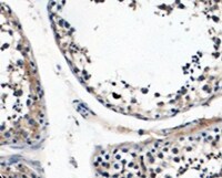
Immunohistochemistry-Paraffin: Nuclear Antigen Antibody (NM106) [NBP2-45273] - Rat Colon stained with Pan Nuclear Antigen Antibody.![Immunohistochemistry-Paraffin: Nuclear Antigen Antibody (NM106) [NBP2-45273]](https://resources.rndsystems.com/images/products/Nuclear-Antigen-Antibody-NM106-Immunohistochemistry-Paraffin-NBP2-45273-img0005.jpg "Immunohistochemistry-Paraffin: Nuclear Antigen Antibody (NM106) [NBP2-45273]")
Immunohistochemistry-Paraffin: Nuclear Antigen Antibody (NM106) [NBP2-45273]
Immunohistochemistry-Paraffin: Nuclear Antigen Antibody (NM106) [NBP2-45273] - Rat Lung stained with Pan Nuclear Antigen Antibody.Applications for Nuclear Antigen Antibody (NM106)
Application
Recommended Usage
Flow Cytometry
1-2 ug/million cells
Immunocytochemistry/ Immunofluorescence
1-2 ug/ml
Immunohistochemistry-Paraffin
1-2 ug/ml
Application Notes
Immunohistochemistry (Formalin-fixed): 1-2ug/ml for 30 minutes at RT. Staining of formalin-fixed tissues requires heating tissue sections in 10mM Tris with 1mM EDTA, pH 9.0, for 45 min at 95C followed by cooling at RT for 20 minutes.
Optimal dilution for a specific application should be determined.
Optimal dilution for a specific application should be determined.
Reviewed Applications
Read 1 review rated 5 using NBP2-45273 in the following applications:
Flow Cytometry Panel Builder
Bio-Techne Knows Flow Cytometry
Save time and reduce costly mistakes by quickly finding compatible reagents using the Panel Builder Tool.
Advanced Features
- Spectra Viewer - Custom analysis of spectra from multiple fluorochromes
- Spillover Popups - Visualize the spectra of individual fluorochromes
- Antigen Density Selector - Match fluorochrome brightness with antigen density

Formulation, Preparation, and Storage
Purification
Protein A or G purified
Formulation
10 mM PBS with 0.05% BSA
Preservative
0.05% Sodium Azide
Concentration
0.2 mg/ml
Shipping
The product is shipped with polar packs. Upon receipt, store it immediately at the temperature recommended below.
Stability & Storage
Store at 4C.
Background: Nuclear Antigen
Alternate Names
Nuclei, Nucleus
Additional Nuclear Antigen Products
Product Documents for Nuclear Antigen Antibody (NM106)
Certificate of Analysis
To download a Certificate of Analysis, please enter a lot or batch number in the search box below.
Product Specific Notices for Nuclear Antigen Antibody (NM106)
This product is for research use only and is not approved for use in humans or in clinical diagnosis. Primary Antibodies are guaranteed for 1 year from date of receipt.
Customer Reviews for Nuclear Antigen Antibody (NM106) (1)
5 out of 5
1 Customer Rating
Have you used Nuclear Antigen Antibody (NM106)?
Submit a review and receive an Amazon gift card!
$25/€18/£15/$25CAN/¥2500 Yen for a review with an image
$10/€7/£6/$10CAN/¥1110 Yen for a review without an image
Submit a review
Customer Images
Showing
1
-
1 of
1 review
Showing All
Filter By:
-
Application: Immunohistochemistry-ParaffinSample Tested: TestisSpecies: MouseVerified Customer | Posted 04/02/2022Mouse testis tissue0.5 ug/ml dilution

There are no reviews that match your criteria.
Protocols
Find general support by application which include: protocols, troubleshooting, illustrated assays, videos and webinars.
- 7-Amino Actinomycin D (7-AAD) Cell Viability Flow Cytometry Protocol
- Antigen Retrieval Protocol (PIER)
- Antigen Retrieval for Frozen Sections Protocol
- Appropriate Fixation of IHC/ICC Samples
- Cellular Response to Hypoxia Protocols
- Chromogenic IHC Staining of Formalin-Fixed Paraffin-Embedded (FFPE) Tissue Protocol
- Chromogenic Immunohistochemistry Staining of Frozen Tissue
- ClariTSA™ Fluorophore Kits
- Detection & Visualization of Antibody Binding
- Extracellular Membrane Flow Cytometry Protocol
- Flow Cytometry Protocol for Cell Surface Markers
- Flow Cytometry Protocol for Staining Membrane Associated Proteins
- Flow Cytometry Staining Protocols
- Flow Cytometry Troubleshooting Guide
- Fluorescent IHC Staining of Frozen Tissue Protocol
- Graphic Protocol for Heat-induced Epitope Retrieval
- Graphic Protocol for the Preparation and Fluorescent IHC Staining of Frozen Tissue Sections
- Graphic Protocol for the Preparation and Fluorescent IHC Staining of Paraffin-embedded Tissue Sections
- Graphic Protocol for the Preparation of Gelatin-coated Slides for Histological Tissue Sections
- ICC Cell Smear Protocol for Suspension Cells
- ICC Immunocytochemistry Protocol Videos
- ICC for Adherent Cells
- IHC Sample Preparation (Frozen sections vs Paraffin)
- Immunocytochemistry (ICC) Protocol
- Immunocytochemistry Troubleshooting
- Immunofluorescence of Organoids Embedded in Cultrex Basement Membrane Extract
- Immunofluorescent IHC Staining of Formalin-Fixed Paraffin-Embedded (FFPE) Tissue Protocol
- Immunohistochemistry (IHC) and Immunocytochemistry (ICC) Protocols
- Immunohistochemistry Frozen Troubleshooting
- Immunohistochemistry Paraffin Troubleshooting
- Intracellular Flow Cytometry Protocol Using Alcohol (Methanol)
- Intracellular Flow Cytometry Protocol Using Detergents
- Intracellular Nuclear Staining Flow Cytometry Protocol Using Detergents
- Intracellular Staining Flow Cytometry Protocol Using Alcohol Permeabilization
- Intracellular Staining Flow Cytometry Protocol Using Detergents to Permeabilize Cells
- Preparing Samples for IHC/ICC Experiments
- Preventing Non-Specific Staining (Non-Specific Binding)
- Primary Antibody Selection & Optimization
- Propidium Iodide Cell Viability Flow Cytometry Protocol
- Protocol for Heat-Induced Epitope Retrieval (HIER)
- Protocol for Liperfluo
- Protocol for Making a 4% Formaldehyde Solution in PBS
- Protocol for VisUCyte™ HRP Polymer Detection Reagent
- Protocol for the Characterization of Human Th22 Cells
- Protocol for the Characterization of Human Th9 Cells
- Protocol for the Fluorescent ICC Staining of Cell Smears - Graphic
- Protocol for the Fluorescent ICC Staining of Cultured Cells on Coverslips - Graphic
- Protocol for the Preparation & Fixation of Cells on Coverslips
- Protocol for the Preparation and Chromogenic IHC Staining of Frozen Tissue Sections
- Protocol for the Preparation and Chromogenic IHC Staining of Frozen Tissue Sections - Graphic
- Protocol for the Preparation and Chromogenic IHC Staining of Paraffin-embedded Tissue Sections
- Protocol for the Preparation and Chromogenic IHC Staining of Paraffin-embedded Tissue Sections - Graphic
- Protocol for the Preparation and Fluorescent ICC Staining of Cells on Coverslips
- Protocol for the Preparation and Fluorescent ICC Staining of Non-adherent Cells
- Protocol for the Preparation and Fluorescent ICC Staining of Stem Cells on Coverslips
- Protocol for the Preparation and Fluorescent IHC Staining of Frozen Tissue Sections
- Protocol for the Preparation and Fluorescent IHC Staining of Paraffin-embedded Tissue Sections
- Protocol for the Preparation of Gelatin-coated Slides for Histological Tissue Sections
- Protocol for the Preparation of a Cell Smear for Non-adherent Cell ICC - Graphic
- Protocol: Annexin V and PI Staining by Flow Cytometry
- Protocol: Annexin V and PI Staining for Apoptosis by Flow Cytometry
- TUNEL and Active Caspase-3 Detection by IHC/ICC Protocol
- The Importance of IHC/ICC Controls
- Troubleshooting Guide: Fluorokine Flow Cytometry Kits
- Troubleshooting Guide: Immunohistochemistry
- View all Protocols, Troubleshooting, Illustrated assays and Webinars
Loading...