Nucleolin Antibody - BSA Free
Novus Biologicals | Catalog # NB600-241
![Immunocytochemistry/ Immunofluorescence: Nucleolin Antibody [NB600-241]](https://resources.rndsystems.com/images/products/Nucleolin-Antibody-Immunocytochemistry-Immunofluorescence-NB600-241-img0008.jpg "Immunocytochemistry/ Immunofluorescence: Nucleolin Antibody [NB600-241]")
Key Product Details
Species Reactivity
Validated:
Cited:
Applications
Validated:
Cited:
Label
Antibody Source
Format
Product Specifications
Immunogen
Localization
Clonality
Host
Isotype
Theoretical MW
Disclaimer note: The observed molecular weight of the protein may vary from the listed predicted molecular weight due to post translational modifications, post translation cleavages, relative charges, and other experimental factors.
Scientific Data Images for Nucleolin Antibody - BSA Free
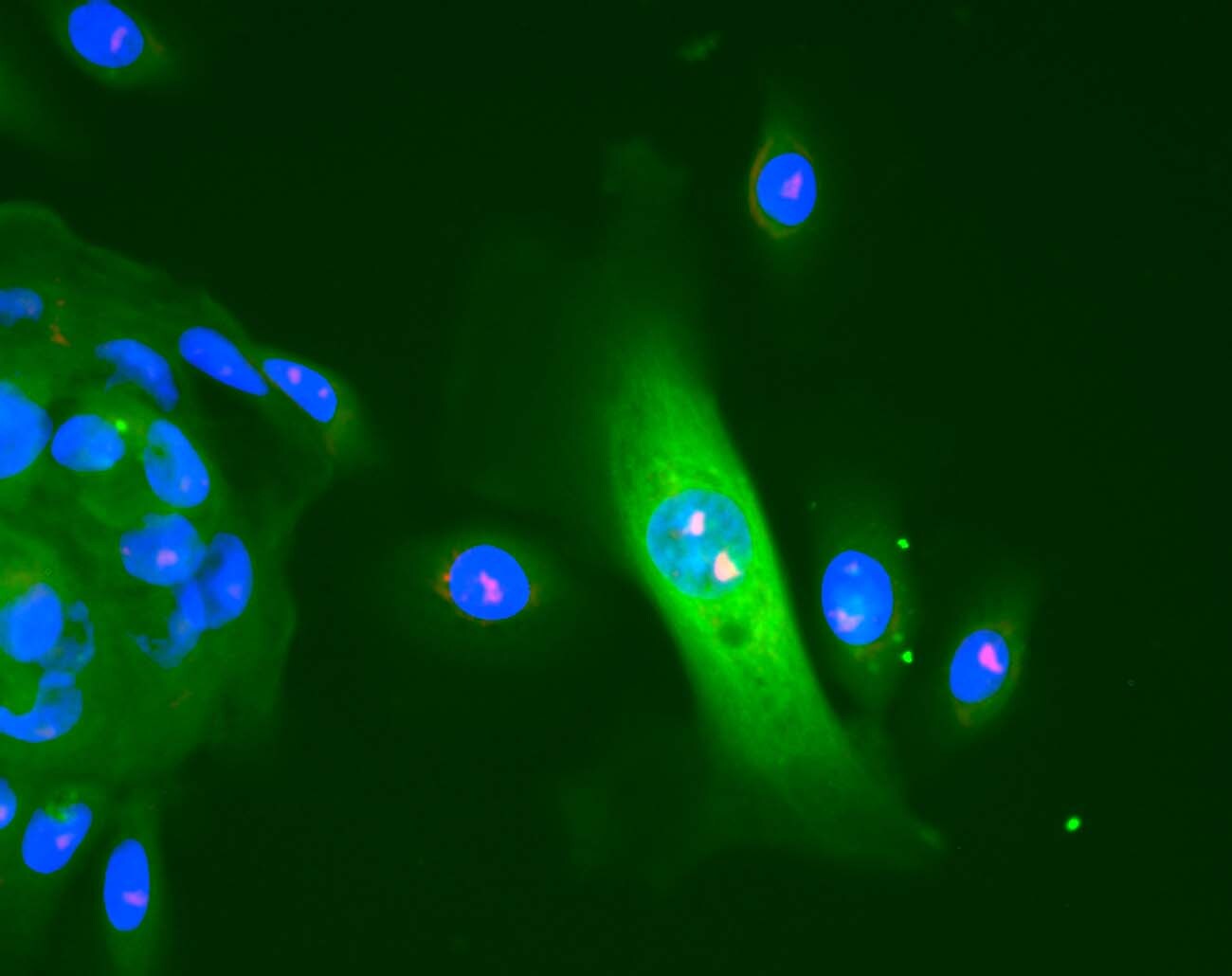
Immunocytochemistry/ Immunofluorescence: Nucleolin Antibody [NB600-241]
Immunocytochemistry/Immunofluorescence: Nucleolin Antibody [NB600-241] - Analysis of Nucleolin MCF-7 breast cancer cells. Image courtsey of product review by Lacey Litchfield.![Simple Western: Nucleolin Antibody [NB600-241]](https://resources.rndsystems.com/images/products/Nucleolin-Antibody-Simple-Western-NB600-241-img0009.jpg "Simple Western: Nucleolin Antibody [NB600-241]")
Simple Western: Nucleolin Antibody [NB600-241]
Simple Western: Nucleolin Antibody [NB600-241] - Image shows a specific band for Nucleolin in 0.5 mg/mL of HeLa lysate. This experiment was performed under reducing conditions using the 12-230 kDa separation system.![Immunocytochemistry/ Immunofluorescence: Nucleolin Antibody [NB600-241]](https://resources.rndsystems.com/images/products/Nucleolin-Antibody-Immunocytochemistry-Immunofluorescence-NB600-241-img0012.jpg "Immunocytochemistry/ Immunofluorescence: Nucleolin Antibody [NB600-241]")
Immunocytochemistry/ Immunofluorescence: Nucleolin Antibody [NB600-241]
Nucleolin-Antibody-Immunocytochemistry-Immunofluorescence-NB600-241-img0012.jpg![Western Blot: Nucleolin Antibody [NB600-241]](https://resources.rndsystems.com/images/products/Nucleolin-Antibody-Western-Blot-NB600-241-img0005.jpg "Western Blot: Nucleolin Antibody [NB600-241]")
Western Blot: Nucleolin Antibody [NB600-241]
Western Blot: Nucleolin Antibody [NB600-241] - Detection of nucleolin in crude PD31 nuclear extracts.![Immunocytochemistry/ Immunofluorescence: Nucleolin Antibody [NB600-241]](https://resources.rndsystems.com/images/products/Nucleolin-Antibody-Immunocytochemistry-Immunofluorescence-NB600-241-img0011.jpg "Immunocytochemistry/ Immunofluorescence: Nucleolin Antibody [NB600-241]")
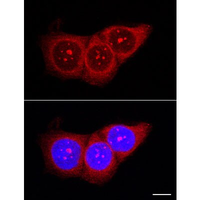
Immunocytochemistry/ Immunofluorescence: Nucleolin Antibody [NB600-241]
Immunocytochemistry/Immunofluorescence: Nucleolin Antibody [NB600-241] - Human prostate epithelial cells stained with anti-nucleolin (red), CFSE (green) retaining cell has increased nucleolin in nucleolus. DAPI (blue). Image from verified customer review.![Immunohistochemistry: Nucleolin Antibody [NB600-241]](https://resources.rndsystems.com/images/products/Nucleolin-Antibody-Immunohistochemistry-NB600-241-img0006.jpg "Immunohistochemistry: Nucleolin Antibody [NB600-241]")
Immunohistochemistry: Nucleolin Antibody [NB600-241]
Immunohistochemistry: Nucleolin Antibody [NB600-241] - Detection of Nucleolin in human tonsil germinal center.![Immunocytochemistry/ Immunofluorescence: Nucleolin Antibody [NB600-241]](https://resources.rndsystems.com/images/products/Nucleolin-Antibody-Immunocytochemistry-Immunofluorescence-NB600-241-img0007.jpg "Immunocytochemistry/ Immunofluorescence: Nucleolin Antibody [NB600-241]")
Immunocytochemistry/ Immunofluorescence: Nucleolin Antibody [NB600-241]
Immunocytochemistry/Immunofluorescence: Nucleolin Antibody [NB600-241] - In-situ immunofluorescent staining of PD31 murine pre-B cells.
Western Blot: Nucleolin Antibody [NB600-241] -
Western Blot: Nucleolin Antibody [NB600-241] - HT-LKO cells recapitulate the hallmarks of lamin A/C deficiency.(a) Quantitative PCR shows a >6-fold reduction (expressed as log2 fold change of ddCt value) of LMNA transcripts in different LKO colonies as compared to HT-WT clones; (b) Quantitative immunofluorescence shows a dramatic reduction of A-type lamin levels in HT-LKO colonies, approximating background levels (dotted white line); (c) Western blot for lamin A/C reveals the absence of both proteins in HT-LKO cells; (d) Immunostaining & nuclear counterstaining of HT-LKO cells reveals their aberrant nuclear morphology, virtual absence of lamin A (green) & local depletion of lamin B (red) (arrowheads) as opposed to HT-WT cells; (e) Time-lapse recordings after H2B-GFP transfection illustrate increased nuclear plasticity of HT-LKO vs. HT-WT cells, as evidenced by their larger projected area (Σ Area) & contour changes (Σ Outline) across time; (f) HT-LKO nuclei have a lower average nuclear circularity (dotted white line indicates a circularity of 0.85) & higher nuclear circularity fluctuations (average + 95% confidence interval) than HT-WT cells; The Y-axis has been cropped for clarity; (g) This translates into a significantly larger coefficient of variation (CoV) for the circularity across time (p < 0.001). (h) Nuclear ruptures occur more frequently in HT-LKO cells than in HT-WT cells (p < 0.001); (i) Representative montages of the NLS channel of a C4 HT-WT & L2 HT-LKO cell, with corresponding NLS/H2B ratio measurements (grey-coded bar plots). The moments of rupture events are indicated as red dots. Bar graphs reflect mean ± standard error (n = number of tracks). Image collected & cropped by CiteAb from the following publication (https://pubmed.ncbi.nlm.nih.gov/27461848), licensed under a CC-BY license. Not internally tested by Novus Biologicals.
Knockdown Validated: Nucleolin Antibody - BSA Free [NB600-241] -
Reduction of COUP-TFII or nucleolin decreases RAR beta 2 transcription in MCF-7 cells.MCF-7 (A) and T47D (B) cells were transfected with control siRNA or an siRNA targeting nucleolin for 48 h. T47D cells were treated with EtOH or 1 uM atRA for 24 h. Q-PCR for nucleolin (NCL) and RARB2. Values are the average of triplicates. C, Western blot showing COUP-TFII and RAR beta 2 expression after transfection with siCOUP-TFII. Values are relative to beta -actin. MCF-7 were transfected with siControl or siCOUP-TFII for 48 h and treated with 1 uM atRA for 6 h. Q-PCR was also performed for RRIG1. P<0.001 * versus control or ** versus atRA. Image collected and cropped by CiteAb from the following open publication (https://pubmed.ncbi.nlm.nih.gov/22693611), licensed under a CC-BY license. Not internally tested by Novus Biologicals.
Immunocytochemistry/ Immunofluorescence: Nucleolin Antibody - BSA Free [NB600-241] -
Endogenous nuclear nucleolin-COUP-TFII interaction in MCF-7 and T47D cells.NE (200 ug protein) from MCF-7 cells (A) and (400 ug protein) from T47D (B) cells were immunoprecipitated with COUP-TFII antibody or with rabbit IgG (negative control), followed by western blot analysis for nucleolin and COUP-TFII. 5% input NE serves as loading control. C, Immunofluorescent staining of endogenous COUP-TFII (green) and nucleolin (red) in the nuclei (Hoechst, blue) of MCF-7 cells. Merged images are shown at the right. Bar is 10 um. D, schematic representation of the N- terminal maltose binding protein (MBP)-tagged recombinant nucleolin proteins used for MBP pull-down assays. MBP was fused to the N-termini of the RNA binding domains (RBD) and/or the arginine/glycine-rich domain (RGG) of nucleolin. E, In vitro transcribed/translated COUP-TFII was incubated with the MBP-nucleolin fragments or MBP. Interacting proteins were captured with amylose resin. Eluted proteins were probed for COUP-TFII (top) and MBP (bottom, control). Image collected and cropped by CiteAb from the following open publication (https://pubmed.ncbi.nlm.nih.gov/22693611), licensed under a CC-BY license. Not internally tested by Novus Biologicals.Applications for Nucleolin Antibody - BSA Free
Chromatin Immunoprecipitation
Electron Microscopy
Immunocytochemistry/ Immunofluorescence
Immunohistochemistry
Immunohistochemistry Whole-Mount
Immunohistochemistry-Frozen
Immunohistochemistry-Paraffin
Simple Western
Western Blot
In Simple Western only 10 - 15 uL of the recommended dilution is used per data point.
See Simple Western Antibody Database for Simple Western validation: Tested in HeLa lysate 0.5 mg/mL, separated by Size, antibody dilution of 1:400, apparent MW was 134 kDa. Separated by Size-Wes, Sally Sue/Peggy Sue.
Reviewed Applications
Read 2 reviews rated 5 using NB600-241 in the following applications:
Formulation, Preparation, and Storage
Purification
Formulation
Format
Preservative
Concentration
Shipping
Stability & Storage
Background: Nucleolin
Alternate Names
Gene Symbol
UniProt
Additional Nucleolin Products
Product Documents for Nucleolin Antibody - BSA Free
Certificate of Analysis
To download a Certificate of Analysis, please enter a lot or batch number in the search box below.
Product Specific Notices for Nucleolin Antibody - BSA Free
This product is for research use only and is not approved for use in humans or in clinical diagnosis. Primary Antibodies are guaranteed for 1 year from date of receipt.
Citations for Nucleolin Antibody - BSA Free
 Powered by Bioz
Customer Reviews for Nucleolin Antibody - BSA Free (2)
Have you used Nucleolin Antibody - BSA Free?
Submit a review and receive an Amazon gift card!
$25/€18/£15/$25CAN/¥2500 Yen for a review with an image
$10/€7/£6/$10CAN/¥1110 Yen for a review without an image
Submit a review
Customer Images
-
Application: ImmunocytochemistrySample Tested: Human prostate cancer cellsSpecies: HumanVerified Customer | Posted 02/09/2017Human prostate epithelial cells stained with anti-nucleolin (red), CFSE (green) retaining cell has increased nucleolin in nucleolus. DAPI (blue).

-
Application: ImmunofluorescenceVerified Customer | Posted 08/22/2012
There are no reviews that match your criteria.
Protocols
View specific protocols for Nucleolin Antibody - BSA Free (NB600-241):
IHC-FFPE sections
I. Deparaffinization:
A. Treat slides with Xylene: 3 changes for 5 minutes each. Drain slides for 10 seconds between changes.
B. Treat slides with 100% Reagent Alcohol: 3 changes for 5 minutes each. Drain slides for 10 seconds between changes.
II. Quench Endogenous Peroxidase:
A. Place slides in peroxidase quenching solution: 15-30 minutes. To Prepare 200 ml of Quenching Solution: Add 3 ml of 30% Hydrogen Peroxide to 200 ml of Methanol.
Use within 4 hours of preparation
B. Place slides in distilled water: 2 changes for 2 minutes each.
III. Retrieve Epitopes:
A. Preheat Citrate Buffer. Place 200 ml of Citrate Buffer Working Solution into container, cover and place into steamer. Heat to 90-96 degrees Celsius.
B. Place rack of slides into hot Citrate Buffer for 20 minutes. Cover.
C. Carefully remove container with slides from steamer and cool on bench, uncovered, for 20 minutes.
D. Slowly add distilled water to further cool for 5 minutes.
E. Rinse slides with distilled water. 2 changes for 2 minutes each.
IV. Immunostaining Procedure:
A. Remove each slide from rack and circle tissue section with a hydrophobic barrier pen (e.g. Liquid Blocker-Super Pap Pen).
B. Flood slide with Wash Solution. Do not allow tissue sections to dry for the rest of the procedure.
C. Drain wash solution and apply 4 drops of Blocking Reagent to each slide and incubate for 15 minutes.
D. Drain Blocking Reagent (do not wash off the Blocking Reagent), apply 200 ul of Primary Antibody solution to each slide, and incubate for 1 hour.
E. Wash slides with Wash Solution: 3 changes for 5 minutes each.
F. Drain wash solution, apply 4 drops of Secondary antibody to each slide and incubate for 1 hour.
G. Wash slides with Wash Solution: 3 changes for 5 minutes each.
H. Drain wash solution, apply 4 drops of DAB Substrate to each slide and develop for 5-10 minutes. Check development with microscope.
I. Wash slides with Wash Solution: 3 changes for 5 minutes each.
J. Drain wash solution, apply 4 drops of Hematoxylin to each slide and stain for 1-3 minutes. Increase time if darker counterstaining is desired.
K. Wash slides with Wash Solution: 2-3 changes for 2 minutes each.
L. Drain wash solution and apply 4 drops of Bluing Solution to each slide for 1-2 minutes.
M. Rinse slides in distilled water.
N. Soak slides in 70% reagent alcohol: 3 minutes with intermittent agitation.
O. Soak slides in 95% reagent alcohol: 2 changes for 3 minutes each with intermittent agitation.
P. Soak slides in 100% reagent alcohol: 3 changes for 3 minutes each with intermittent agitation. Drain slides for 10 seconds between each change.
Q. Soak slides in Xylene: 3 changes for 3 minutes each with intermittent agitation. Drain slides for 10 seconds between each change.
R. Apply 2-3 drops of non-aqueous mounting media to each slide and mount coverslip.
S. Lay slides on a flat surface to dry prior to viewing under microscope.
NOTES:
-Use treated slides (e.g. HistoBond) to assure adherence of FFPE sections to slide.
-Prior to deparaffinization, heat slides overnight in a 60 degrees Celsius oven.
-All steps in which Xylene is used should be performed in a fume hood.
-For Epitope Retrieval, a microwave or pressure cooker may be substituted for the steamer method. Adjust times as necessary depending on conditions.
-For the initial IHC run with a new primary antibody, test tissues with and without Epitope Retrieval. In some instances, Epitope Retrieval may not be necessary.
-200 ul is the recommended maximum volume to apply to a slide for full coverage. Using more than 200 ul may allow solutions to wick off the slide and create drying artifacts. For small tissue sections less than 200 ul may be used.
-5 minutes of development with DAB Substrate should be sufficient. Do not develop for more than 10 minutes. If 5 minutes of development causes background staining, further dilution of the primary antibody may be necessary.
-Hematoxylin should produce a light nuclear counterstain so as not to obscure the DAB staining. Counterstain for 1-1.5 minutes for nuclear antigens. Counterstain for 2-3 minutes for cytoplasmic and membranous antigens. If darker counterstaining is desired increase time (up to 10 minutes).
Find general support by application which include: protocols, troubleshooting, illustrated assays, videos and webinars.
- Antigen Retrieval Protocol (PIER)
- Antigen Retrieval for Frozen Sections Protocol
- Appropriate Fixation of IHC/ICC Samples
- Cellular Response to Hypoxia Protocols
- ChIP Protocol Video
- Chromatin Immunoprecipitation (ChIP) Protocol
- Chromatin Immunoprecipitation Protocol
- Chromogenic IHC Staining of Formalin-Fixed Paraffin-Embedded (FFPE) Tissue Protocol
- Chromogenic Immunohistochemistry Staining of Frozen Tissue
- ClariTSA™ Fluorophore Kits
- Detection & Visualization of Antibody Binding
- Fluorescent IHC Staining of Frozen Tissue Protocol
- Graphic Protocol for Heat-induced Epitope Retrieval
- Graphic Protocol for the Preparation and Fluorescent IHC Staining of Frozen Tissue Sections
- Graphic Protocol for the Preparation and Fluorescent IHC Staining of Paraffin-embedded Tissue Sections
- Graphic Protocol for the Preparation of Gelatin-coated Slides for Histological Tissue Sections
- ICC Cell Smear Protocol for Suspension Cells
- ICC Immunocytochemistry Protocol Videos
- ICC for Adherent Cells
- IHC Sample Preparation (Frozen sections vs Paraffin)
- Immunocytochemistry (ICC) Protocol
- Immunocytochemistry Troubleshooting
- Immunofluorescence of Organoids Embedded in Cultrex Basement Membrane Extract
- Immunofluorescent IHC Staining of Formalin-Fixed Paraffin-Embedded (FFPE) Tissue Protocol
- Immunohistochemistry (IHC) and Immunocytochemistry (ICC) Protocols
- Immunohistochemistry Frozen Troubleshooting
- Immunohistochemistry Paraffin Troubleshooting
- Preparing Samples for IHC/ICC Experiments
- Preventing Non-Specific Staining (Non-Specific Binding)
- Primary Antibody Selection & Optimization
- Protocol for Heat-Induced Epitope Retrieval (HIER)
- Protocol for Making a 4% Formaldehyde Solution in PBS
- Protocol for VisUCyte™ HRP Polymer Detection Reagent
- Protocol for the Fluorescent ICC Staining of Cell Smears - Graphic
- Protocol for the Fluorescent ICC Staining of Cultured Cells on Coverslips - Graphic
- Protocol for the Preparation & Fixation of Cells on Coverslips
- Protocol for the Preparation and Chromogenic IHC Staining of Frozen Tissue Sections
- Protocol for the Preparation and Chromogenic IHC Staining of Frozen Tissue Sections - Graphic
- Protocol for the Preparation and Chromogenic IHC Staining of Paraffin-embedded Tissue Sections
- Protocol for the Preparation and Chromogenic IHC Staining of Paraffin-embedded Tissue Sections - Graphic
- Protocol for the Preparation and Fluorescent ICC Staining of Cells on Coverslips
- Protocol for the Preparation and Fluorescent ICC Staining of Non-adherent Cells
- Protocol for the Preparation and Fluorescent ICC Staining of Stem Cells on Coverslips
- Protocol for the Preparation and Fluorescent IHC Staining of Frozen Tissue Sections
- Protocol for the Preparation and Fluorescent IHC Staining of Paraffin-embedded Tissue Sections
- Protocol for the Preparation of Gelatin-coated Slides for Histological Tissue Sections
- Protocol for the Preparation of a Cell Smear for Non-adherent Cell ICC - Graphic
- R&D Systems Quality Control Western Blot Protocol
- TUNEL and Active Caspase-3 Detection by IHC/ICC Protocol
- The Importance of IHC/ICC Controls
- Troubleshooting Guide: Immunohistochemistry
- Troubleshooting Guide: Western Blot Figures
- Western Blot Conditions
- Western Blot Protocol
- Western Blot Protocol for Cell Lysates
- Western Blot Troubleshooting
- Western Blot Troubleshooting Guide
- View all Protocols, Troubleshooting, Illustrated assays and Webinars
FAQs for Nucleolin Antibody - BSA Free
-
Q: Have you tested NB600-241 on nucleus fraction or cytoplasmic fraction? Do you have any publication or data could support the signal only appear on nucleus?
A: Yes, it has been tested in nuclear extracts. Furthermore, it appears that a customer detected nucleolin in both the cytomplasm and the nucleus. Nucleolin appears to reside in both the cytoplasm and the nucleus (UniProt P19338) so it would be impossible to create an antibody that only detects nucleolin in just the nucleus. Our 10+ publications using this antibody can also be found on our website. These papers may provide further insight beyond our internal testing.