Numb Antibody - BSA Free
Novus Biologicals | Catalog # NB500-178
Key Product Details
Species Reactivity
Validated:
Cited:
Predicted:
Applications
Validated:
Cited:
Label
Antibody Source
Format
Product Specifications
Immunogen
Reactivity Notes
Localization
Specificity
Clonality
Host
Isotype
Scientific Data Images for Numb Antibody - BSA Free
![Immunocytochemistry/ Immunofluorescence: Numb Antibody [NB500-178]](https://resources.rndsystems.com/images/products/Numb-Antibody-Immunocytochemistry-Immunofluorescence-NB500-178-img0009.jpg "Immunocytochemistry/ Immunofluorescence: Numb Antibody [NB500-178]")
Immunocytochemistry/ Immunofluorescence: Numb Antibody [NB500-178]
Immunocytochemistry/Immunofluorescence: Numb Antibody [NB500-178] - Rat FR cells were fixed in 4% paraformaldehyde for 10 minutes and permeabilized in 0.05% Triton X-100 in PBS for 5 minutes. The cells were incubated with (NB500-178) at 1ug/ml overnight at 4C and detected with an anti-rabbit DyLight 488 (Green) at a 1:1000 dilution for 60 minutes. Nuclei were counterstained with DAPI (Blue). Cells were imaged using a 40X objective.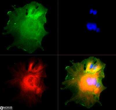
![Immunocytochemistry/ Immunofluorescence: Numb Antibody [NB500-178]](https://resources.rndsystems.com/images/products/Numb-Antibody-Immunocytochemistry-Immunofluorescence-NB500-178-img0007.jpg "Immunocytochemistry/ Immunofluorescence: Numb Antibody [NB500-178]")
Immunocytochemistry/ Immunofluorescence: Numb Antibody [NB500-178]
Immunocytochemistry/Immunofluorescence: Numb Antibody [NB500-178] - HepG2 cells were fixed in 4% paraformaldehyde for 10 minutes and permeabilized in 0.05% Triton X-100 in PBS for 5 minutes. The cells were incubated with Numb Antibody (NB500-178) at 1ug/ml overnight at 4C and detected with an anti-rabbit DyLight 488 (Green) at a 1:1000 dilution for 60 minutes. Nuclei were counterstained with DAPI (Blue). Cells were imaged using a 40X objective.![Simple Western: Numb Antibody [NB500-178]](https://resources.rndsystems.com/images/products/Numb-Antibody-Simple-Western-NB500-178-img0006.jpg "Simple Western: Numb Antibody [NB500-178]")
Simple Western: Numb Antibody [NB500-178]
Simple Western: Numb Antibody [NB500-178] - Simple Western lane view shows a specific band for NUMB in 0.5 mg/ml of A431 lysate. This experiment was performed under reducing conditions using the 12-230 kDa separation system.
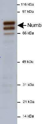
Western Blot: Numb Antibody - BSA Free [NB500-178] -
Characterisation of LNX1 interacting proteins.(A) Schematic diagram of the domain structure of LNX1p80 and LNX2 showing the RING and four PDZ domains. N represents the NUMB-binding NPAY/NPAF motif. (B) The ability of the indicated proteins to interact with transfected GFP-tagged LNX constructs was assessed in HEK 293 cells. For each interacting protein, top panels show western blots of cell lysates (Lys), while the bottom panels show the output of a GFP “pull down" assay (PD). In the panels on the left, the specificity of interactions for LNX1 versus LNX2 was assessed, while on the right the interaction site on LNX1 was mapped to individual protein domains. Binding of endogenous proteins to LNX was assessed for liprin alpha -1, KIF7 and NUMB. For the other proteins, interactions of transfected HA or GST epitope-tagged proteins were assessed. For AKAP13, the mapping to LNX domains was performed in two separate experiments. Successful expression and pull down of GFP-tagged LNX proteins was verified in all assays and representative “pull down" blots probed for GFP are shown. n = 2–3. Image collected and cropped by CiteAb from the following open publication (https://pubmed.ncbi.nlm.nih.gov/29121065), licensed under a CC-BY license. Not internally tested by Novus Biologicals.Applications for Numb Antibody - BSA Free
Immunocytochemistry/ Immunofluorescence
Simple Western
Western Blot
In Simple Western only 10 - 15 uL of the recommended dilution is used per data point.
See Simple Western Antibody Database for Simple Western validation: Tested in A431 lysate 0.5 mg/mL, separated by Size, antibody dilution of 1:200, apparent MW was 83 kDa. Separated by Size-Wes, Sally Sue/Peggy Sue.
Formulation, Preparation, and Storage
Purification
Formulation
Format
Preservative
Concentration
Shipping
Stability & Storage
Background: Numb
Alternate Names
Gene Symbol
Additional Numb Products
Product Documents for Numb Antibody - BSA Free
Certificate of Analysis
To download a Certificate of Analysis, please enter a lot or batch number in the search box below.
Product Specific Notices for Numb Antibody - BSA Free
This product is for research use only and is not approved for use in humans or in clinical diagnosis. Primary Antibodies are guaranteed for 1 year from date of receipt.
Related Research Areas
Citations for Numb Antibody - BSA Free
 Powered by Bioz
Customer Reviews for Numb Antibody - BSA Free
There are currently no reviews for this product. Be the first to review Numb Antibody - BSA Free and earn rewards!
Have you used Numb Antibody - BSA Free?
Submit a review and receive an Amazon gift card!
$25/€18/£15/$25CAN/¥2500 Yen for a review with an image
$10/€7/£6/$10CAN/¥1110 Yen for a review without an image
Submit a review
Protocols
View specific protocols for Numb Antibody - BSA Free (NB500-178):
Western Blot Protocol
1. Perform SDS-PAGE (4-12%) on samples to be analyzed, loading 20ug of total protein per lane.
2. Transfer proteins to Nitrocellulose according to the instructions provided by the manufacturer of the transfer apparatus.
3. Stain the blot using ponceau S for 1-2 minutes to access the transfer of proteins onto the nitrocellulose membrane. Rinse the blot in water to remove excess stain and mark the lane locations and locations of molecular weight markers using a pencil.
4. Rinse the blot in TBS for approximately 5 minutes.
5. Block the membrane using 5% non-fat dry milk in TBS for 1 hour.
6. Dilute the rabbit anti-Numb primary antibody (NB 500-178) in blocking buffer and incubate 2 hours at room temperature.
7. Wash the membrane in water for 5 minutes and apply the diluted rabbit-IgG HRP-conjugated secondary antibody in blocking buffer (as per manufacturer's instructions) and incubate 1 hour at room temperature.
8. Wash the blot in TBS containing 0.05-0.1% Tween-20 for 10-20 minutes.
9. Wash the blot in type I water for an additional 10-20 minutes (this step can be repeated as required to reduce background).
10. Apply the detection reagent of choice in accordance with the manufacturer's instructions (Amersham's ECL is the standard reagent used at Novus Biologicals).
Note: Tween-20 can be added to the blocking buffer at a final concentration of 0.05-0.2%, provided it does not interfere with antibody-antigen binding.
Find general support by application which include: protocols, troubleshooting, illustrated assays, videos and webinars.
- Appropriate Fixation of IHC/ICC Samples
- Cellular Response to Hypoxia Protocols
- ClariTSA™ Fluorophore Kits
- Detection & Visualization of Antibody Binding
- ICC Cell Smear Protocol for Suspension Cells
- ICC Immunocytochemistry Protocol Videos
- ICC for Adherent Cells
- Immunocytochemistry (ICC) Protocol
- Immunocytochemistry Troubleshooting
- Immunofluorescence of Organoids Embedded in Cultrex Basement Membrane Extract
- Immunohistochemistry (IHC) and Immunocytochemistry (ICC) Protocols
- Preparing Samples for IHC/ICC Experiments
- Preventing Non-Specific Staining (Non-Specific Binding)
- Primary Antibody Selection & Optimization
- Protocol for VisUCyte™ HRP Polymer Detection Reagent
- Protocol for the Fluorescent ICC Staining of Cell Smears - Graphic
- Protocol for the Fluorescent ICC Staining of Cultured Cells on Coverslips - Graphic
- Protocol for the Preparation and Fluorescent ICC Staining of Cells on Coverslips
- Protocol for the Preparation and Fluorescent ICC Staining of Non-adherent Cells
- Protocol for the Preparation and Fluorescent ICC Staining of Stem Cells on Coverslips
- Protocol for the Preparation of a Cell Smear for Non-adherent Cell ICC - Graphic
- R&D Systems Quality Control Western Blot Protocol
- TUNEL and Active Caspase-3 Detection by IHC/ICC Protocol
- The Importance of IHC/ICC Controls
- Troubleshooting Guide: Western Blot Figures
- Western Blot Conditions
- Western Blot Protocol
- Western Blot Protocol for Cell Lysates
- Western Blot Troubleshooting
- Western Blot Troubleshooting Guide
- View all Protocols, Troubleshooting, Illustrated assays and Webinars
FAQs for Numb Antibody - BSA Free
-
Q: I have looked at the data sheet for NB500-178 and do not see any reference to IP, but for some reason it was listed on our data sheet. Do you by chance have any reports of attempted use of the antibody in IP or any historical information regarding a listing for IP that might be helpful?
A: I pulled data on this target and I am not showing any notes of it being removed or ever being tested in that application. So unfortunately I have nothing to provide.
-
Q: I'm using your NUMB Antibody, cat# NB500-178. Does it pick up Numb Like protein also?
A: We performed an alignment of the immunogen sequence against the numb like protein for mouse based on UniProt. We show an approximately 50% similarity between the two sequences. As a result, we do not believe this antibody will recognize Numb-like protein. However, we have to stipulate that the lab has not tested the binding between the numb like protein and this antibody directly. Therefore, we cannot say with certainty whether the numb like protein will also be detected. Especially due to the fact that numb like protein has a similar molecular weight to one of the isoforms of the numb protein this antibody targets making it more difficult to analyze. Based on the sequence analysis information, it appears unlikely that the antibody detects numb like protein.
-
Q: I have looked at the data sheet for NB500-178 and do not see any reference to IP, but for some reason it was listed on our data sheet. Do you by chance have any reports of attempted use of the antibody in IP or any historical information regarding a listing for IP that might be helpful?
A: I pulled data on this target and I am not showing any notes of it being removed or ever being tested in that application. So unfortunately I have nothing to provide.
-
Q: I'm using your NUMB Antibody, cat# NB500-178. Does it pick up Numb Like protein also?
A: We performed an alignment of the immunogen sequence against the numb like protein for mouse based on UniProt. We show an approximately 50% similarity between the two sequences. As a result, we do not believe this antibody will recognize Numb-like protein. However, we have to stipulate that the lab has not tested the binding between the numb like protein and this antibody directly. Therefore, we cannot say with certainty whether the numb like protein will also be detected. Especially due to the fact that numb like protein has a similar molecular weight to one of the isoforms of the numb protein this antibody targets making it more difficult to analyze. Based on the sequence analysis information, it appears unlikely that the antibody detects numb like protein.
Associated Pathways
