Occludin Antibody - BSA Free
Novus Biologicals | Catalog # NBP1-87402
![Western Blot: Occludin Antibody [NBP1-87402]](https://resources.rndsystems.com/images/products/Occludin-Antibody-Western-Blot-NBP1-87402-img0020.jpg "Western Blot: Occludin Antibody [NBP1-87402]")
Loading...
Key Product Details
Validated by
Knockout/Knockdown, Orthogonal Validation
Species Reactivity
Validated:
Human
Cited:
Human, Mouse, Rat
Applications
Validated:
Immunohistochemistry, Immunohistochemistry-Paraffin, Western Blot, Knockdown Validated
Cited:
Immunohistochemistry, Immunohistochemistry-Paraffin, Western Blot, Immunocytochemistry/ Immunofluorescence, IF/IHC
Label
Unconjugated
Antibody Source
Polyclonal Rabbit IgG
Format
BSA Free
Loading...
Product Specifications
Immunogen
This antibody was developed against Recombinant Protein corresponding to amino acids: DKEHIYDEQPPNVEEWVKNVSAGTQDVPSPPSDYVERVDSPMAYSSNGKVNDKRFYPESSYKSTPVPEVVQELPLTSPVDDFRQPRYSSGGNFETPSKRAPAKGRAGRSKRTEQDHYETDYTTGGESCDELEED
Reactivity Notes
Rat reactivity reported in scientific literature (PMID: 31715313). Mouse reactivity reported in (PMID: 30685438), and a verified customer review.
Marker
Tight Junctions Marker
Clonality
Polyclonal
Host
Rabbit
Isotype
IgG
Scientific Data Images for Occludin Antibody - BSA Free
![Western Blot: Occludin Antibody [NBP1-87402]](https://resources.rndsystems.com/images/products/Occludin-Antibody-Western-Blot-NBP1-87402-img0009.jpg "Western Blot: Occludin Antibody [NBP1-87402]")
![Immunohistochemistry-Paraffin: Occludin Antibody [NBP1-87402]](https://resources.rndsystems.com/images/products/Occludin-Antibody-Immunohistochemistry-Paraffin-NBP1-87402-img0019.jpg "Immunohistochemistry-Paraffin: Occludin Antibody [NBP1-87402]")
Immunohistochemistry-Paraffin: Occludin Antibody [NBP1-87402]
Immunohistochemistry-Paraffin: Occludin Antibody [NBP1-87402] - Staining of human prostate shows negative membranous positivity in glandular cells as expected.![Immunohistochemistry-Paraffin: Occludin Antibody [NBP1-87402]](https://resources.rndsystems.com/images/products/Occludin-Antibody-Immunohistochemistry-Paraffin-NBP1-87402-img0016.jpg "Immunohistochemistry-Paraffin: Occludin Antibody [NBP1-87402]")
Immunohistochemistry-Paraffin: Occludin Antibody [NBP1-87402]
Immunohistochemistry-Paraffin: Occludin Antibody [NBP1-87402] - Staining of human breast cancer shows moderate membranous positivity in tumor cells.![Immunohistochemistry-Paraffin: Occludin Antibody [NBP1-87402]](https://resources.rndsystems.com/images/products/Occludin-Antibody-Immunohistochemistry-Paraffin-NBP1-87402-img0017.jpg "Immunohistochemistry-Paraffin: Occludin Antibody [NBP1-87402]")
Immunohistochemistry-Paraffin: Occludin Antibody [NBP1-87402]
Immunohistochemistry-Paraffin: Occludin Antibody [NBP1-87402] - Staining of human prostate cancer shows moderate membranous positivity in tumor cells.![Immunohistochemistry-Paraffin: Occludin Antibody [NBP1-87402]](https://resources.rndsystems.com/images/products/Occludin-Antibody-Immunohistochemistry-Paraffin-NBP1-87402-img0018.jpg "Immunohistochemistry-Paraffin: Occludin Antibody [NBP1-87402]")
Immunohistochemistry-Paraffin: Occludin Antibody [NBP1-87402]
Immunohistochemistry-Paraffin: Occludin Antibody [NBP1-87402] - Staining of human stomach shows moderate membranous positivity in glandular cells.
Western Blot: Occludin Antibody - BSA Free [NBP1-87402] -
KD improved vascular hyperpermeability and reduced vascular stiffness and leakage in type 2 diabetic mice. (A, B) The Evans blue injection and haematoxylin and eosin staining of abdominal aorta were performed for different groups, original magnification, ×10 (haematoxylin and eosin staining). Scale bar, 50 μm (haematoxylin and eosin staining). (C–H) The protein levels of vascular PES1, VEGF, VE‐cadherin, Ang‐1 and Occludin were detected by Immunoblotting. Data are represented as mean +/- SEM, each assay was performed independently three times (n = 12 per group). KD (ketogenic diet), SD (standard diet). **p < 0.01 C57BL/6J‐KD versus C57BL/6J‐SD, ***p < 0.001 C57BL/6J‐KD versus C57BL/6J‐SD, #p < 0.05 db/db‐KD versus db/db‐SD, ##p < 0.01 db/db‐KD versus db/db‐SD, ###p < 0.001 db/db‐KD versus db/db‐SD, +p < 0.05 C57BL/J‐SD versus db/db‐SD, ++p < 0.01 C57BL/J‐SD versus db/db‐SD, +++p < 0.001 C57BL/J‐SD versus db/db‐SD, &&p < 0.01 C57BL/6J‐KD versus db/db‐KD, &&&p < 0.001 C57BL/6J‐KD versus db/db‐KD (anova, Student–Newman–Keuls q‐test). Image collected and cropped by CiteAb from the following open publication (https://pubmed.ncbi.nlm.nih.gov/37060584), licensed under a CC-BY license. Not internally tested by Novus Biologicals.
Western Blot: Occludin Antibody - BSA Free [NBP1-87402] -
Pes1 knockout in mice decreased vascular permeability. (A, B) The Evans blue injection and haematoxylin and eosin staining of abdominal aorta were conducted in different groups, original magnification, ×10 (haematoxylin and eosin staining). Scale bar, 50 μm (haematoxylin and eosin staining). (C–F) The protein levels of vascular PES1, VEGF, VE‐cadherin, Ang‐1 and Occludin were measured by Immunoblotting. Data were represented as mean +/- SEM, each assay was performed independently three times. **p < 0.01, ***p < 0.001 compared with control (Student's t‐test). Image collected and cropped by CiteAb from the following open publication (https://pubmed.ncbi.nlm.nih.gov/37060584), licensed under a CC-BY license. Not internally tested by Novus Biologicals.
Western Blot: Occludin Antibody - BSA Free [NBP1-87402] -
beta ‐HB treatment impaired the increment of paracellular permeability by in vitro supplementation of Pes1. (A, B) The protein levels of PES1, VEGF, VE‐cadherin, Ang‐1 and Occludin in MVECs were detected by immunoblotting after Flag‐Pes1 plus beta ‐HB treatment. (C, D) Shown are immunofluorescence images of Flag‐Pes1 plus beta ‐HB‐treated MVECs for Occludin and VE‐cadherin expression and localizations, scale bar represents 20 μm. The nuclei were stained with DAPI. (E) Exhibited is the paracellular permeability in the cultured MVECs in different groups. Data were represented as mean +/- SEM, each experiment was performed independently three times. **p < 0.01, ***p < 0.001 compared with control (anova, Student–Newman–Keuls q‐test). Image collected and cropped by CiteAb from the following open publication (https://pubmed.ncbi.nlm.nih.gov/37060584), licensed under a CC-BY license. Not internally tested by Novus Biologicals.
Western Blot: Occludin Antibody - BSA Free [NBP1-87402] -
beta ‐HB treatment reduced vascular endothelial paracellular permeability in vitro. (A, B) The protein levels of PES1, VEGF, VE‐cadherin, Ang‐1 and Occludin in MVECs were detected by immunoblotting after 2 mM beta ‐HB treatment for 24 h. (C–H) Displayed are immunofluorescence images of beta ‐HB‐treated MVECs for VE‐cadherin, VEGF and PES1 expression and localizations, scale bar represents 20 μm. The nuclei were stained with DAPI. (I) Exhibited is the paracellular permeability in the cultured MVECs under different treatments. Ctrl (Control), beta ‐HB ( beta ‐hydroxybutyric acid). Data were represented as mean +/- SEM, each experiment was performed independently three times. **p < 0.01, ***p < 0.001 compared with control (Student's t‐test). Image collected and cropped by CiteAb from the following open publication (https://pubmed.ncbi.nlm.nih.gov/37060584), licensed under a CC-BY license. Not internally tested by Novus Biologicals.
Western Blot: Occludin Antibody - BSA Free [NBP1-87402] -
PLX3397 reduces tight junction expressionA, BWestern blot of lysates from PLX3397 treated b.End3 cells for tight junction proteins ZO‐1, Occludin and Claudin‐5 (A). The horizontal line indicates untreated cells, with increasing PLX3397 concentrations (5, 10, 20 μM). Corresponding densitometry is given in (B). (One‐way ANOVA with Dunnett’s post‐test for multiple comparisons, *P < 0.05, **P < 0.005, ***P < 0.0005, n = 3 independent experiments, error bars indicate SEM)CGene expression changes at 24 (top) and 48 (bottom) h in PLX3397 treated b.End3 cells shown by qPCR for Tjp1, Ocln and Cldn5 (*P < 0.05, **P < 0.006, n = 3 independent experiments one‐way ANOVA with Dunnett’s post‐test, error bars indicate SEM).DqPCR analysis of tight junction and CSF‐1R pathway gene expression changes at 24 h in PLX3397 treated MBECs (one‐way ANOVA with Dunnett’s post‐test for multiple comparisons, **P < 0.009, n = 3 independent experiments, error bars indicate SEM).EFITC‐4kDA transwell permeability assay of primary mouse microvascular endothelial cells (MBECs) treated for 24 h with PLX3397 at indicated doses (one‐way ANOVA with Dunnett’s correction, n = 3 technical replicates for flux assay, one‐way ANOVA with Dunnett’s post‐test for multiple comparisons, **P = 0.0012, error bars indicate SEM). Image collected and cropped by CiteAb from the following open publication (https://pubmed.ncbi.nlm.nih.gov/33350588), licensed under a CC-BY license. Not internally tested by Novus Biologicals.
Western Blot: Occludin Antibody - BSA Free [NBP1-87402] -
In vitro knockdown of Pes1 lowered the paracellular permeability of MVECs. (A, B) The protein levels of PES1, VEGF, VE‐cadherin, Ang‐1 and Occludin in MVECs were detected by immunoblotting after Pes1‐siRNA treatment. (C, D) Shown are immunofluorescence images of Pes1‐siRNA‐treated MVECs for Occludin and VE‐cadherin expression and localizations, scale bar represents 20 μm. The nuclei were stained with DAPI. (E) Exhibited is the paracellular permeability in the cultured MVECs in different groups. Data were represented as mean +/- SEM, each experiment was performed independently three times. **p < 0.01, ***p < 0.001 compared with control (Student's t‐test). Image collected and cropped by CiteAb from the following open publication (https://pubmed.ncbi.nlm.nih.gov/37060584), licensed under a CC-BY license. Not internally tested by Novus Biologicals.Applications for Occludin Antibody - BSA Free
Application
Recommended Usage
Immunohistochemistry
1:200 - 1:500
Immunohistochemistry-Paraffin
1:200 - 1:500
Western Blot
0.04 - 0.4 ug/mL
Application Notes
Reviews and literature that use this antibody in ICC/IF are from a previous lot. This antibody is not suitable for ICC/IF usage. IHC-Paraffin, HIER pH 6 retrieval is recommended
Reviewed Applications
Read 4 reviews rated 4 using NBP1-87402 in the following applications:
Formulation, Preparation, and Storage
Purification
Affinity purified
Formulation
PBS (pH 7.2) and 40% Glycerol
Format
BSA Free
Preservative
0.02% Sodium Azide
Concentration
Concentrations vary lot to lot. See vial label for concentration. If unlisted please contact technical services.
Shipping
The product is shipped with polar packs. Upon receipt, store it immediately at the temperature recommended below.
Stability & Storage
Store at 4C short term. Aliquot and store at -20C long term. Avoid freeze-thaw cycles.
Background: Occludin
Alternate Names
BLCPMG, OCLN
Gene Symbol
OCLN
Additional Occludin Products
Product Documents for Occludin Antibody - BSA Free
Certificate of Analysis
To download a Certificate of Analysis, please enter a lot or batch number in the search box below.
Product Specific Notices for Occludin Antibody - BSA Free
This product is for research use only and is not approved for use in humans or in clinical diagnosis. Primary Antibodies are guaranteed for 1 year from date of receipt.
Citations for Occludin Antibody - BSA Free
 Powered by Bioz
Customer Reviews for Occludin Antibody - BSA Free (4)
4 out of 5
4 Customer Ratings
Have you used Occludin Antibody - BSA Free?
Submit a review and receive an Amazon gift card!
$25/€18/£15/$25CAN/¥2500 Yen for a review with an image
$10/€7/£6/$10CAN/¥1110 Yen for a review without an image
Submit a review
Customer Images
Showing
1
-
4 of
4 reviews
Showing All
Filter By:
-
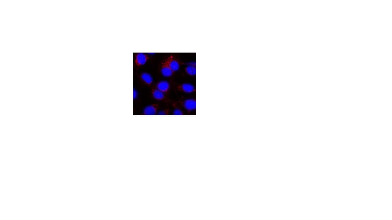
Application: ImmunocytochemistrySample Tested: IEC-6 rat intestinal epithelial cellsSpecies: RatVerified Customer | Posted 04/06/2022Occludin expression in IEC-6 cells

-
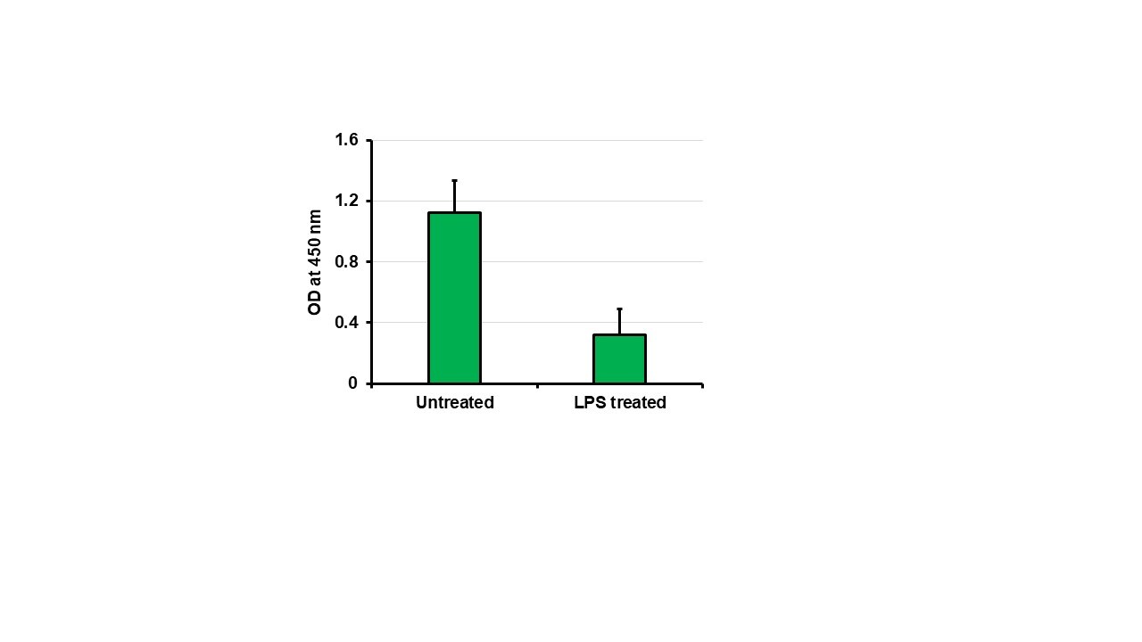
Application: ELISASample Tested: rat intestineSpecies: RatVerified Customer | Posted 06/01/2021Occludin levels in untreated and LPS treated rat intestinal tissues.
-
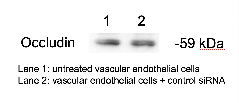
Application: Western BlotSample Tested: Primary lung endothelial cellsSpecies: MouseVerified Customer | Posted 06/15/2017Lane 1: untreated vascular endothelial cells Lane 2: vascular endothelial cells + control siRNAMembrane was blocked in 5% milk in TBST. Primary antibody dilution was 1:1000 and the dilution for the secondary ab was 1:5000.
-
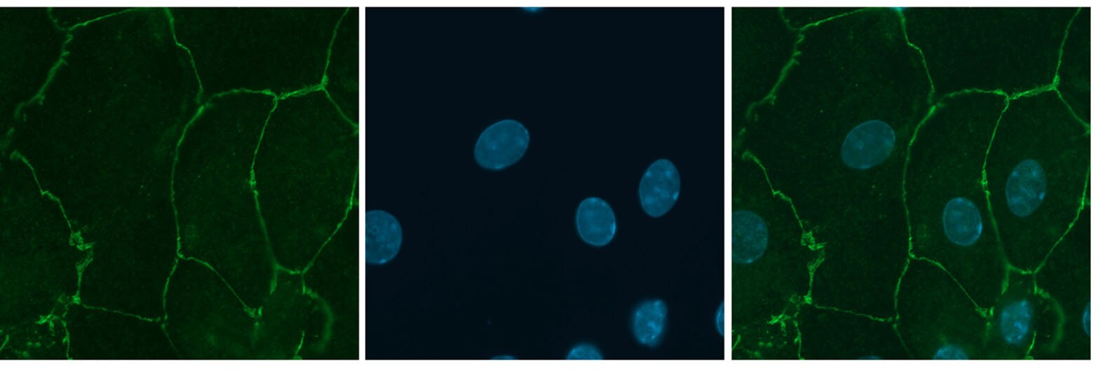
Application: ImmunofluorescenceSample Tested: Mouse choroid plexus cellsSpecies: MouseVerified Customer | Posted 04/06/2015Occludin staining in murine choroid plexus cells at 1:50 dilution (Methanol fixation)
There are no reviews that match your criteria.
Protocols
Find general support by application which include: protocols, troubleshooting, illustrated assays, videos and webinars.
- Antigen Retrieval Protocol (PIER)
- Antigen Retrieval for Frozen Sections Protocol
- Appropriate Fixation of IHC/ICC Samples
- Cellular Response to Hypoxia Protocols
- Chromogenic IHC Staining of Formalin-Fixed Paraffin-Embedded (FFPE) Tissue Protocol
- Chromogenic Immunohistochemistry Staining of Frozen Tissue
- ClariTSA™ Fluorophore Kits
- Detection & Visualization of Antibody Binding
- Fluorescent IHC Staining of Frozen Tissue Protocol
- Graphic Protocol for Heat-induced Epitope Retrieval
- Graphic Protocol for the Preparation and Fluorescent IHC Staining of Frozen Tissue Sections
- Graphic Protocol for the Preparation and Fluorescent IHC Staining of Paraffin-embedded Tissue Sections
- Graphic Protocol for the Preparation of Gelatin-coated Slides for Histological Tissue Sections
- IHC Sample Preparation (Frozen sections vs Paraffin)
- Immunofluorescent IHC Staining of Formalin-Fixed Paraffin-Embedded (FFPE) Tissue Protocol
- Immunohistochemistry (IHC) and Immunocytochemistry (ICC) Protocols
- Immunohistochemistry Frozen Troubleshooting
- Immunohistochemistry Paraffin Troubleshooting
- Preparing Samples for IHC/ICC Experiments
- Preventing Non-Specific Staining (Non-Specific Binding)
- Primary Antibody Selection & Optimization
- Protocol for Heat-Induced Epitope Retrieval (HIER)
- Protocol for Making a 4% Formaldehyde Solution in PBS
- Protocol for VisUCyte™ HRP Polymer Detection Reagent
- Protocol for the Preparation & Fixation of Cells on Coverslips
- Protocol for the Preparation and Chromogenic IHC Staining of Frozen Tissue Sections
- Protocol for the Preparation and Chromogenic IHC Staining of Frozen Tissue Sections - Graphic
- Protocol for the Preparation and Chromogenic IHC Staining of Paraffin-embedded Tissue Sections
- Protocol for the Preparation and Chromogenic IHC Staining of Paraffin-embedded Tissue Sections - Graphic
- Protocol for the Preparation and Fluorescent IHC Staining of Frozen Tissue Sections
- Protocol for the Preparation and Fluorescent IHC Staining of Paraffin-embedded Tissue Sections
- Protocol for the Preparation of Gelatin-coated Slides for Histological Tissue Sections
- R&D Systems Quality Control Western Blot Protocol
- TUNEL and Active Caspase-3 Detection by IHC/ICC Protocol
- The Importance of IHC/ICC Controls
- Troubleshooting Guide: Immunohistochemistry
- Troubleshooting Guide: Western Blot Figures
- Western Blot Conditions
- Western Blot Protocol
- Western Blot Protocol for Cell Lysates
- Western Blot Troubleshooting
- Western Blot Troubleshooting Guide
- View all Protocols, Troubleshooting, Illustrated assays and Webinars
Loading...
Associated Pathways


