ODF2 Antibody (1A1) - Azide and BSA Free
Novus Biologicals | Catalog # H00004957-M01
![Western Blot: ODF2 Antibody (1A1) [H00004957-M01]](https://resources.rndsystems.com/images/products/ODF2-Antibody-1A1-Western-Blot-H00004957-M01-img0006.jpg "Western Blot: ODF2 Antibody (1A1) [H00004957-M01]")
Loading...
Key Product Details
Species Reactivity
Validated:
Human, C. elegans
Cited:
Human, Mouse, Nematode - Caenorhabditis elegans
Applications
Validated:
Immunohistochemistry, Western Blot, ELISA, Immunocytochemistry/ Immunofluorescence
Cited:
Western Blot, Immunocytochemistry/ Immunofluorescence, IF/IHC
Label
Unconjugated
Antibody Source
Monoclonal Mouse IgG2a Kappa Clone # 1A1
Format
Azide and BSA Free
Loading...
Product Specifications
Immunogen
ODF2 (NP_002531.3, 706 a.a. ~ 804 a.a) partial recombinant protein with GST tag. MW of the GST tag alone is 26 KDa. KEHALSKERAAQNKILDLETQLSRTKTELSQLRRSRDDADRRYQSRLQDLKDRLEQSESTNRSMQNYVQFLKSSYANVFGDGPYSTFLTSSPIRSRSPP
Reactivity Notes
C. elegans reactivity reported in scientific literature (PMID: 24231678).
Specificity
ODF2 - outer dense fiber of sperm tails 2 (1A1)
Clonality
Monoclonal
Host
Mouse
Isotype
IgG2a Kappa
Description
Quality control test: Antibody Reactive Against Recombinant Protein.
Scientific Data Images for ODF2 Antibody (1A1) - Azide and BSA Free
Western Blot: ODF2 Antibody (1A1) [H00004957-M01]
Western Blot: ODF2 Antibody (1A1) [H00004957-M01] - Detection against Immunogen (36.63 KDa).![Immunocytochemistry/ Immunofluorescence: ODF2 Antibody (1A1) [H00004957-M01]](https://resources.rndsystems.com/images/products/ODF2-Antibody-1A1-Immunocytochemistry-Immunofluorescence-H00004957-M01-img0007.jpg "Immunocytochemistry/ Immunofluorescence: ODF2 Antibody (1A1) [H00004957-M01]")
Immunocytochemistry/ Immunofluorescence: ODF2 Antibody (1A1) [H00004957-M01]
ODF2-Antibody-1A1-Immunocytochemistry-Immunofluorescence-H00004957-M01-img0007.jpg![Immunocytochemistry/ Immunofluorescence: ODF2 Antibody (1A1) [H00004957-M01]](https://resources.rndsystems.com/images/products/ODF2-Antibody-1A1-Immunocytochemistry-Immunofluorescence-H00004957-M01-img0005.jpg "Immunocytochemistry/ Immunofluorescence: ODF2 Antibody (1A1) [H00004957-M01]")
Immunocytochemistry/ Immunofluorescence: ODF2 Antibody (1A1) [H00004957-M01]
Immunocytochemistry/Immunofluorescence: ODF2 Antibody (1A1) [H00004957-M01] - Co-staining of ODF2 (Cy3), DAPI and r-tubulin (Alexa Fluor 488) in RPE1 cell line. ODF2 stains the subdistal appendage of mother centriole. Cells were fixed with cold methanol, 10min. Blocked with 3% BSA for 30min at room temperature. ODF2 antibody was used at 1:100. Image from verified customer review.![Immunocytochemistry/ Immunofluorescence: ODF2 Antibody (1A1) [H00004957-M01]](https://resources.rndsystems.com/images/products/ODF2-Antibody-1A1-Immunocytochemistry-Immunofluorescence-H00004957-M01-img0004.jpg "Immunocytochemistry/ Immunofluorescence: ODF2 Antibody (1A1) [H00004957-M01]")
Immunocytochemistry/ Immunofluorescence: ODF2 Antibody (1A1) [H00004957-M01]
Immunocytochemistry/Immunofluorescence: ODF2 Antibody (1A1) [H00004957-M01] - Analysis of monoclonal antibody to ODF2 on HeLa cell. Antibody concentration 10 ug/ml. [H00004957-M01] -")
Immunocytochemistry/ Immunofluorescence: ODF2 Antibody (1A1) [H00004957-M01] -
Immunocytochemistry/ Immunofluorescence: ODF2 Antibody (1A1) [H00004957-M01] - The localization & function of DYF-19 are highly conservedEither endogenous (a) or overexpressed (b) FBF1, the mammalian homolog of worm DYF-19, localizes specifically on one centriole with a ring-like pattern in IMCD3 cells. c, FBF1 localizes at the ciliary base, above the basal body. d, In IMCD3 cells, FBF1 localizes above rootlet & subdistal appendage protein ODF2 & completely colocalizes with distal appendage protein CEP164. e, Immuno-EM demonstrates that FBF1 localizes specifically to distal appendages of mother centrioles. f–h, Knock-down of FBF1 leads to severely truncated cilia in most RNAi-treated hTERT-RPE cells. Data are represented as mean of 3 independent experiments (n=200) & error bars indicate s.d. Significant differences were identified by the Student’s t-test. *P<0.001. i & j, The IFT-B component IFT88, but not the IFT-A component IFT140, enters the truncated cilia of FBF1-knockdown hTERT-RPE cells. Arrows indicate the tips of truncated cilia. k, Endogenous IFT54 immunoprecipitates with FBF1 in hTERT-RPE cells. l, HEK293 cells were transiently transfected with FLAG-HA-tagged FBF1, & 48 hours later, cells were subjected to immunoprecipitation using normal mouse IgG (mIgG) or anti-IFT54 antibody. 50 μg protein were loaded into each lane. Bars: c, 1μm; e, 200 nm; others, 20 μm. Image collected & cropped by CiteAb from the following publication (https://pubmed.ncbi.nlm.nih.gov/24231678), licensed under a CC-BY license. Not internally tested by Novus Biologicals.Applications for ODF2 Antibody (1A1) - Azide and BSA Free
Application
Recommended Usage
Immunocytochemistry/ Immunofluorescence
1:10-1:500
Immunohistochemistry
1:10-1:500
Western Blot
1:500
Application Notes
Antibody reactivity against Recombinant Protein with GST tag on ELISA and WB. GST tag alone is used as a negative control. ICC/IF usage reported in scientific literature. IHC usage reported in scientific literature (PMID: 24231678).
Reviewed Applications
Read 2 reviews rated 5 using H00004957-M01 in the following applications:
Formulation, Preparation, and Storage
Purification
IgG purified
Formulation
In 1x PBS, pH 7.4
Format
Azide and BSA Free
Preservative
No Preservative
Concentration
Concentrations vary lot to lot. See vial label for concentration. If unlisted please contact technical services.
Shipping
The product is shipped with polar packs. Upon receipt, store it immediately at the temperature recommended below.
Stability & Storage
Aliquot and store at -20C or -80C. Avoid freeze-thaw cycles.
Background: ODF2
Alternate Names
Cenexin, FLJ44866, MGC111096, MGC9034, ODF2/1, ODF2/2, ODF8484-kD, outer dense fiber of sperm tails 2, outer dense fiber protein 2, sperm tail structural protein
Entrez Gene IDs
4957 (Human)
Gene Symbol
ODF2
UniProt
Additional ODF2 Products
Product Documents for ODF2 Antibody (1A1) - Azide and BSA Free
Certificate of Analysis
To download a Certificate of Analysis, please enter a lot or batch number in the search box below.
Product Specific Notices for ODF2 Antibody (1A1) - Azide and BSA Free
This product is produced by and distributed for Abnova, a company based in Taiwan.
This product is for research use only and is not approved for use in humans or in clinical diagnosis. Primary Antibodies are guaranteed for 1 year from date of receipt.
Citations for ODF2 Antibody (1A1) - Azide and BSA Free
 Powered by Bioz
Customer Reviews for ODF2 Antibody (1A1) - Azide and BSA Free (2)
5 out of 5
2 Customer Ratings
Have you used ODF2 Antibody (1A1) - Azide and BSA Free?
Submit a review and receive an Amazon gift card!
$25/€18/£15/$25CAN/¥2500 Yen for a review with an image
$10/€7/£6/$10CAN/¥1110 Yen for a review without an image
Submit a review
Customer Images
Showing
1
-
2 of
2 reviews
Showing All
Filter By:
-
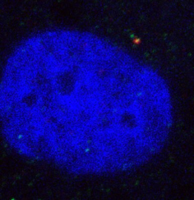
Application: ImmunofluorescenceSample Tested: RPE1 cells and hTERT RPE-1 cell line from ATCCSpecies: HumanVerified Customer | Posted 04/09/2019Co-staining of ODF2 (Cy3), DAPI and r-tubulin (Alexa Fluor 488) in RPE1 cell line. Cells were fixed with cold methanol. ODF2 antibody was used at 1:100. ODF2 stains the subdistal appendage of mother centriole.Fix by cold MEOH for 10 mins. Block with 3% BSA for 0.5 hours at room temperature. Dilute the ab at 1:100 in 3% BSA and incubate with cells for 2 hours at room temperature.

-
Application: ImmunocytochemistrySample Tested: hTERT RPE-1 cell line from ATCCSpecies: HumanVerified Customer | Posted 08/25/2010
There are no reviews that match your criteria.
Protocols
Find general support by application which include: protocols, troubleshooting, illustrated assays, videos and webinars.
- Antigen Retrieval Protocol (PIER)
- Antigen Retrieval for Frozen Sections Protocol
- Appropriate Fixation of IHC/ICC Samples
- Cellular Response to Hypoxia Protocols
- Chromogenic IHC Staining of Formalin-Fixed Paraffin-Embedded (FFPE) Tissue Protocol
- Chromogenic Immunohistochemistry Staining of Frozen Tissue
- ClariTSA™ Fluorophore Kits
- Detection & Visualization of Antibody Binding
- ELISA Sample Preparation & Collection Guide
- ELISA Troubleshooting Guide
- Fluorescent IHC Staining of Frozen Tissue Protocol
- Graphic Protocol for Heat-induced Epitope Retrieval
- Graphic Protocol for the Preparation and Fluorescent IHC Staining of Frozen Tissue Sections
- Graphic Protocol for the Preparation and Fluorescent IHC Staining of Paraffin-embedded Tissue Sections
- Graphic Protocol for the Preparation of Gelatin-coated Slides for Histological Tissue Sections
- How to Run an R&D Systems DuoSet ELISA
- How to Run an R&D Systems Quantikine ELISA
- How to Run an R&D Systems Quantikine™ QuicKit™ ELISA
- ICC Cell Smear Protocol for Suspension Cells
- ICC Immunocytochemistry Protocol Videos
- ICC for Adherent Cells
- IHC Sample Preparation (Frozen sections vs Paraffin)
- Immunocytochemistry (ICC) Protocol
- Immunocytochemistry Troubleshooting
- Immunofluorescence of Organoids Embedded in Cultrex Basement Membrane Extract
- Immunofluorescent IHC Staining of Formalin-Fixed Paraffin-Embedded (FFPE) Tissue Protocol
- Immunohistochemistry (IHC) and Immunocytochemistry (ICC) Protocols
- Immunohistochemistry Frozen Troubleshooting
- Immunohistochemistry Paraffin Troubleshooting
- Preparing Samples for IHC/ICC Experiments
- Preventing Non-Specific Staining (Non-Specific Binding)
- Primary Antibody Selection & Optimization
- Protocol for Heat-Induced Epitope Retrieval (HIER)
- Protocol for Making a 4% Formaldehyde Solution in PBS
- Protocol for VisUCyte™ HRP Polymer Detection Reagent
- Protocol for the Fluorescent ICC Staining of Cell Smears - Graphic
- Protocol for the Fluorescent ICC Staining of Cultured Cells on Coverslips - Graphic
- Protocol for the Preparation & Fixation of Cells on Coverslips
- Protocol for the Preparation and Chromogenic IHC Staining of Frozen Tissue Sections
- Protocol for the Preparation and Chromogenic IHC Staining of Frozen Tissue Sections - Graphic
- Protocol for the Preparation and Chromogenic IHC Staining of Paraffin-embedded Tissue Sections
- Protocol for the Preparation and Chromogenic IHC Staining of Paraffin-embedded Tissue Sections - Graphic
- Protocol for the Preparation and Fluorescent ICC Staining of Cells on Coverslips
- Protocol for the Preparation and Fluorescent ICC Staining of Non-adherent Cells
- Protocol for the Preparation and Fluorescent ICC Staining of Stem Cells on Coverslips
- Protocol for the Preparation and Fluorescent IHC Staining of Frozen Tissue Sections
- Protocol for the Preparation and Fluorescent IHC Staining of Paraffin-embedded Tissue Sections
- Protocol for the Preparation of Gelatin-coated Slides for Histological Tissue Sections
- Protocol for the Preparation of a Cell Smear for Non-adherent Cell ICC - Graphic
- Quantikine HS ELISA Kit Assay Principle, Alkaline Phosphatase
- Quantikine HS ELISA Kit Principle, Streptavidin-HRP Polymer
- R&D Systems Quality Control Western Blot Protocol
- Sandwich ELISA (Colorimetric) – Biotin/Streptavidin Detection Protocol
- Sandwich ELISA (Colorimetric) – Direct Detection Protocol
- TUNEL and Active Caspase-3 Detection by IHC/ICC Protocol
- The Importance of IHC/ICC Controls
- Troubleshooting Guide: ELISA
- Troubleshooting Guide: Immunohistochemistry
- Troubleshooting Guide: Western Blot Figures
- Western Blot Conditions
- Western Blot Protocol
- Western Blot Protocol for Cell Lysates
- Western Blot Troubleshooting
- Western Blot Troubleshooting Guide
- View all Protocols, Troubleshooting, Illustrated assays and Webinars
Loading...