
Loading...
Key Product Details
Species Reactivity
Mouse, Rat
Applications
Immunohistochemistry, Immunohistochemistry-Paraffin, Western Blot
Label
Unconjugated
Antibody Source
Polyclonal Goat IgG
Loading...
Product Specifications
Immunogen
A synthetic peptide from the cytoplasmic domain of mouse P2X3/P2RX3 conjugated to blue carrier protein was used as the antigen. The peptide is homologous in rat.
Clonality
Polyclonal
Host
Goat
Isotype
IgG
Applications for P2X3/P2RX3 Antibody
Application
Recommended Usage
Immunohistochemistry
1:300-1:2000
Immunohistochemistry-Paraffin
1:300-1:2000
Western Blot
1:300-1:2000
Reviewed Applications
Read 1 review rated 1 using NBP2-14838 in the following applications:
Formulation, Preparation, and Storage
Purification
Unpurified
Reconstitution
Reconstitute in 0.1 ml of sterile water. Centrifuge to remove any insoluble material. Glycerol may be added (1:1) for additional stability. Please note the sample size is provided in reconstituted format.
Formulation
Lyophilized from whole antisera
Preservative
No Preservative
Concentration
This product is unpurified. The exact concentration of antibody is not quantifiable.
Shipping
The product is shipped with polar packs. Upon receipt, store it immediately at the temperature recommended below.
Stability & Storage
Store at 4C short term. Aliquot and store at -20C long term. Avoid freeze-thaw cycles.
Calculators
Background: P2X3/P2RX3
Long Name
P2X Purinergic Receptor 3
Alternate Names
P2RX3
Entrez Gene IDs
5023 (Human)
Gene Symbol
P2RX1
Additional P2X3/P2RX3 Products
Product Documents for P2X3/P2RX3 Antibody
Certificate of Analysis
To download a Certificate of Analysis, please enter a lot or batch number in the search box below.
Product Specific Notices for P2X3/P2RX3 Antibody
This product is for research use only and is not approved for use in humans or in clinical diagnosis. Primary Antibodies are guaranteed for 1 year from date of receipt.
Customer Reviews for P2X3/P2RX3 Antibody (1)
1 out of 5
1 Customer Rating
Have you used P2X3/P2RX3 Antibody?
Submit a review and receive an Amazon gift card!
$25/€18/£15/$25CAN/¥2500 Yen for a review with an image
$10/€7/£6/$10CAN/¥1110 Yen for a review without an image
Submit a review
Customer Images
Showing
1
-
1 of
1 review
Showing All
Filter By:
-
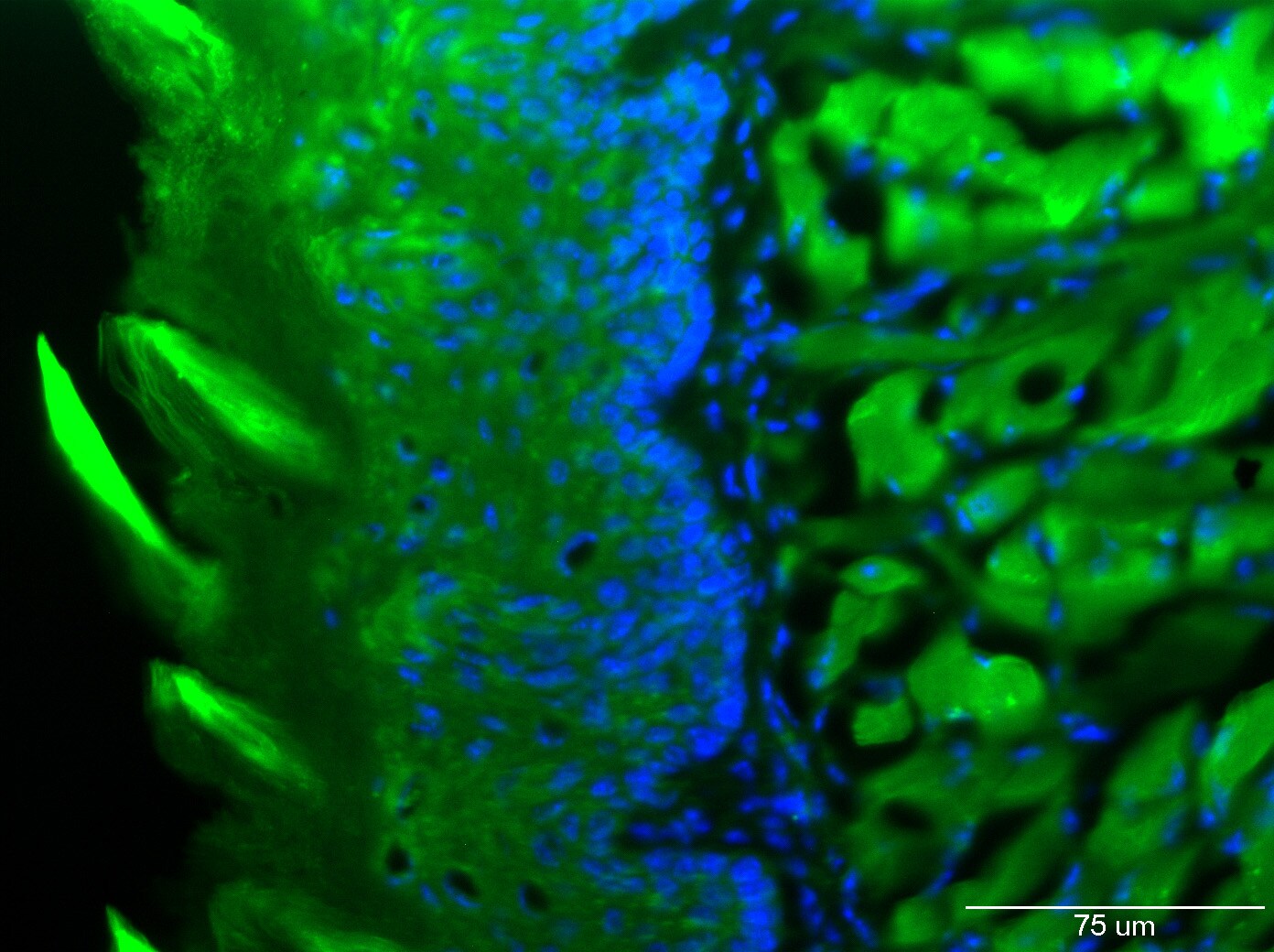
Application: Immunohistochemistry-FrozenSample Tested: mouse tongueSpecies: MouseVerified Customer | Posted 02/10/2020Mouse tongue, 20xTissue: Frozen mouse tongue Postfixed in 4% PFA Cyroprotected in 20% sucrose Cryosectioned at 8 um Protocol: · Rinse sections with PBS 3 x 3 min · Fix in ice-cold acetone – 5 min · Rinse 3X3 PBS · Block with 2% donkey serum in PBS · Primary antibody – overnight at 4 deg C o P2X3 at 1:10, 1:50, 1:100 or negative control/no antibody o Mix= 0.05% Triton + 0.2% carrageenan in PBS · Rinse PBS 3x3 min · Secondary antibody: Incubate 1 hour at RT o Alexa fluor donkey anti-goat 488 1:500 o Mix=0.05% Triton + 0.2% carrageenan in PBS Protect from light · Rinse PBS 3X3 min · Counterstain with DAPI – 5 min at room temp · Rinse PBS 3x3 min
 Bio-Techne ResponseThis review was submitted through the legacy Novus Innovators Program, reflecting a new species or application tested on a primary antibody.
There are no reviews that match your criteria.
Protocols
Find general support by application which include: protocols, troubleshooting, illustrated assays, videos and webinars.
- Antigen Retrieval Protocol (PIER)
- Antigen Retrieval for Frozen Sections Protocol
- Appropriate Fixation of IHC/ICC Samples
- Cellular Response to Hypoxia Protocols
- Chromogenic IHC Staining of Formalin-Fixed Paraffin-Embedded (FFPE) Tissue Protocol
- Chromogenic Immunohistochemistry Staining of Frozen Tissue
- ClariTSA™ Fluorophore Kits
- Detection & Visualization of Antibody Binding
- Fluorescent IHC Staining of Frozen Tissue Protocol
- Graphic Protocol for Heat-induced Epitope Retrieval
- Graphic Protocol for the Preparation and Fluorescent IHC Staining of Frozen Tissue Sections
- Graphic Protocol for the Preparation and Fluorescent IHC Staining of Paraffin-embedded Tissue Sections
- Graphic Protocol for the Preparation of Gelatin-coated Slides for Histological Tissue Sections
- IHC Sample Preparation (Frozen sections vs Paraffin)
- Immunofluorescent IHC Staining of Formalin-Fixed Paraffin-Embedded (FFPE) Tissue Protocol
- Immunohistochemistry (IHC) and Immunocytochemistry (ICC) Protocols
- Immunohistochemistry Frozen Troubleshooting
- Immunohistochemistry Paraffin Troubleshooting
- Preparing Samples for IHC/ICC Experiments
- Preventing Non-Specific Staining (Non-Specific Binding)
- Primary Antibody Selection & Optimization
- Protocol for Heat-Induced Epitope Retrieval (HIER)
- Protocol for Making a 4% Formaldehyde Solution in PBS
- Protocol for VisUCyte™ HRP Polymer Detection Reagent
- Protocol for the Preparation & Fixation of Cells on Coverslips
- Protocol for the Preparation and Chromogenic IHC Staining of Frozen Tissue Sections
- Protocol for the Preparation and Chromogenic IHC Staining of Frozen Tissue Sections - Graphic
- Protocol for the Preparation and Chromogenic IHC Staining of Paraffin-embedded Tissue Sections
- Protocol for the Preparation and Chromogenic IHC Staining of Paraffin-embedded Tissue Sections - Graphic
- Protocol for the Preparation and Fluorescent IHC Staining of Frozen Tissue Sections
- Protocol for the Preparation and Fluorescent IHC Staining of Paraffin-embedded Tissue Sections
- Protocol for the Preparation of Gelatin-coated Slides for Histological Tissue Sections
- R&D Systems Quality Control Western Blot Protocol
- TUNEL and Active Caspase-3 Detection by IHC/ICC Protocol
- The Importance of IHC/ICC Controls
- Troubleshooting Guide: Immunohistochemistry
- Troubleshooting Guide: Western Blot Figures
- Western Blot Conditions
- Western Blot Protocol
- Western Blot Protocol for Cell Lysates
- Western Blot Troubleshooting
- Western Blot Troubleshooting Guide
- View all Protocols, Troubleshooting, Illustrated assays and Webinars
Loading...