Key Product Details
Species Reactivity
Human, Canine, Equine, Feline, Rhesus Macaque
Applications
Immunohistochemistry-Paraffin, Immunohistochemistry-Frozen, Western Blot, Protein Array
Label
Unconjugated
Antibody Source
Monoclonal Mouse IgG1 Lambda Clone # PAX5/3735
Loading...
Product Specifications
Immunogen
A recombinant fragment (around aa 235-382) of human PAX5 protein (exact sequence is proprietary) (Uniprot: Q02548)
Reactivity Notes
Feline and Equine reactivity reported from a verified customer review.
Localization
Nuclear
Marker
Early B-Cell Marker
Specificity
The specificity of this monoclonal antibody to its intended target was validated by HuProtTM Array, containing more than 21,000, full-length human proteins. The PAX5 gene is a member of the paired box (PAX)family of transcription factors. The central feature of this gene family is a novel, highly conservedDNA-binding domain, known as the paired box. The PAX proteins are important regulators in early development, and alterations in the expression of their genes are thought to contribute toneoplastic transformation. The PAX5 gene encodes theB-celllineage specific activator protein (BSAP) that is expressed at early, but not late stages of B-cell differentiation. Its expression has also been detected in developing CNS and testis; therefore, PAX5 gene product may not only play an important role in B-cell differentiation, but also in neural development and spermatogenesis.
Clonality
Monoclonal
Host
Mouse
Isotype
IgG1 Lambda
Theoretical MW
42 kDa.
Disclaimer note: The observed molecular weight of the protein may vary from the listed predicted molecular weight due to post translational modifications, post translation cleavages, relative charges, and other experimental factors.
Disclaimer note: The observed molecular weight of the protein may vary from the listed predicted molecular weight due to post translational modifications, post translation cleavages, relative charges, and other experimental factors.
Description
200ug/ml of antibody purified from Bioreactor Concentrate by Protein A or G. Prepared in 10 mM PBS with 0.05% BSA & 0.05% azide. Also available WITHOUT BSA & azide at 1.0 mg/ml. (NBP3-08418)
Antibody with azide - store at 2 to 8C. Antibody without azide - store at -20 to -80C.
Antibody with azide - store at 2 to 8C. Antibody without azide - store at -20 to -80C.
Scientific Data Images for Pax5/BSAP Antibody (PAX5/3735)
![Western Blot: Pax5/BSAP Antibody (PAX5/3735) [NBP3-07581]](https://resources.rndsystems.com/images/products/Pax5-BSAP-Antibody-PAX5-3735-Western-Blot-NBP3-07581-img0004.jpg "Western Blot: Pax5/BSAP Antibody (PAX5/3735) [NBP3-07581]")
Western Blot: Pax5/BSAP Antibody (PAX5/3735) [NBP3-07581]
Western Blot: Pax5/BSAP Antibody (PAX5/3735) [NBP3-07581] - Western Blot Analysis of Raji cell lysate using Pax5/BSAP Mouse Monoclonal Antibody (PAX5/3735).![Immunohistochemistry-Paraffin: Pax5/BSAP Antibody (PAX5/3735) [NBP3-07581]](https://resources.rndsystems.com/images/products/Pax5-BSAP-Antibody-PAX5-3735-Immunohistochemistry-Paraffin-NBP3-07581-img0003.jpg "Immunohistochemistry-Paraffin: Pax5/BSAP Antibody (PAX5/3735) [NBP3-07581]")
Immunohistochemistry-Paraffin: Pax5/BSAP Antibody (PAX5/3735) [NBP3-07581]
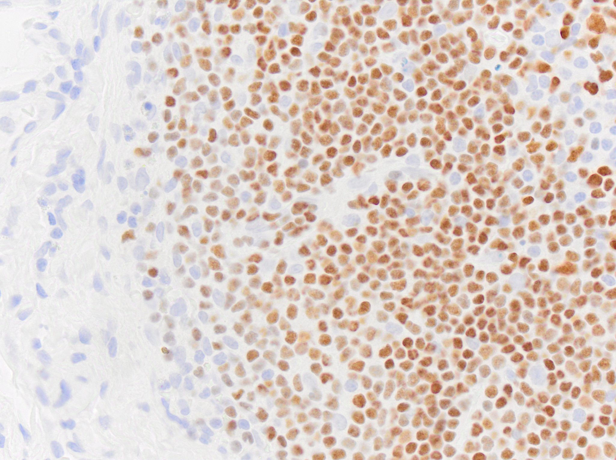
Immunohistochemistry-Paraffin: Pax5/BSAP Antibody (PAX5/3735) [NBP3-07581] - Formalin-fixed, paraffin-embedded human Tonsil stained with Pax5/BSAP Mouse Monoclonal Antibody (Pax5/BSAP/3735).![Immunohistochemistry-Paraffin: Pax5/BSAP Antibody (PAX5/3735) [NBP3-07581]](https://resources.rndsystems.com/images/products/Pax5-BSAP-Antibody-PAX5-3735-Immunohistochemistry-Paraffin-NBP3-07581-img0002.jpg "Immunohistochemistry-Paraffin: Pax5/BSAP Antibody (PAX5/3735) [NBP3-07581]")
Immunohistochemistry-Paraffin: Pax5/BSAP Antibody (PAX5/3735) [NBP3-07581]
Immunohistochemistry-Paraffin: Pax5/BSAP Antibody (PAX5/3735) [NBP3-07581] - Formalin-fixed, paraffin-embedded human Tonsil stained with Pax5/BSAP Mouse Monoclonal Antibody (Pax5/BSAP/3735).![Protein Array: Pax5/BSAP Antibody (PAX5/3735) [NBP3-07581]](https://resources.rndsystems.com/images/products/Pax5-BSAP-Antibody-PAX5-3735-Protein-Array-NBP3-07581-img0001.jpg "Protein Array: Pax5/BSAP Antibody (PAX5/3735) [NBP3-07581]")
Protein Array: Pax5/BSAP Antibody (PAX5/3735) [NBP3-07581]
Protein Array: Pax5/BSAP Antibody (PAX5/3735) [NBP3-07581] - Analysis of Protein Array containing more than 21,000 full-length human proteins using Pax5/BSAP Mouse Monoclonal Antibody (Pax5/BSAP/3735). [NBP3-07581] -")
Immunohistochemistry-paraffin: Mouse Monoclonal Pax5/BSAP Antibody (PAX5/3735) [NBP3-07581] -
Immunohistochemistry-paraffin: Mouse Monoclonal Pax5/BSAP Antibody (PAX5/3735) [NBP3-07581] - Pax5/BSAP immunoreactivity in a dog lymph node. Primary was used at 0.5ug/mL and was incubated with the tissue section for 30m at room temperature. Secondary was Horse Anti-Mouse HRP polymer. Image from a verified customer review. [NBP3-07581] -")
Immunohistochemistry-paraffin: Mouse Monoclonal Pax5/BSAP Antibody (PAX5/3735) [NBP3-07581] -
Immunohistochemistry-paraffin: Mouse Monoclonal Pax5/BSAP Antibody (PAX5/3735) [NBP3-07581] - Pax5/BSAP immunoreactivity in equine skin. Primary was used at 0.5ug/mL and was incubated with the tissue section for 30m at room temperature. Secondary was Horse Anti-Mouse HRP polymer. Image from a verified customer review. [NBP3-07581] -")
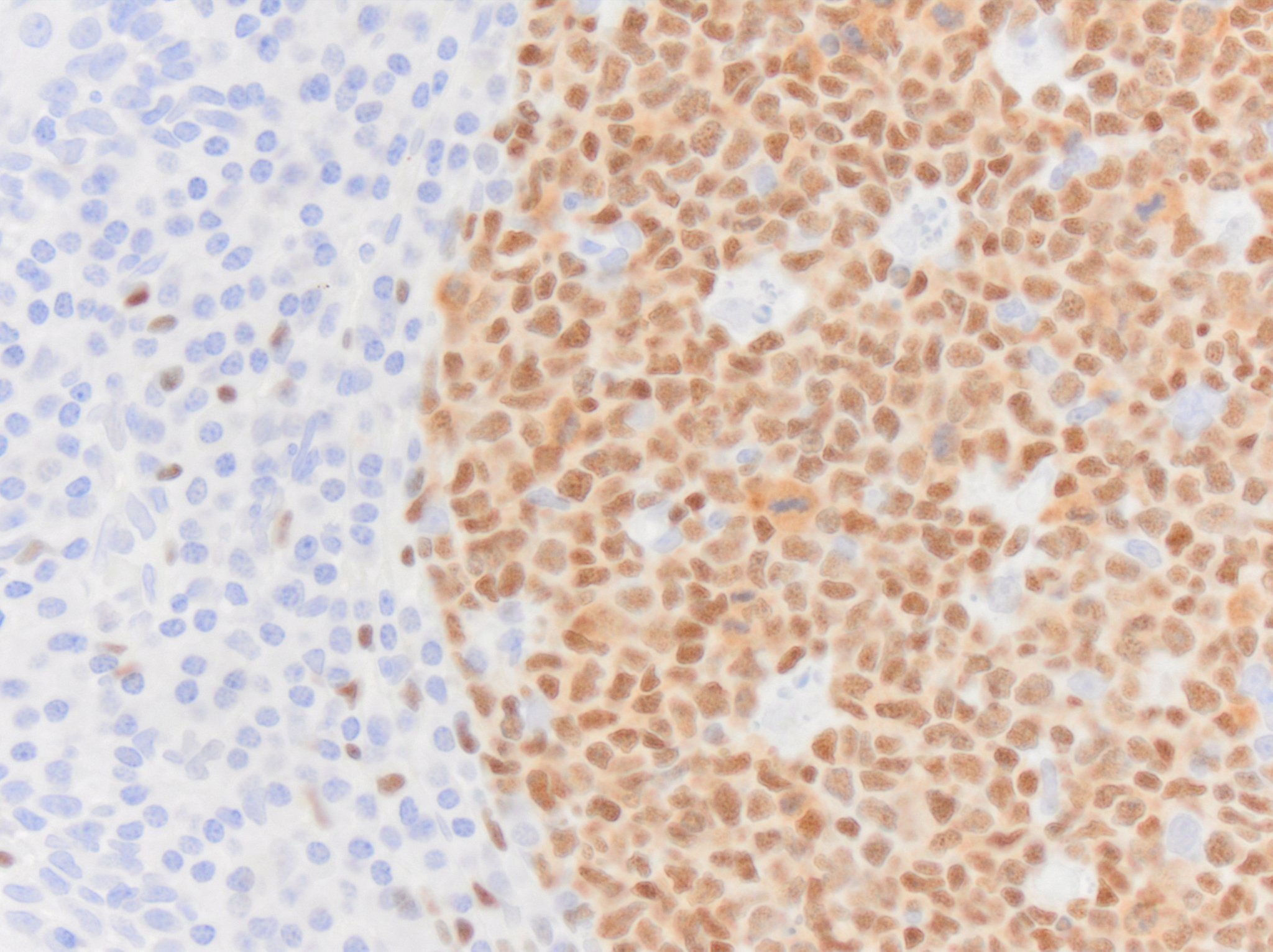
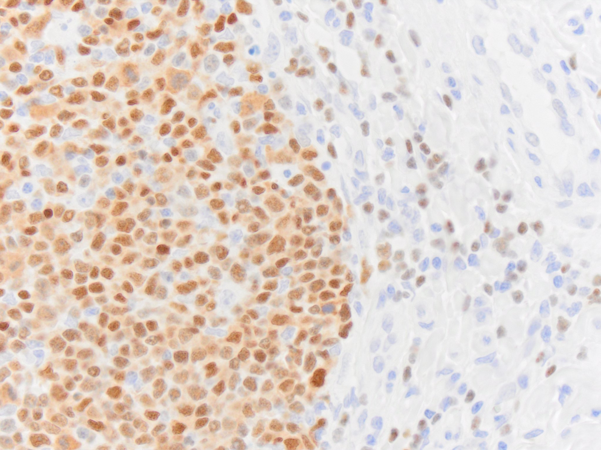
Immunohistochemistry-paraffin: Mouse Monoclonal Pax5/BSAP Antibody (PAX5/3735) [NBP3-07581] -
Immunohistochemistry-paraffin: Mouse Monoclonal Pax5/BSAP Antibody (PAX5/3735) [NBP3-07581] - Pax5/BSAP immunoreactivity in human tonsil. Primary was used at 0.5ug/mL and was incubated with the tissue section for 30m at room temperature. Secondary was Horse Anti-Mouse HRP polymer. Image from a verified customer review. [NBP3-07581] -")
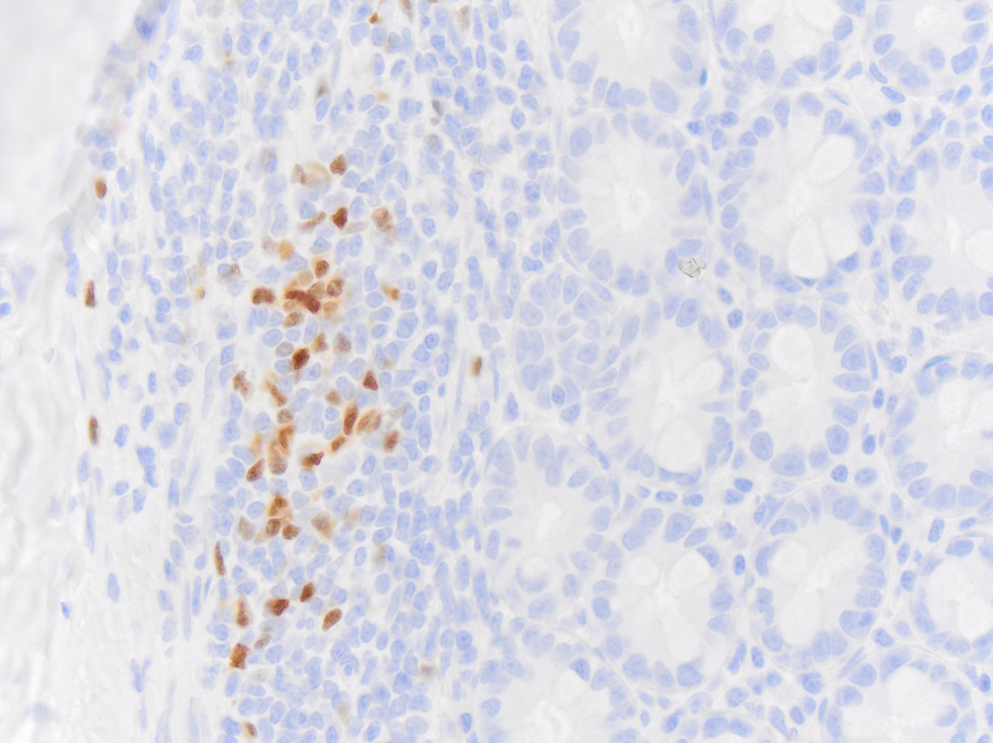
Immunohistochemistry-paraffin: Mouse Monoclonal Pax5/BSAP Antibody (PAX5/3735) [NBP3-07581] -
Immunohistochemistry-paraffin: Mouse Monoclonal Pax5/BSAP Antibody (PAX5/3735) [NBP3-07581] - Pax5/BSAP immunoreactivity in feline intestine. Primary was used at 0.5ug/mL and was incubated with the tissue section for 30m at room temperature. Secondary was Horse Anti-Mouse HRP polymer. Image from a verified customer review. [NBP3-07581] -")
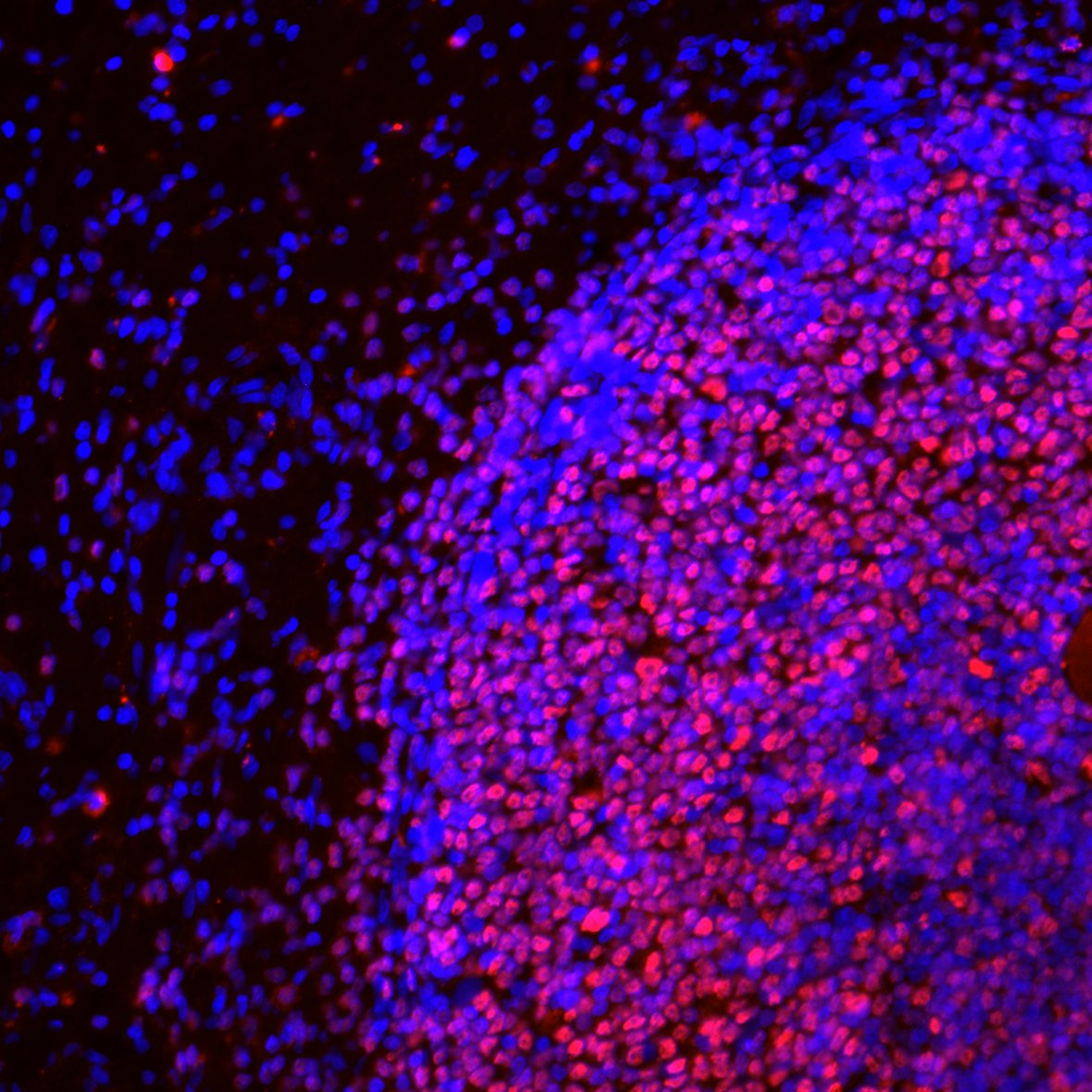
Immunohistochemistry-Frozen: Mouse Monoclonal Pax5/BSAP Antibody (PAX5/3735) [NBP3-07581] -
Immunohistochemistry-Frozen: Mouse Monoclonal Pax5/BSAP Antibody (PAX5/3735) [NBP3-07581] - Immunoreactivity was observed with this Pax5 antibody (red) in Rhesus macaque spleen tissues. Nuclei counterstained with DAPI (blue). Primary antibody dilution used: 1:100. Image from a verified customer review.![Pax5/BSAP Antibody (PAX5/3735) Immunohistochemistry-Paraffin: Pax5/BSAP Antibody (PAX5/3735) [NBP3-07581] -](https://resources.rndsystems.com/images/products/nbp3-07581_mouse-monoclonal-pax5-bsap-antibody-pax5-3735-29520251641227.jpg "Immunohistochemistry-Paraffin: Pax5/BSAP Antibody (PAX5/3735) [NBP3-07581] -")
Immunohistochemistry-Paraffin: Pax5/BSAP Antibody (PAX5/3735) [NBP3-07581] -
Formalin-fixed, paraffin-embedded human Tonsil stained with Pax5/BSAP Antibody (PAX5/3735).![Pax5/BSAP Antibody (PAX5/3735) Immunohistochemistry-Paraffin: Pax5/BSAP Antibody (PAX5/3735) [NBP3-07581] -](https://resources.rndsystems.com/images/products/nbp3-07581_mouse-monoclonal-pax5-bsap-antibody-pax5-3735-295202516392817.jpg "Immunohistochemistry-Paraffin: Pax5/BSAP Antibody (PAX5/3735) [NBP3-07581] -")
Immunohistochemistry-Paraffin: Pax5/BSAP Antibody (PAX5/3735) [NBP3-07581] -
Formalin-fixed, paraffin-embedded human Tonsil stained with Pax5/BSAP Antibody (PAX5/3735).Applications for Pax5/BSAP Antibody (PAX5/3735)
Application
Recommended Usage
Immunohistochemistry-Frozen
Validated for Immunohistochemistry-Frozen from a verified customer review.
Immunohistochemistry-Paraffin
1-2 ug/ml
Application Notes
Immunohistochemistry (Formalin-fixed): 1-2ug/ml for 30 min at RT. Staining of formalin-fixed tissues requires heating tissue sections in 10mM Tris with 1mM EDTA, pH 9.0, for 45 min at 95C followed by cooling at RT for 20 minutes.
Optimal dilution for a specific application should be determined.
Optimal dilution for a specific application should be determined.
Reviewed Applications
Read 5 reviews rated 5 using NBP3-07581 in the following applications:
Formulation, Preparation, and Storage
Purification
Protein A or G purified
Formulation
10 mM PBS with 0.05% BSA
Preservative
0.05% Sodium Azide
Concentration
0.2 mg/ml
Shipping
The product is shipped with polar packs. Upon receipt, store it immediately at the temperature recommended below.
Stability & Storage
Store at 4C.
Background: Pax5/BSAP
Long Name
Paired Box Gene 5/B-cell Specific Activator Protein
Alternate Names
BSAP
Gene Symbol
PAX5
UniProt
Additional Pax5/BSAP Products
Product Documents for Pax5/BSAP Antibody (PAX5/3735)
Certificate of Analysis
To download a Certificate of Analysis, please enter a lot or batch number in the search box below.
Product Specific Notices for Pax5/BSAP Antibody (PAX5/3735)
This product is for research use only and is not approved for use in humans or in clinical diagnosis. Primary Antibodies are guaranteed for 1 year from date of receipt.
Related Research Areas
Customer Reviews for Pax5/BSAP Antibody (PAX5/3735) (5)
5 out of 5
5 Customer Ratings
Have you used Pax5/BSAP Antibody (PAX5/3735)?
Submit a review and receive an Amazon gift card!
$25/€18/£15/$25CAN/¥2500 Yen for a review with an image
$10/€7/£6/$10CAN/¥1110 Yen for a review without an image
Submit a review
Customer Images
Showing
1
-
5 of
5 reviews
Showing All
Filter By:
-
Application: Immunohistochemistry-FrozenSample Tested: spleenSpecies: Rhesus MacaqueVerified Customer | Posted 04/18/2024Immunoreactivity was observed with this Pax5 antibody (red) in monkey spleen tissues. Nuclei conterstained with DAPI (blue). Primary antibody dilution used: 1:100.

-
Application: Immunohistochemistry-ParaffinSample Tested: Adult intestine and IntestineSpecies: FelineVerified Customer | Posted 10/13/2023Pax5 NBP3-07581 immunoreactivity in cat intestine. Primary was used at 0.5ug/mL and was incubated with the tissue section for 30m at room temperature. Secondary was Horse Anti-Mouse HRP polymer.Requires heat-induced epitope retrieval.
Bio-Techne ResponseThis review was submitted through the legacy Novus Innovators Program, reflecting a new species or application tested on a primary antibody.
-
Application: Immunohistochemistry-ParaffinSample Tested: human tonsilSpecies: HumanVerified Customer | Posted 10/13/2023Pax5 NBP3-07581 immunoreactivity in human tonsil. Primary was used at 0.5ug/mL and was incubated with the tissue section for 30m at room temperature. Secondary was Horse Anti-Mouse HRP polymer.
-
Application: Immunohistochemistry-ParaffinSample Tested: SkinSpecies: EquineVerified Customer | Posted 10/13/2023Pax5 (NBP3-07581) immunoreactivity in horse skin. Primary was used at 0.5ug/mL and was incubated with the tissue section for 30m at room temperature. Secondary was Horse Anti-Mouse HRP polymer.
Bio-Techne ResponseThis review was submitted through the legacy Novus Innovators Program, reflecting a new species or application tested on a primary antibody.
-
Application: Immunohistochemistry-ParaffinSample Tested: Lymph NodeSpecies: CanineVerified Customer | Posted 10/13/2023Pax5 (NBP3-07581) immunoreactivity in a dog lymph node. Primary was used at 0.5ug/mL and was incubated with the tissue section for 30m at room temperature. Secondary was Horse Anti-Mouse HRP polymer.Requires heat-induced epitope retrieval. I found that both citrate and EDTA buffer works, but the signal is better with EDTA.
There are no reviews that match your criteria.
Protocols
Find general support by application which include: protocols, troubleshooting, illustrated assays, videos and webinars.
- Antigen Retrieval Protocol (PIER)
- Antigen Retrieval for Frozen Sections Protocol
- Appropriate Fixation of IHC/ICC Samples
- Cellular Response to Hypoxia Protocols
- Chromogenic IHC Staining of Formalin-Fixed Paraffin-Embedded (FFPE) Tissue Protocol
- Chromogenic Immunohistochemistry Staining of Frozen Tissue
- ClariTSA™ Fluorophore Kits
- Detection & Visualization of Antibody Binding
- Fluorescent IHC Staining of Frozen Tissue Protocol
- Graphic Protocol for Heat-induced Epitope Retrieval
- Graphic Protocol for the Preparation and Fluorescent IHC Staining of Frozen Tissue Sections
- Graphic Protocol for the Preparation and Fluorescent IHC Staining of Paraffin-embedded Tissue Sections
- Graphic Protocol for the Preparation of Gelatin-coated Slides for Histological Tissue Sections
- IHC Sample Preparation (Frozen sections vs Paraffin)
- Immunofluorescent IHC Staining of Formalin-Fixed Paraffin-Embedded (FFPE) Tissue Protocol
- Immunohistochemistry (IHC) and Immunocytochemistry (ICC) Protocols
- Immunohistochemistry Frozen Troubleshooting
- Immunohistochemistry Paraffin Troubleshooting
- Preparing Samples for IHC/ICC Experiments
- Preventing Non-Specific Staining (Non-Specific Binding)
- Primary Antibody Selection & Optimization
- Protocol for Heat-Induced Epitope Retrieval (HIER)
- Protocol for Making a 4% Formaldehyde Solution in PBS
- Protocol for VisUCyte™ HRP Polymer Detection Reagent
- Protocol for the Preparation & Fixation of Cells on Coverslips
- Protocol for the Preparation and Chromogenic IHC Staining of Frozen Tissue Sections
- Protocol for the Preparation and Chromogenic IHC Staining of Frozen Tissue Sections - Graphic
- Protocol for the Preparation and Chromogenic IHC Staining of Paraffin-embedded Tissue Sections
- Protocol for the Preparation and Chromogenic IHC Staining of Paraffin-embedded Tissue Sections - Graphic
- Protocol for the Preparation and Fluorescent IHC Staining of Frozen Tissue Sections
- Protocol for the Preparation and Fluorescent IHC Staining of Paraffin-embedded Tissue Sections
- Protocol for the Preparation of Gelatin-coated Slides for Histological Tissue Sections
- R&D Systems Quality Control Western Blot Protocol
- TUNEL and Active Caspase-3 Detection by IHC/ICC Protocol
- The Importance of IHC/ICC Controls
- Troubleshooting Guide: Immunohistochemistry
- Troubleshooting Guide: Western Blot Figures
- Western Blot Conditions
- Western Blot Protocol
- Western Blot Protocol for Cell Lysates
- Western Blot Troubleshooting
- Western Blot Troubleshooting Guide
- View all Protocols, Troubleshooting, Illustrated assays and Webinars
Loading...