PAX8 Antibody - BSA Free
Novus Biologicals | Catalog # NBP1-32440
![Immunocytochemistry/ Immunofluorescence: PAX8 Antibody [NBP1-32440]](https://resources.rndsystems.com/images/products/PAX8-Antibody-Immunocytochemistry-Immunofluorescence-NBP1-32440-img0020.jpg "Immunocytochemistry/ Immunofluorescence: PAX8 Antibody [NBP1-32440]")
Loading...
Key Product Details
Species Reactivity
Validated:
Human, Mouse, Rat, Canine
Cited:
Human, Mouse
Predicted:
Xenopus (91%). Backed by our 100% Guarantee.
Applications
Validated:
Immunohistochemistry, Immunohistochemistry-Paraffin, Western Blot, Immunocytochemistry/ Immunofluorescence, Simple Western, Immunoprecipitation, Chromatin Immunoprecipitation (ChIP), Single Cell Western
Cited:
Immunohistochemistry, Immunohistochemistry-Paraffin, Western Blot, Immunocytochemistry/ Immunofluorescence, Immunoprecipitation, Chromatin Immunoprecipitation (ChIP), Chemotaxis, Chip Cytometry, IF/IHC
Label
Unconjugated
Antibody Source
Polyclonal Rabbit IgG
Format
BSA Free
Loading...
Product Specifications
Immunogen
Recombinant protein encompassing a sequence within the center region of human PAX8. The exact sequence is proprietary.
Reactivity Notes
Xenopus laevis (91%).
Localization
Nucleus
Clonality
Polyclonal
Host
Rabbit
Isotype
IgG
Theoretical MW
48 kDa.
Disclaimer note: The observed molecular weight of the protein may vary from the listed predicted molecular weight due to post translational modifications, post translation cleavages, relative charges, and other experimental factors.
Disclaimer note: The observed molecular weight of the protein may vary from the listed predicted molecular weight due to post translational modifications, post translation cleavages, relative charges, and other experimental factors.
Scientific Data Images for PAX8 Antibody - BSA Free
Immunocytochemistry/ Immunofluorescence: PAX8 Antibody [NBP1-32440]
PAX8-Antibody-Immunocytochemistry-Immunofluorescence-NBP1-32440-img0020.jpg![Immunohistochemistry-Paraffin: PAX8 Antibody [NBP1-32440]](https://resources.rndsystems.com/images/products/PAX8-Antibody-Immunohistochemistry-Paraffin-NBP1-32440-img0021.jpg "Immunohistochemistry-Paraffin: PAX8 Antibody [NBP1-32440]")
Immunohistochemistry-Paraffin: PAX8 Antibody [NBP1-32440]
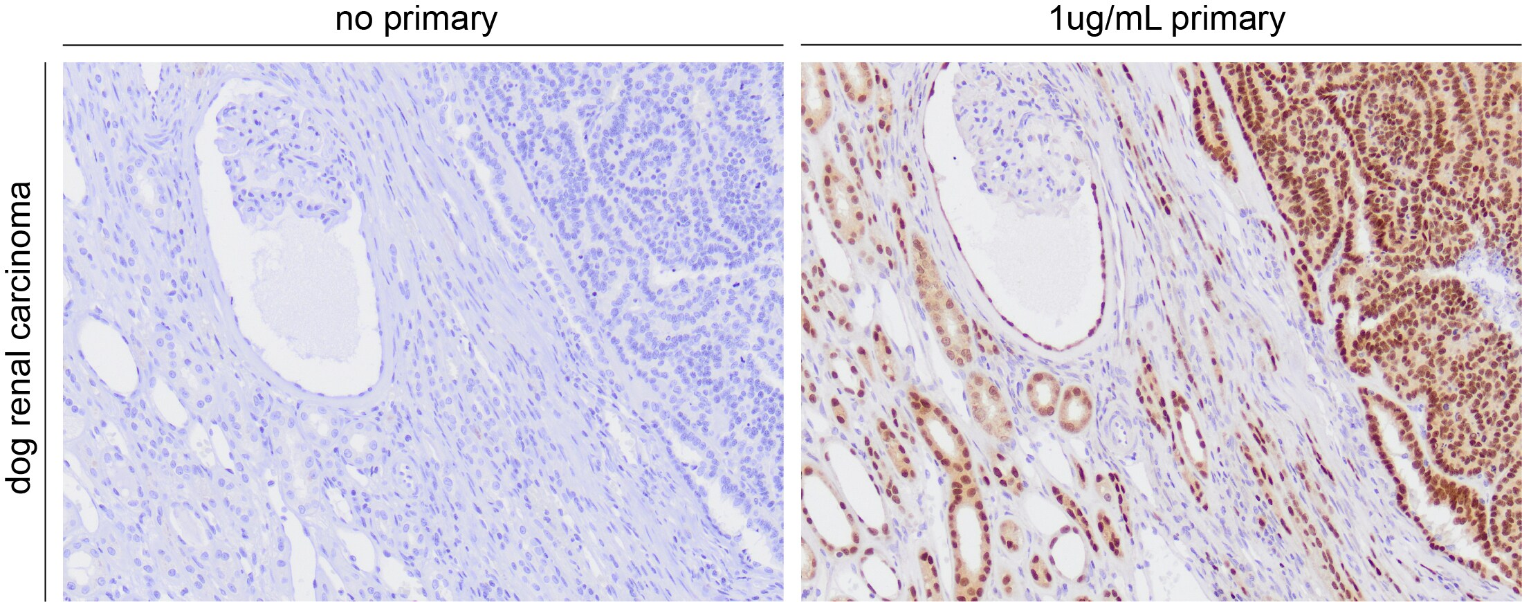
Immunohistochemistry-Paraffin: PAX8 Antibody [NBP1-32440] - Pax8 immunoreactivity in canine renal carcinoma. Primary antibody incubated with tissue for 1h at room temperature. IHC-P image submitted by a verified customer review.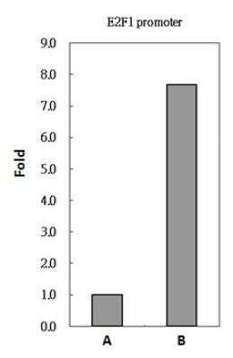
Chromatin Immunoprecipitation: PAX8 Antibody [NBP1-32440] - Sample: 293T whole cell lysate/extract A. 5 ug preimmune rabbit IgG, B. 5 ug of PAX8 antibody The precipitated DNA was detected by PCR with primer set targeting to E2F1 promoter.
![Immunocytochemistry/ Immunofluorescence: PAX8 Antibody [NBP1-32440]](https://resources.rndsystems.com/images/products/PAX8-Antibody-Immunocytochemistry-Immunofluorescence-NBP1-32440-img0011.jpg "Immunocytochemistry/ Immunofluorescence: PAX8 Antibody [NBP1-32440]")
Immunocytochemistry/ Immunofluorescence: PAX8 Antibody [NBP1-32440]
Immunocytochemistry/Immunofluorescence: PAX8 Antibody [NBP1-32440] - Paraformaldehyde-fixed A549, using antibody at 1:200 dilution.![Immunohistochemistry-Paraffin: PAX8 Antibody [NBP1-32440]](https://resources.rndsystems.com/images/products/PAX8-Antibody-Immunohistochemistry-Paraffin-NBP1-32440-img0015.jpg "Immunohistochemistry-Paraffin: PAX8 Antibody [NBP1-32440]")
Immunohistochemistry-Paraffin: PAX8 Antibody [NBP1-32440]
Immunohistochemistry-Paraffin: PAX8 Antibody [NBP1-32440] - Rat thyroid gland. PAX8 antibody dilution: 1:500. Antigen Retrieval: Trilogy™ (EDTA based, pH 8.0) buffer, 15min.![Immunohistochemistry-Paraffin: PAX8 Antibody [NBP1-32440]](https://resources.rndsystems.com/images/products/PAX8-Antibody-Immunohistochemistry-Paraffin-NBP1-32440-img0016.jpg "Immunohistochemistry-Paraffin: PAX8 Antibody [NBP1-32440]")
Immunohistochemistry-Paraffin: PAX8 Antibody [NBP1-32440]
Immunohistochemistry-Paraffin: PAX8 Antibody [NBP1-32440] - DLD1 xenograft. PAX8 antibody diluted at 1:500. Antigen Retrieval: Trilogy™ (EDTA based, pH 8.0) buffer, 15min.![Immunohistochemistry-Paraffin: PAX8 Antibody [NBP1-32440]](https://resources.rndsystems.com/images/products/PAX8-Antibody-Immunohistochemistry-Paraffin-NBP1-32440-img0018.jpg "Immunohistochemistry-Paraffin: PAX8 Antibody [NBP1-32440]")
Immunohistochemistry-Paraffin: PAX8 Antibody [NBP1-32440]
Immunohistochemistry-Paraffin: PAX8 Antibody [NBP1-32440] - Mouse liver. PAX8 antibody diluted at 1:500. Antigen Retrieval: Citrate buffer, pH 6.0, 15 min.![Immunoprecipitation: PAX8 Antibody [NBP1-32440]](https://resources.rndsystems.com/images/products/PAX8-Antibody-Immunoprecipitation-NBP1-32440-img0013.jpg "Immunoprecipitation: PAX8 Antibody [NBP1-32440]")
Immunoprecipitation: PAX8 Antibody [NBP1-32440]
Immunoprecipitation: PAX8 Antibody [NBP1-32440] - Sample: 1000 ug 293T whole cell lysate/extract A. 40 ug 293T whole cell lysate/extract, B. Control with 2 ug of preimmune rabbit IgG, C. Immunoprecipitation of PAX8 protein by 2 ug of PAX8 antibody 10% SDS-PAGE gel.
Immunocytochemistry/ Immunofluorescence: PAX8 Antibody [NBP1-32440] -
Immunocytochemistry/ Immunofluorescence: PAX8 Antibody [NBP1-32440] - Differentiation of human iPSC clone IV towards renal commitment.(a) Representative immunofluorescence images of co-staining for Lhx1/Osr1 up to day 6. (b) Images of co-staining of Pax2/Six2, Pax8/Six2 & Six2/Wt1 from day 6 to 12. (c) Expression of renal progenitor markers such as CD24, Claudin1 & GGT1 from day 0 to 19. Nuclei are stained with DAPI (blue). Scale bars: 50 μm (a, b), 20 μm (c). (d) Gene expression analysis for renal progenitor markers at different points in time. Image collected & cropped by CiteAb from the following publication (https://www.nature.com/articles/srep08826), licensed under a CC-BY license. Not internally tested by Novus Biologicals.
Immunohistochemistry-Paraffin: PAX8 Antibody [NBP1-32440] -
Immunohistochemistry-Paraffin: PAX8 Antibody [NBP1-32440] - PDX model establishment from ccRCC injection. a Hematoxylin & Eosin staining of human tumor engrafted in murine models. A epresentative image of kidney PDX reported. Microscope Nikon Eclipse E1000 10X & 20X (b) Graph reporting % of patients who engrafted when orthotopically injected in mice & distributed following grading (3 G1; 7 G2; 13 G3; 7 G4). Eight mice for each patient injected & all 18 tumors on 30 which evaluated as engrafted developed tumor masses on ≥ of 6 mice. Tumors declared unable to engraft did not produce, at all, tumor masses. c Representative images (1 × 0.63) of PDXs excised 90 days after injection by Stereomicroscope (Olympus SZX10,XCX50). One representative image for G2, G3 & G4 types reported. d Representative images (1 × 0.63 & 1.25) of aberrant neo-angiogenesis formation in PDXs by Stereomicroscope (Olympus SZX10, XCX50 camera). e Hematoxylin & Eosin staining of PDXs versus parental primary patients. One representative tumor for each grade (G2, G3, G4) reported. Staining executed on OCT frozen samples. f Hematoxylin & Eosin & anti-PAX8, CD10, Vimentin & EMA staining reported on formalin-fixed & paraffin- embedded parental primary, metastatic tissues & PDXs. Primary, metastatic & PDX tissues obtained & shown from one representative patient. Microscope Nikon Eclipse 55i, magnification 20×. g Histogram showing number of engrafting tumor populations evaluated over 30 injected patient samples & correlated w/ patient recurrence frequency calculated as development of metastases after surgery. Orange color represents recurrent (n = 7) & metastatic (n = 3) patients in engrafted group (n = 18). Pink color represents recurrent patients (n = 2) in non-engrafted group (n = 12) for a total of 30 injected samples Image collected & cropped by CiteAb from following publication (https://pubmed.ncbi.nlm.nih.gov/30185225), licensed under a CC-BY license. Not internally tested by Novus Biologicals.
Western Blot: PAX8 Antibody [NBP1-32440] -
Western Blot: PAX8 Antibody [NBP1-32440] - Non-transfected (-) and transfected (+) 293T whole cell extracts (30 ug) were separated by 10% SDS-PAGE, and the membrane was blotted with PAX8 antibody (NBP1-32440) diluted at 1:10000. The HRP-conjugated anti-rabbit IgG antibody was used to detect the primary antibody.
Immunohistochemistry-Paraffin: PAX8 Antibody [NBP1-32440] -
Immunohistochemistry-Paraffin: PAX8 Antibody [NBP1-32440] - PAX8 antibody detects PAX8 protein at nucleus by immunohistochemical analysis.Sample: Paraffin-embedded dog kidney.
PAX8 stained by PAX8 antibody (NBP1-32440) diluted at 1:2000.
Antigen Retrieval: Citrate buffer, pH 6.0, 15 min

Western Blot: PAX8 Antibody [NBP1-32440] -
Western Blot: PAX8 Antibody [NBP1-32440] - Whole cell extract (30 ug) was separated by 10% SDS-PAGE, and the membrane was blotted with PAX8 antibody (NBP1-32440) diluted at 1:500. The HRP-conjugated anti-rabbit IgG antibody was used to detect the primary antibody.
Western Blot: PAX8 Antibody [NBP1-32440] -
293T whole cell and nuclear extracts (30 ug) were separated by 10% SDS-PAGE, and the membrane was blotted with PAX8 antibody (NBP1-32440) diluted at 1:1000.
Western Blot: PAX8 Antibody [NBP1-32440] -
Various whole cell extracts (30 ug) were separated by 10% SDS-PAGE, and the membrane was blotted with PAX8 antibody (NBP1-32440) diluted at 1:1000. The HRP-conjugated anti-rabbit IgG antibody was used to detect the primary antibody. Corresponding RNA expression data for the same cell lines are based on Human Protein Atlas program.Applications for PAX8 Antibody - BSA Free
Application
Recommended Usage
Immunocytochemistry/ Immunofluorescence
1:100 - 1:1000
Immunohistochemistry
1:100 - 1:1000
Immunohistochemistry-Paraffin
1:100 - 1:1000
Immunoprecipitation
1:500 - 1:1000
Simple Western
1:10
Single Cell Western
50 ug/mL
Western Blot
1:5000-1:20000
Application Notes
In Simple Western only 10 - 15 uL of the recommended dilution is used per data point.
See Simple Western Antibody Database for Simple Western validation: Tested in Hek293 lysate 0.5 mg/mL, separated by Size, antibody dilution of 1:10, apparent MW was 60 kDa. Separated by Size-Wes, Sally Sue/Peggy Sue.
SCW is validated using 786-O cells.
See Simple Western Antibody Database for Simple Western validation: Tested in Hek293 lysate 0.5 mg/mL, separated by Size, antibody dilution of 1:10, apparent MW was 60 kDa. Separated by Size-Wes, Sally Sue/Peggy Sue.
SCW is validated using 786-O cells.
Reviewed Applications
Read 1 review rated 4 using NBP1-32440 in the following applications:
Formulation, Preparation, and Storage
Purification
Antigen Affinity-purified
Formulation
PBS, 20% Glycerol
Format
BSA Free
Preservative
0.025% Proclin 300
Concentration
Concentrations vary lot to lot. See vial label for concentration. If unlisted please contact technical services.
Shipping
The product is shipped with polar packs. Upon receipt, store it immediately at the temperature recommended below.
Stability & Storage
Aliquot and store at -20C or -80C. Avoid freeze-thaw cycles.
Background: PAX8
Alternate Names
paired box 8, paired box gene 8, paired box protein Pax-8, paired domain gene 8
Gene Symbol
PAX8
Additional PAX8 Products
Product Documents for PAX8 Antibody - BSA Free
Certificate of Analysis
To download a Certificate of Analysis, please enter a lot or batch number in the search box below.
Product Specific Notices for PAX8 Antibody - BSA Free
This product is for research use only and is not approved for use in humans or in clinical diagnosis. Primary Antibodies are guaranteed for 1 year from date of receipt.
Citations for PAX8 Antibody - BSA Free
 Powered by Bioz
Customer Reviews for PAX8 Antibody - BSA Free (1)
4 out of 5
1 Customer Rating
Have you used PAX8 Antibody - BSA Free?
Submit a review and receive an Amazon gift card!
$25/€18/£15/$25CAN/¥2500 Yen for a review with an image
$10/€7/£6/$10CAN/¥1110 Yen for a review without an image
Submit a review
Customer Images
Showing
1
-
1 of
1 review
Showing All
Filter By:
-
Application: Immunohistochemistry-ParaffinSample Tested: kidneySpecies: CanineVerified Customer | Posted 10/29/2021Pax8 immunoreactivity in canine renal carcinoma. Primary incubated with tissue for 1h at room temperature.
 Bio-Techne ResponseThis review was submitted through the legacy Novus Innovators Program, reflecting a new species or application tested on a primary antibody.
There are no reviews that match your criteria.
Protocols
Find general support by application which include: protocols, troubleshooting, illustrated assays, videos and webinars.
- Antigen Retrieval Protocol (PIER)
- Antigen Retrieval for Frozen Sections Protocol
- Appropriate Fixation of IHC/ICC Samples
- Cellular Response to Hypoxia Protocols
- ChIP Protocol Video
- Chromatin Immunoprecipitation (ChIP) Protocol
- Chromatin Immunoprecipitation Protocol
- Chromogenic IHC Staining of Formalin-Fixed Paraffin-Embedded (FFPE) Tissue Protocol
- Chromogenic Immunohistochemistry Staining of Frozen Tissue
- ClariTSA™ Fluorophore Kits
- Detection & Visualization of Antibody Binding
- Fluorescent IHC Staining of Frozen Tissue Protocol
- Graphic Protocol for Heat-induced Epitope Retrieval
- Graphic Protocol for the Preparation and Fluorescent IHC Staining of Frozen Tissue Sections
- Graphic Protocol for the Preparation and Fluorescent IHC Staining of Paraffin-embedded Tissue Sections
- Graphic Protocol for the Preparation of Gelatin-coated Slides for Histological Tissue Sections
- ICC Cell Smear Protocol for Suspension Cells
- ICC Immunocytochemistry Protocol Videos
- ICC for Adherent Cells
- IHC Sample Preparation (Frozen sections vs Paraffin)
- Immunocytochemistry (ICC) Protocol
- Immunocytochemistry Troubleshooting
- Immunofluorescence of Organoids Embedded in Cultrex Basement Membrane Extract
- Immunofluorescent IHC Staining of Formalin-Fixed Paraffin-Embedded (FFPE) Tissue Protocol
- Immunohistochemistry (IHC) and Immunocytochemistry (ICC) Protocols
- Immunohistochemistry Frozen Troubleshooting
- Immunohistochemistry Paraffin Troubleshooting
- Immunoprecipitation Protocol
- Preparing Samples for IHC/ICC Experiments
- Preventing Non-Specific Staining (Non-Specific Binding)
- Primary Antibody Selection & Optimization
- Protocol for Heat-Induced Epitope Retrieval (HIER)
- Protocol for Making a 4% Formaldehyde Solution in PBS
- Protocol for VisUCyte™ HRP Polymer Detection Reagent
- Protocol for the Fluorescent ICC Staining of Cell Smears - Graphic
- Protocol for the Fluorescent ICC Staining of Cultured Cells on Coverslips - Graphic
- Protocol for the Preparation & Fixation of Cells on Coverslips
- Protocol for the Preparation and Chromogenic IHC Staining of Frozen Tissue Sections
- Protocol for the Preparation and Chromogenic IHC Staining of Frozen Tissue Sections - Graphic
- Protocol for the Preparation and Chromogenic IHC Staining of Paraffin-embedded Tissue Sections
- Protocol for the Preparation and Chromogenic IHC Staining of Paraffin-embedded Tissue Sections - Graphic
- Protocol for the Preparation and Fluorescent ICC Staining of Cells on Coverslips
- Protocol for the Preparation and Fluorescent ICC Staining of Non-adherent Cells
- Protocol for the Preparation and Fluorescent ICC Staining of Stem Cells on Coverslips
- Protocol for the Preparation and Fluorescent IHC Staining of Frozen Tissue Sections
- Protocol for the Preparation and Fluorescent IHC Staining of Paraffin-embedded Tissue Sections
- Protocol for the Preparation of Gelatin-coated Slides for Histological Tissue Sections
- Protocol for the Preparation of a Cell Smear for Non-adherent Cell ICC - Graphic
- R&D Systems Quality Control Western Blot Protocol
- TUNEL and Active Caspase-3 Detection by IHC/ICC Protocol
- The Importance of IHC/ICC Controls
- Troubleshooting Guide: Immunohistochemistry
- Troubleshooting Guide: Western Blot Figures
- Western Blot Conditions
- Western Blot Protocol
- Western Blot Protocol for Cell Lysates
- Western Blot Troubleshooting
- Western Blot Troubleshooting Guide
- View all Protocols, Troubleshooting, Illustrated assays and Webinars
Loading...