Pepsinogen A Antibody (961902) - Azide and BSA Free
Novus Biologicals | Catalog # MAB9350
![Western Blot: Pepsinogen A Antibody (961902) [MAB9350]](https://resources.rndsystems.com/images/products/Pepsinogen-A-Antibody-961902-Western-Blot-MAB9350-img0001.jpg "Western Blot: Pepsinogen A Antibody (961902) [MAB9350]")
Key Product Details
Species Reactivity
Human
Applications
Immunohistochemistry, Immunohistochemistry-Paraffin, Western Blot
Label
Unconjugated
Antibody Source
Monoclonal Rat IgG1 Clone # 961902
Format
Azide and BSA Free
Loading...
Product Specifications
Immunogen
Mouse myeloma cell line NS0-derived recombinant human Pepsinogen A4/PGA4
Ile16-Ala388
Accession # P00790
Ile16-Ala388
Accession # P00790
Clonality
Monoclonal
Host
Rat
Isotype
IgG1
Scientific Data Images for Pepsinogen A Antibody (961902) - Azide and BSA Free
Western Blot: Pepsinogen A Antibody (961902) [MAB9350]
Western Blot: Pepsinogen A Antibody (961902) [MAB9350] - Analysis of human stomach tissue lysate with 2 ug/ml of hPGA-4 Monoclonal Antibody followed by HAF005. A specific band was detected for hPGA-4 at approximately45 kDA (as indicated). This experiment was conducted under reducing conditions and using Immunoblot Buffer Group 1.![Immunohistochemistry: Pepsinogen A Antibody (961902) [MAB9350]](https://resources.rndsystems.com/images/products/Pepsinogen-A-Antibody-961902-Immunohistochemistry-MAB9350-img0004.jpg "Immunohistochemistry: Pepsinogen A Antibody (961902) [MAB9350]")
Immunohistochemistry: Pepsinogen A Antibody (961902) [MAB9350]
Immunohistochemistry: Pepsinogen A Antibody (961902) [MAB9350] - Pepsinogen A was detected in immersion fixed paraffin-embedded sections of human stomach using Pepsinogen A Monoclonal Antibody at 1.7 ug/ml. Tissue was stained using Rat IgG VisUCyte HRP Polymer Antibody (brown) and counterstained with hematoxylin (blue). Specific staining was localized to the cytoplasm in epithelial cells.Applications for Pepsinogen A Antibody (961902) - Azide and BSA Free
Application
Recommended Usage
Immunohistochemistry
1 - 2 ug/ml
Immunohistochemistry-Paraffin
1 - 2 ug/ml
Western Blot
2 ug/ml
Reviewed Applications
Read 1 review rated 5 using MAB9350 in the following applications:
Formulation, Preparation, and Storage
Purification
Protein A or G purified
Formulation
PBS
Format
Azide and BSA Free
Preservative
No Preservative
Concentration
Please see the vial label for concentration. If unlisted please contact technical services.
Shipping
The product is shipped with polar packs. Upon receipt, store it immediately at the temperature recommended below.
Stability & Storage
Store at -20C. Avoid freeze-thaw cycles.
Background: Pepsinogen A
Alternate Names
EC 3.4.23, EC 3.4.23.1, FLJ58952, FLJ77962, pepsin A, pepsinogen 4, group I (pepsinogen A), pepsinogen A4, PGA3, PGA5
Gene Symbol
PGA4
Additional Pepsinogen A Products
Product Documents for Pepsinogen A Antibody (961902) - Azide and BSA Free
Certificate of Analysis
To download a Certificate of Analysis, please enter a lot or batch number in the search box below.
Product Specific Notices for Pepsinogen A Antibody (961902) - Azide and BSA Free
This product is for research use only and is not approved for use in humans or in clinical diagnosis. Primary Antibodies are guaranteed for 1 year from date of receipt.
Related Research Areas
Customer Reviews for Pepsinogen A Antibody (961902) - Azide and BSA Free (1)
5 out of 5
1 Customer Rating
Have you used Pepsinogen A Antibody (961902) - Azide and BSA Free?
Submit a review and receive an Amazon gift card!
$25/€18/£15/$25CAN/¥2500 Yen for a review with an image
$10/€7/£6/$10CAN/¥1110 Yen for a review without an image
Submit a review
Customer Images
Showing
1
-
1 of
1 review
Showing All
Filter By:
-
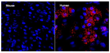
Application: ImmunocytochemistrySample Tested: Stomach tissueSpecies: Mouse and HumanVerified Customer | Posted 06/11/2017We tested this antibody in both mouse and human stomach tissue. This antibody DOESN'T react to the mouse pepsinogen A. It is good for human tissue. Positive staining(red) can be observed in human tissue.

There are no reviews that match your criteria.
Protocols
Find general support by application which include: protocols, troubleshooting, illustrated assays, videos and webinars.
- Antigen Retrieval Protocol (PIER)
- Antigen Retrieval for Frozen Sections Protocol
- Appropriate Fixation of IHC/ICC Samples
- Cellular Response to Hypoxia Protocols
- Chromogenic IHC Staining of Formalin-Fixed Paraffin-Embedded (FFPE) Tissue Protocol
- Chromogenic Immunohistochemistry Staining of Frozen Tissue
- ClariTSA™ Fluorophore Kits
- Detection & Visualization of Antibody Binding
- Fluorescent IHC Staining of Frozen Tissue Protocol
- Graphic Protocol for Heat-induced Epitope Retrieval
- Graphic Protocol for the Preparation and Fluorescent IHC Staining of Frozen Tissue Sections
- Graphic Protocol for the Preparation and Fluorescent IHC Staining of Paraffin-embedded Tissue Sections
- Graphic Protocol for the Preparation of Gelatin-coated Slides for Histological Tissue Sections
- IHC Sample Preparation (Frozen sections vs Paraffin)
- Immunofluorescent IHC Staining of Formalin-Fixed Paraffin-Embedded (FFPE) Tissue Protocol
- Immunohistochemistry (IHC) and Immunocytochemistry (ICC) Protocols
- Immunohistochemistry Frozen Troubleshooting
- Immunohistochemistry Paraffin Troubleshooting
- Preparing Samples for IHC/ICC Experiments
- Preventing Non-Specific Staining (Non-Specific Binding)
- Primary Antibody Selection & Optimization
- Protocol for Heat-Induced Epitope Retrieval (HIER)
- Protocol for Making a 4% Formaldehyde Solution in PBS
- Protocol for VisUCyte™ HRP Polymer Detection Reagent
- Protocol for the Preparation & Fixation of Cells on Coverslips
- Protocol for the Preparation and Chromogenic IHC Staining of Frozen Tissue Sections
- Protocol for the Preparation and Chromogenic IHC Staining of Frozen Tissue Sections - Graphic
- Protocol for the Preparation and Chromogenic IHC Staining of Paraffin-embedded Tissue Sections
- Protocol for the Preparation and Chromogenic IHC Staining of Paraffin-embedded Tissue Sections - Graphic
- Protocol for the Preparation and Fluorescent IHC Staining of Frozen Tissue Sections
- Protocol for the Preparation and Fluorescent IHC Staining of Paraffin-embedded Tissue Sections
- Protocol for the Preparation of Gelatin-coated Slides for Histological Tissue Sections
- R&D Systems Quality Control Western Blot Protocol
- TUNEL and Active Caspase-3 Detection by IHC/ICC Protocol
- The Importance of IHC/ICC Controls
- Troubleshooting Guide: Immunohistochemistry
- Troubleshooting Guide: Western Blot Figures
- Western Blot Conditions
- Western Blot Protocol
- Western Blot Protocol for Cell Lysates
- Western Blot Troubleshooting
- Western Blot Troubleshooting Guide
- View all Protocols, Troubleshooting, Illustrated assays and Webinars
Loading...