PER1 Antibody - BSA Free
Novus Biologicals | Catalog # NBP2-24589
![Western Blot: PER1 Antibody [NBP2-24589]](https://resources.rndsystems.com/images/products/PER1-Antibody-Western-Blot-NBP2-24589-img0002.jpg "Western Blot: PER1 Antibody [NBP2-24589]")
Loading...
Key Product Details
Species Reactivity
Validated:
Human, Mouse, Bovine, Canine, Equine, Primate
Cited:
Mouse, Amphibian
Predicted:
Rat (93%). Backed by our 100% Guarantee.
Applications
Validated:
Knockout Validated, Immunohistochemistry, Immunohistochemistry-Paraffin, Western Blot, Immunoblotting, Simple Western
Cited:
Knockout Validated, Western Blot, Immunoblotting, Simple Western
Label
Unconjugated
Antibody Source
Polyclonal Rabbit IgG
Format
BSA Free
Loading...
Product Specifications
Immunogen
A portion of amino acids 1150-1200 of human PER1 was used as the immunogen.
Reactivity Notes
Mouse reactivity reported in scientific literature (PMID: 29937374).
Clonality
Polyclonal
Host
Rabbit
Isotype
IgG
Scientific Data Images for PER1 Antibody - BSA Free
Western Blot: PER1 Antibody [NBP2-24589]
Western Blot: PER1 Antibody [NBP2-24589] - Analysis of PER1 in HEPG2 cell lysates in the 1) absence and 2) presence of immunizing peptide using NBP2-24589. Goat anti-rabbit Ig HRP secondary antibody and PicoTect ECL substrate solution were used for this test.![Immunohistochemistry-Paraffin: PER1 Antibody [NBP2-24589]](https://resources.rndsystems.com/images/products/PER1-Antibody-Immunohistochemistry-Paraffin-NBP2-24589-img0001.jpg "Immunohistochemistry-Paraffin: PER1 Antibody [NBP2-24589]")
Immunohistochemistry-Paraffin: PER1 Antibody [NBP2-24589]
Immunohistochemistry-Paraffin: PER1 Antibody [NBP2-24589] - Human brain tissue using an isotype control (top) and NBP2-24589 (bottom) at 5 ug/ml.Applications for PER1 Antibody - BSA Free
Application
Recommended Usage
Immunoblotting
reported in scientific literature (PMID 29937374)
Immunohistochemistry-Paraffin
5 ug/ml
Knockout Validated
reported in scientific literature (Hamilton et al)
Simple Western
1:1000 - 1:5000
Western Blot
5-10 ug/ml
Application Notes
See Simple Western Antibody Database for Simple Western validation: Tested in C57BL/6 liver whole-cell lysate (CT15), separated by Size, antibody dilution of 1:1000, 1:2000, 1:5000, apparent MW was 146 kDa
Reviewed Applications
Read 2 reviews rated 1.5 using NBP2-24589 in the following applications:
Formulation, Preparation, and Storage
Purification
Immunogen affinity purified
Formulation
PBS
Format
BSA Free
Preservative
0.05% Sodium Azide
Concentration
1.0 mg/ml
Shipping
The product is shipped with polar packs. Upon receipt, store it immediately at the temperature recommended below.
Stability & Storage
Store at 4C short term. Aliquot and store at -20C long term. Avoid freeze-thaw cycles.
Background: PER1
Long Name
Period Circadian Protein 1
Alternate Names
PERIOD1, RIGUI
Gene Symbol
PER1
Additional PER1 Products
Product Documents for PER1 Antibody - BSA Free
Certificate of Analysis
To download a Certificate of Analysis, please enter a lot or batch number in the search box below.
Product Specific Notices for PER1 Antibody - BSA Free
This product is for research use only and is not approved for use in humans or in clinical diagnosis. Primary Antibodies are guaranteed for 1 year from date of receipt.
Citations for PER1 Antibody - BSA Free
 Powered by Bioz
Customer Reviews for PER1 Antibody - BSA Free (2)
1.5 out of 5
2 Customer Ratings
Have you used PER1 Antibody - BSA Free?
Submit a review and receive an Amazon gift card!
$25/€18/£15/$25CAN/¥2500 Yen for a review with an image
$10/€7/£6/$10CAN/¥1110 Yen for a review without an image
Submit a review
Customer Images
Showing
1
-
2 of
2 reviews
Showing All
Filter By:
-
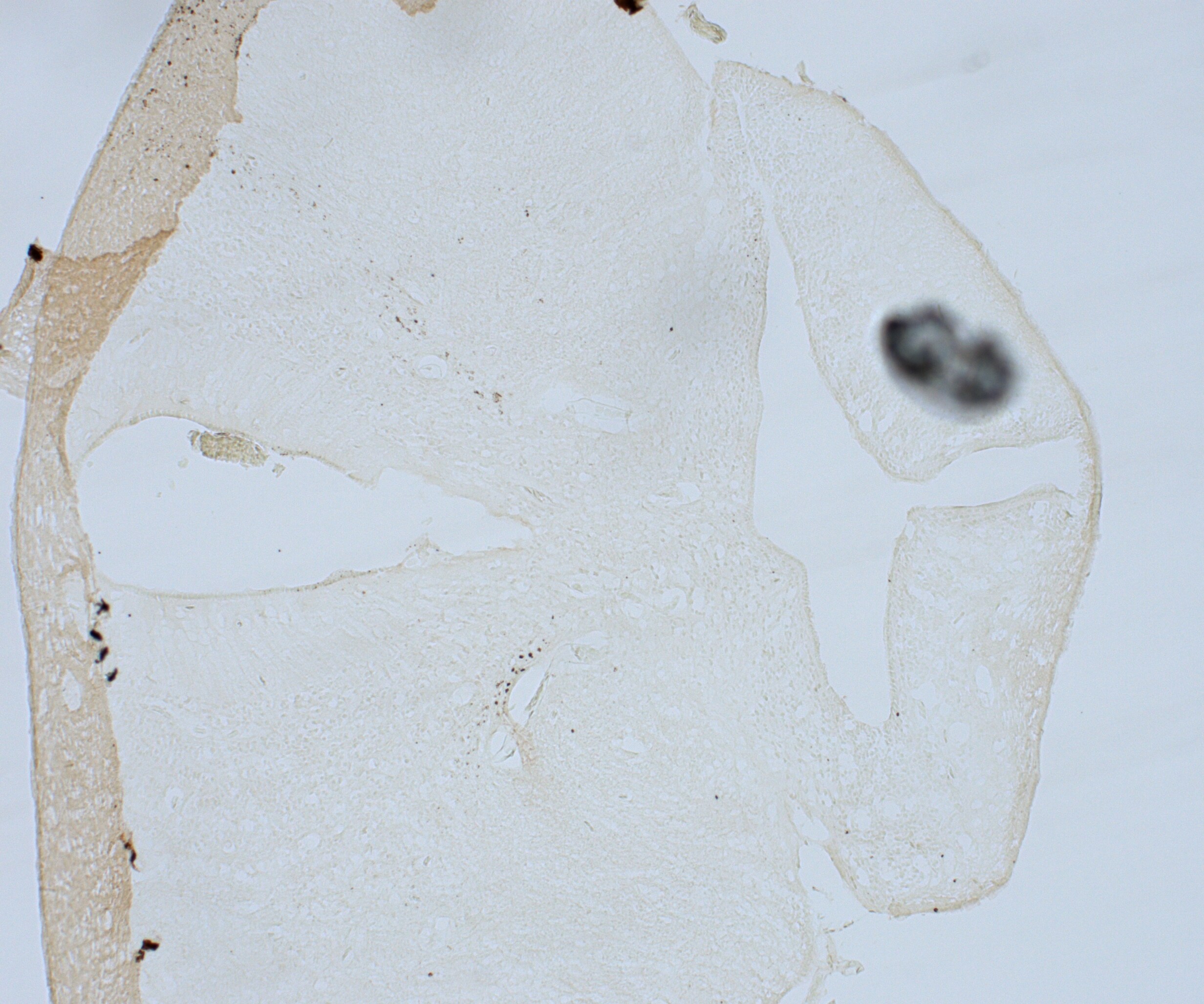
Application: Immunohistochemistry-FrozenSample Tested: brain sectionSpecies: OtherVerified Customer | Posted 09/11/202320um section including hypothalamus and SCN of Hyla cinerea. Staining using DAB
 Bio-Techne ResponseThis review was submitted through the legacy Novus Innovators Program, reflecting a new species or application tested on a primary antibody.
 -
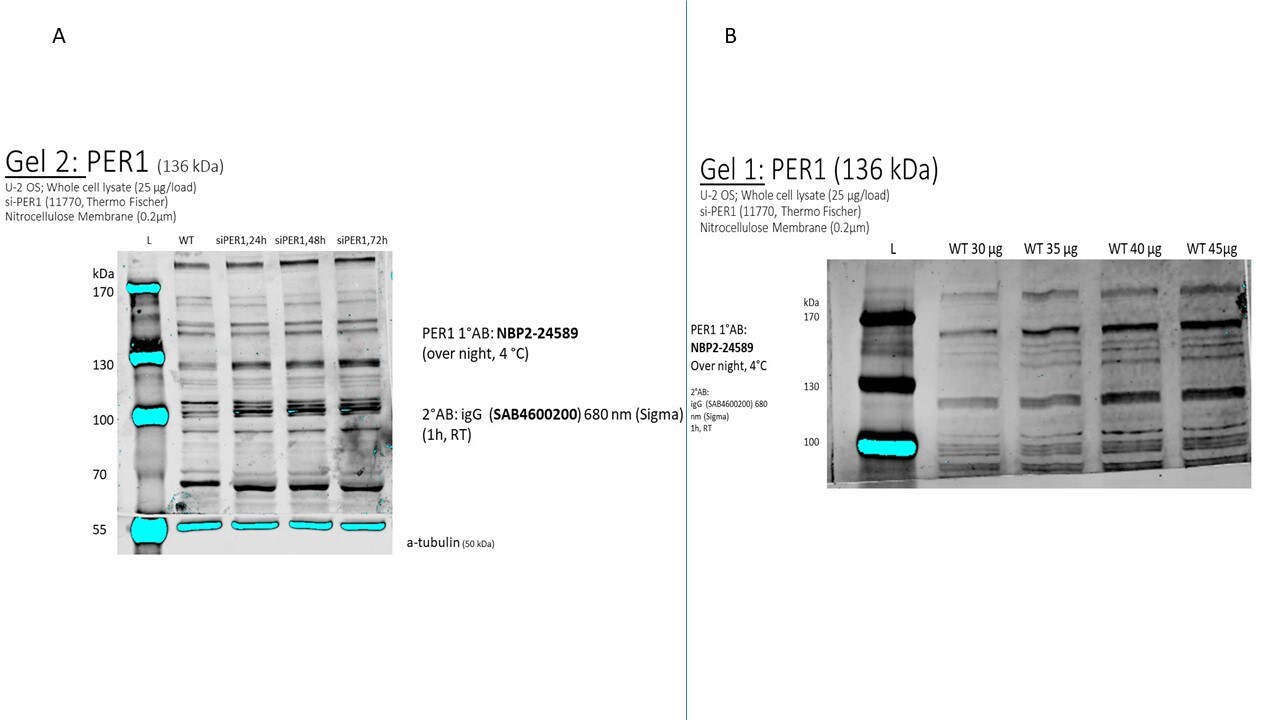
Application: Western BlotSample Tested: U-2 OS cellsSpecies: HumanVerified Customer | Posted 02/21/2019siRNA transfection optimisation: whole cell lysate of U-2 OS cells a) siRNA knockdown of PER1 (load 25 µg/well) b) wt U-2 OS cells with different protein amounts/well 1°AB over night, 4 °C 2°AB 1h, RT Nitrocellulose Membraneunspecific bands with no obvious band of correct size
There are no reviews that match your criteria.
Protocols
Find general support by application which include: protocols, troubleshooting, illustrated assays, videos and webinars.
- Antigen Retrieval Protocol (PIER)
- Antigen Retrieval for Frozen Sections Protocol
- Appropriate Fixation of IHC/ICC Samples
- Cellular Response to Hypoxia Protocols
- Chromogenic IHC Staining of Formalin-Fixed Paraffin-Embedded (FFPE) Tissue Protocol
- Chromogenic Immunohistochemistry Staining of Frozen Tissue
- ClariTSA™ Fluorophore Kits
- Detection & Visualization of Antibody Binding
- Fluorescent IHC Staining of Frozen Tissue Protocol
- Graphic Protocol for Heat-induced Epitope Retrieval
- Graphic Protocol for the Preparation and Fluorescent IHC Staining of Frozen Tissue Sections
- Graphic Protocol for the Preparation and Fluorescent IHC Staining of Paraffin-embedded Tissue Sections
- Graphic Protocol for the Preparation of Gelatin-coated Slides for Histological Tissue Sections
- IHC Sample Preparation (Frozen sections vs Paraffin)
- Immunofluorescent IHC Staining of Formalin-Fixed Paraffin-Embedded (FFPE) Tissue Protocol
- Immunohistochemistry (IHC) and Immunocytochemistry (ICC) Protocols
- Immunohistochemistry Frozen Troubleshooting
- Immunohistochemistry Paraffin Troubleshooting
- Preparing Samples for IHC/ICC Experiments
- Preventing Non-Specific Staining (Non-Specific Binding)
- Primary Antibody Selection & Optimization
- Protocol for Heat-Induced Epitope Retrieval (HIER)
- Protocol for Making a 4% Formaldehyde Solution in PBS
- Protocol for VisUCyte™ HRP Polymer Detection Reagent
- Protocol for the Preparation & Fixation of Cells on Coverslips
- Protocol for the Preparation and Chromogenic IHC Staining of Frozen Tissue Sections
- Protocol for the Preparation and Chromogenic IHC Staining of Frozen Tissue Sections - Graphic
- Protocol for the Preparation and Chromogenic IHC Staining of Paraffin-embedded Tissue Sections
- Protocol for the Preparation and Chromogenic IHC Staining of Paraffin-embedded Tissue Sections - Graphic
- Protocol for the Preparation and Fluorescent IHC Staining of Frozen Tissue Sections
- Protocol for the Preparation and Fluorescent IHC Staining of Paraffin-embedded Tissue Sections
- Protocol for the Preparation of Gelatin-coated Slides for Histological Tissue Sections
- R&D Systems Quality Control Western Blot Protocol
- TUNEL and Active Caspase-3 Detection by IHC/ICC Protocol
- The Importance of IHC/ICC Controls
- Troubleshooting Guide: Immunohistochemistry
- Troubleshooting Guide: Western Blot Figures
- Western Blot Conditions
- Western Blot Protocol
- Western Blot Protocol for Cell Lysates
- Western Blot Troubleshooting
- Western Blot Troubleshooting Guide
- View all Protocols, Troubleshooting, Illustrated assays and Webinars
Loading...