PGD2 Synthase/PTGDS Antibody - BSA Free
Novus Biologicals | Catalog # NBP1-79280
![Western Blot: PGD2 Synthase/PTGDS Antibody [NBP1-79280]](https://resources.rndsystems.com/images/products/PGD2-Synthase-PTGDS-Antibody-Western-Blot-NBP1-79280-img0004.jpg "Western Blot: PGD2 Synthase/PTGDS Antibody [NBP1-79280]")
Key Product Details
Species Reactivity
Applications
Label
Antibody Source
Format
Product Specifications
Immunogen
Clonality
Host
Isotype
Theoretical MW
Disclaimer note: The observed molecular weight of the protein may vary from the listed predicted molecular weight due to post translational modifications, post translation cleavages, relative charges, and other experimental factors.
Description
Scientific Data Images for PGD2 Synthase/PTGDS Antibody - BSA Free
Western Blot: PGD2 Synthase/PTGDS Antibody [NBP1-79280]
Western Blot: PGD2 Synthase/PTGDS Antibody [NBP1-79280] - Titration: 1 ug/ml Positive Control: HepG2 cell lysate.![Immunohistochemistry-Paraffin: PGD2 Synthase/PTGDS Antibody [NBP1-79280]](https://resources.rndsystems.com/images/products/PGD2-Synthase-PTGDS-Antibody-Immunohistochemistry-Paraffin-NBP1-79280-img0005.jpg "Immunohistochemistry-Paraffin: PGD2 Synthase/PTGDS Antibody [NBP1-79280]")
Immunohistochemistry-Paraffin: PGD2 Synthase/PTGDS Antibody [NBP1-79280]
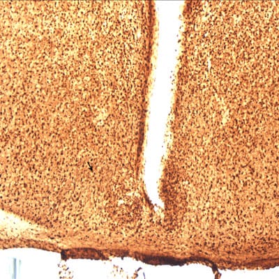
Immunohistochemistry-Paraffin: PGD2 Synthase/PTGDS Antibody [NBP1-79280] - Staining of mouse brain. Image provided by Allison Brager - Department of Neurobiology, Morehouse School of Medicine.![Immunohistochemistry-Paraffin: PGD2 Synthase/PTGDS Antibody [NBP1-79280]](https://resources.rndsystems.com/images/products/PGD2-Synthase-PTGDS-Antibody-Immunohistochemistry-Paraffin-NBP1-79280-img0003.jpg "Immunohistochemistry-Paraffin: PGD2 Synthase/PTGDS Antibody [NBP1-79280]")
Immunohistochemistry-Paraffin: PGD2 Synthase/PTGDS Antibody [NBP1-79280]
Immunohistochemistry-Paraffin: PGD2 Synthase/PTGDS Antibody [NBP1-79280] - Human kidney Tissue, antibody concentration 4-8ug/ml. Cells with positive label: renal corpuscle cells (indicated with arrows) 400X magnification.
Western Blot: PGD2 Synthase/PTGDS Antibody - BSA Free [NBP1-79280] -
Human monocytes and monocyte-derived macrophages constitutively express hPGDS, which is upregulated in parallel to COX-2 by LPS/IFN-gamma. (A) Peripheral blood monocytes were either directly activated or first differentiated into monocyte-derived macrophages (MDM). hPGDS expression was evaluated by flow cytometry, microscopy or Western blotting at indicated time points. (B) As evaluated by flow cytometry, MDM express higher levels of hPGDS compared to monocytes in PBMC (n = 10). (C) Peripheral blood monocytes and (D) MDM express high basal hPGDS levels as determined by fluorescence microscopy (n = 3 donors, scale bar 50 um). COX-2 and hPGDS densitometric results obtained from (E) monocyte and (F) macrophage lysates by Western blotting were normalized to GAPDH values and subsequently to unstimulated controls (n = 7–8). Two-way ANOVA for repeated measurements with Dunnetts’s post hoc test (log-transformed), * p < 0.05, ** p < 0.01, *** p < 0.001, **** p < 0.0001. Image collected and cropped by CiteAb from the following open publication (https://pubmed.ncbi.nlm.nih.gov/34769126), licensed under a CC-BY license. Not internally tested by Novus Biologicals.Applications for PGD2 Synthase/PTGDS Antibody - BSA Free
Immunohistochemistry
Immunohistochemistry-Paraffin
Western Blot
Reviewed Applications
Read 1 review rated 5 using NBP1-79280 in the following applications:
Formulation, Preparation, and Storage
Purification
Formulation
Format
Preservative
Concentration
Shipping
Stability & Storage
Background: PGD2 Synthase/PTGDS
Long Name
Alternate Names
Gene Symbol
UniProt
Additional PGD2 Synthase/PTGDS Products
Product Documents for PGD2 Synthase/PTGDS Antibody - BSA Free
Certificate of Analysis
To download a Certificate of Analysis, please enter a lot or batch number in the search box below.
Product Specific Notices for PGD2 Synthase/PTGDS Antibody - BSA Free
This product is for research use only and is not approved for use in humans or in clinical diagnosis. Primary Antibodies are guaranteed for 1 year from date of receipt.
Citations for PGD2 Synthase/PTGDS Antibody - BSA Free
 Powered by Bioz
Customer Reviews for PGD2 Synthase/PTGDS Antibody - BSA Free (1)
Have you used PGD2 Synthase/PTGDS Antibody - BSA Free?
Submit a review and receive an Amazon gift card!
$25/€18/£15/$25CAN/¥2500 Yen for a review with an image
$10/€7/£6/$10CAN/¥1110 Yen for a review without an image
Submit a review
Customer Images
-
Application: Immunohistochemistry-ParaffinSample Tested: mouse brainSpecies: MouseVerified Customer | Posted 08/08/2013

There are no reviews that match your criteria.
Protocols
Find general support by application which include: protocols, troubleshooting, illustrated assays, videos and webinars.
- Antigen Retrieval Protocol (PIER)
- Antigen Retrieval for Frozen Sections Protocol
- Appropriate Fixation of IHC/ICC Samples
- Cellular Response to Hypoxia Protocols
- Chromogenic IHC Staining of Formalin-Fixed Paraffin-Embedded (FFPE) Tissue Protocol
- Chromogenic Immunohistochemistry Staining of Frozen Tissue
- ClariTSA™ Fluorophore Kits
- Detection & Visualization of Antibody Binding
- Fluorescent IHC Staining of Frozen Tissue Protocol
- Graphic Protocol for Heat-induced Epitope Retrieval
- Graphic Protocol for the Preparation and Fluorescent IHC Staining of Frozen Tissue Sections
- Graphic Protocol for the Preparation and Fluorescent IHC Staining of Paraffin-embedded Tissue Sections
- Graphic Protocol for the Preparation of Gelatin-coated Slides for Histological Tissue Sections
- IHC Sample Preparation (Frozen sections vs Paraffin)
- Immunofluorescent IHC Staining of Formalin-Fixed Paraffin-Embedded (FFPE) Tissue Protocol
- Immunohistochemistry (IHC) and Immunocytochemistry (ICC) Protocols
- Immunohistochemistry Frozen Troubleshooting
- Immunohistochemistry Paraffin Troubleshooting
- Preparing Samples for IHC/ICC Experiments
- Preventing Non-Specific Staining (Non-Specific Binding)
- Primary Antibody Selection & Optimization
- Protocol for Heat-Induced Epitope Retrieval (HIER)
- Protocol for Making a 4% Formaldehyde Solution in PBS
- Protocol for VisUCyte™ HRP Polymer Detection Reagent
- Protocol for the Preparation & Fixation of Cells on Coverslips
- Protocol for the Preparation and Chromogenic IHC Staining of Frozen Tissue Sections
- Protocol for the Preparation and Chromogenic IHC Staining of Frozen Tissue Sections - Graphic
- Protocol for the Preparation and Chromogenic IHC Staining of Paraffin-embedded Tissue Sections
- Protocol for the Preparation and Chromogenic IHC Staining of Paraffin-embedded Tissue Sections - Graphic
- Protocol for the Preparation and Fluorescent IHC Staining of Frozen Tissue Sections
- Protocol for the Preparation and Fluorescent IHC Staining of Paraffin-embedded Tissue Sections
- Protocol for the Preparation of Gelatin-coated Slides for Histological Tissue Sections
- R&D Systems Quality Control Western Blot Protocol
- TUNEL and Active Caspase-3 Detection by IHC/ICC Protocol
- The Importance of IHC/ICC Controls
- Troubleshooting Guide: Immunohistochemistry
- Troubleshooting Guide: Western Blot Figures
- Western Blot Conditions
- Western Blot Protocol
- Western Blot Protocol for Cell Lysates
- Western Blot Troubleshooting
- Western Blot Troubleshooting Guide
- View all Protocols, Troubleshooting, Illustrated assays and Webinars
FAQs for PGD2 Synthase/PTGDS Antibody - BSA Free
-
Q: We have selected NBP1-79280 as the PTGDS antibody for ICH-P in our research. We noticed that IHC-P HIER pH6 retrieval is recommended for NBP1-81291, but there is no recommended retrieval method for NBP1-79280. So we want to ask that whether it is unnecessary to perform any retrieval method for NBP1-79280 in IHC-P protocol for the human brain tissues?
A:
If an antigen retrieval method is not mentioned on the datasheet, it may be that it is not required. (The testing of this particular antibody is carried out for us by an external company, and so unfortunately I do not have all the details of the protocols used.) However, you may choose to carry out the antigen retrieval anyway, as it will only enhance the signal. We recommend the citrate buffer (pH 6) method described in our antigen retrieval protocol.