![Western Blot: PIBF1 Antibody [NBP2-19823]](https://resources.rndsystems.com/images/products/PIBF1-Antibody-Western-Blot-NBP2-19823-img0001.jpg "Western Blot: PIBF1 Antibody [NBP2-19823]")
Loading...
Key Product Details
Species Reactivity
Validated:
Human
Cited:
Human
Applications
Validated:
Western Blot, Immunocytochemistry/ Immunofluorescence
Cited:
Immunocytochemistry/ Immunofluorescence
Label
Unconjugated
Antibody Source
Polyclonal Rabbit IgG
Loading...
Product Specifications
Immunogen
Recombinant protein encompassing a sequence within the C-terminus region of human PIBF1. The exact sequence is proprietary.
Clonality
Polyclonal
Host
Rabbit
Isotype
IgG
Theoretical MW
90 kDa.
Disclaimer note: The observed molecular weight of the protein may vary from the listed predicted molecular weight due to post translational modifications, post translation cleavages, relative charges, and other experimental factors.
Disclaimer note: The observed molecular weight of the protein may vary from the listed predicted molecular weight due to post translational modifications, post translation cleavages, relative charges, and other experimental factors.
Scientific Data Images for PIBF1 Antibody
Western Blot: PIBF1 Antibody [NBP2-19823]
Western Blot: PIBF1 Antibody [NBP2-19823] - Sample (30 ug of whole cell lysate) A: HCT116 7. 5% SDS PAGE gel, diluted at 1:1000.![Immunocytochemistry/ Immunofluorescence: PIBF1 Antibody [NBP2-19823]](https://resources.rndsystems.com/images/products/PIBF1-Antibody-Immunocytochemistry-Immunofluorescence-NBP2-19823-img0002.jpg "Immunocytochemistry/ Immunofluorescence: PIBF1 Antibody [NBP2-19823]")
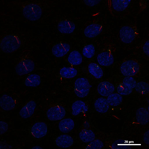
Immunocytochemistry/ Immunofluorescence: PIBF1 Antibody [NBP2-19823]
Immunocytochemistry/Immunofluorescence: PIBF1 Antibody [NBP2-19823] - Analysis of methanol-fixed HCT116, using antibody at 1:500 dilution.Applications for PIBF1 Antibody
Application
Recommended Usage
Immunocytochemistry/ Immunofluorescence
1:100-1:1000
Western Blot
1:500-1:3000
Reviewed Applications
Read 2 reviews rated 4 using NBP2-19823 in the following applications:
Formulation, Preparation, and Storage
Purification
Antigen Affinity-purified
Formulation
PBS, 1% BSA, 20% Glycerol
Preservative
0.01% Thimerosal
Concentration
Concentrations vary lot to lot. See vial label for concentration. If unlisted please contact technical services.
Shipping
The product is shipped with polar packs. Upon receipt, store it immediately at the temperature recommended below.
Stability & Storage
Aliquot and store at -20C or -80C. Avoid freeze-thaw cycles.
Background: PIBF1
Long Name
Progesterone Immunomodulatory Binding Factor 1
Alternate Names
C13orf24, chromosome 13 open reading frame 24, KIAA1008, PIBF, progesterone immunomodulatory binding factor 1, progesterone-induced blocking factor 1, progesterone-induced-blocking factor 1, RP11-505F3.1
Gene Symbol
PIBF1
UniProt
Additional PIBF1 Products
Product Documents for PIBF1 Antibody
Certificate of Analysis
To download a Certificate of Analysis, please enter a lot or batch number in the search box below.
Product Specific Notices for PIBF1 Antibody
This product is for research use only and is not approved for use in humans or in clinical diagnosis. Primary Antibodies are guaranteed for 1 year from date of receipt.
⚠ WARNING: This product can expose you to chemicals including mercury, which is known to the State of California to cause reproductive toxicity with developmental effects. For more information go to www.P65Warnings.ca.gov.Citations for PIBF1 Antibody
 Powered by Bioz
Customer Reviews for PIBF1 Antibody (2)
4 out of 5
2 Customer Ratings
Have you used PIBF1 Antibody?
Submit a review and receive an Amazon gift card!
$25/€18/£15/$25CAN/¥2500 Yen for a review with an image
$10/€7/£6/$10CAN/¥1110 Yen for a review without an image
Submit a review
Customer Images

Showing
1
-
2 of
2 reviews
Showing All
Filter By:
-
Application: Western BlotSample Tested: IMCD3, hTertRPE1, HDF, SHSY5Y whole cell lysatesSpecies: HumanVerified Customer | Posted 10/30/2013Rabbit anti-PIBF1

-
Application: ImmunofluorescenceSample Tested: Mouse inner medullary collecting duct (IMCD3) cell lineSpecies: MouseVerified Customer | Posted 10/14/2013
There are no reviews that match your criteria.
Protocols
Find general support by application which include: protocols, troubleshooting, illustrated assays, videos and webinars.
- Appropriate Fixation of IHC/ICC Samples
- Cellular Response to Hypoxia Protocols
- ClariTSA™ Fluorophore Kits
- Detection & Visualization of Antibody Binding
- ICC Cell Smear Protocol for Suspension Cells
- ICC Immunocytochemistry Protocol Videos
- ICC for Adherent Cells
- Immunocytochemistry (ICC) Protocol
- Immunocytochemistry Troubleshooting
- Immunofluorescence of Organoids Embedded in Cultrex Basement Membrane Extract
- Immunohistochemistry (IHC) and Immunocytochemistry (ICC) Protocols
- Preparing Samples for IHC/ICC Experiments
- Preventing Non-Specific Staining (Non-Specific Binding)
- Primary Antibody Selection & Optimization
- Protocol for VisUCyte™ HRP Polymer Detection Reagent
- Protocol for the Fluorescent ICC Staining of Cell Smears - Graphic
- Protocol for the Fluorescent ICC Staining of Cultured Cells on Coverslips - Graphic
- Protocol for the Preparation and Fluorescent ICC Staining of Cells on Coverslips
- Protocol for the Preparation and Fluorescent ICC Staining of Non-adherent Cells
- Protocol for the Preparation and Fluorescent ICC Staining of Stem Cells on Coverslips
- Protocol for the Preparation of a Cell Smear for Non-adherent Cell ICC - Graphic
- R&D Systems Quality Control Western Blot Protocol
- TUNEL and Active Caspase-3 Detection by IHC/ICC Protocol
- The Importance of IHC/ICC Controls
- Troubleshooting Guide: Western Blot Figures
- Western Blot Conditions
- Western Blot Protocol
- Western Blot Protocol for Cell Lysates
- Western Blot Troubleshooting
- Western Blot Troubleshooting Guide
- View all Protocols, Troubleshooting, Illustrated assays and Webinars
Loading...