PKM1 Antibody - BSA Free
Novus Biologicals | Catalog # NBP2-14833
![Western Blot: PKM1 AntibodyBSA Free [NBP2-14833]](https://resources.rndsystems.com/images/products/PKM1-Antibody-Western-Blot-NBP2-14833-img0008.jpg "Western Blot: PKM1 AntibodyBSA Free [NBP2-14833]")
Key Product Details
Validated by
Species Reactivity
Validated:
Cited:
Applications
Validated:
Cited:
Label
Antibody Source
Format
Product Specifications
Immunogen
Reactivity Notes
Localization
Specificity
Clonality
Host
Isotype
Scientific Data Images for PKM1 Antibody - BSA Free
Western Blot: PKM1 AntibodyBSA Free [NBP2-14833]
PKM1-Antibody-Western-Blot-NBP2-14833-img0008.jpg![Immunohistochemistry-Paraffin: PKM1 Antibody - BSA Free [NBP2-14833]](https://resources.rndsystems.com/images/products/PKM1-Antibody-Immunohistochemistry-Paraffin-NBP2-14833-img0002.jpg "Immunohistochemistry-Paraffin: PKM1 Antibody - BSA Free [NBP2-14833]")
Immunohistochemistry-Paraffin: PKM1 Antibody - BSA Free [NBP2-14833]
Immunohistochemistry-Paraffin: PKM1 Antibody [NBP2-14833] - PKM1 antibody was tested in human breast cancer using DAB with hematoxylin counterstain.![Western Blot: PKM1 AntibodyBSA Free [NBP2-14833]](https://resources.rndsystems.com/images/products/PKM1-Antibody-Western-Blot-NBP2-14833-img0003.jpg "Western Blot: PKM1 AntibodyBSA Free [NBP2-14833]")
Western Blot: PKM1 AntibodyBSA Free [NBP2-14833]
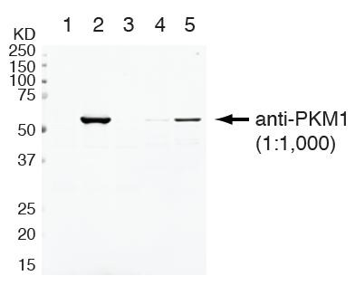
Western Blot: PKM1 Antibody [NBP2-14833] - PKM1 antibody was tested in 1. human skeletal muscle 2. mouse skeletal muscle 3. human brain 4. mouse brain and 5. human heart cell lysate.![Western Blot: PKM1 AntibodyBSA Free [NBP2-14833]](https://resources.rndsystems.com/images/products/PKM1-Antibody-Western-Blot-NBP2-14833-img0004.jpg "Western Blot: PKM1 AntibodyBSA Free [NBP2-14833]")
Western Blot: PKM1 AntibodyBSA Free [NBP2-14833]
Western Blot: PKM1 Antibody [NBP2-14833] - Detection of PKM1 on HEK293T and U-87 cells. Lane 1: HEK293T cells (major isoform is PKM2, human). Lane 2: HEK293T cells with plasmid overexpressing mouse PKM1. Lane 3: HEK293T cells with plasmid overexpressing mouse PKM2. Lane 4: U-87 cells with control shRNA (major isoform is PKM2, human). Lane 5: U-87 cells with PTB/A1/A2 shRNAs (major isoform is PKM1, human). Photo courtesy of product review by verified customer.![Western Blot: PKM1 AntibodyBSA Free [NBP2-14833]](https://resources.rndsystems.com/images/products/PKM1-Antibody-Western-Blot-NBP2-14833-img0006.jpg "Western Blot: PKM1 AntibodyBSA Free [NBP2-14833]")
![Western Blot: PKM1 AntibodyBSA Free [NBP2-14833]](https://resources.rndsystems.com/images/products/PKM1-Antibody-Western-Blot-NBP2-14833-img0007.jpg "Western Blot: PKM1 AntibodyBSA Free [NBP2-14833]")
Western Blot: PKM1 AntibodyBSA Free [NBP2-14833]
PKM1-Antibody-Western-Blot-NBP2-14833-img0007.jpg![Simple Western: PKM1 AntibodyBSA Free [NBP2-14833]](https://resources.rndsystems.com/images/products/PKM1-Antibody-Simple-Western-NBP2-14833-img0005.jpg "Simple Western: PKM1 AntibodyBSA Free [NBP2-14833]")
Simple Western: PKM1 AntibodyBSA Free [NBP2-14833]
Simple Western: PKM1 Antibody [NBP2-14833] - Simple Western lane view shows a specific band for PKM1 in 0.5 mg/ml of Human Brain lysate. This experiment was performed under reducing conditions using the 12-230 kDa separation system.
Immunohistochemistry-Paraffin: PKM1 Antibody - BSA Free [NBP2-14833] -
Immunohistochemistry-Paraffin: PKM1 Antibody - BSA Free [NBP2-14833] - Expression of PKM1 & PKM2 in clinical colorectal cancer samples.(a) The protein expression of PKM1 & PKM2 in clinical specimens of cancer tumor (T) & the adjacent normal tissues (N) is shown. PKM1 & PKM2 were detected by Western blotting in under the same experimental conditions at the same time. The full-length blots are presented in Supplementary Figure S3b. (b–d) Immunohistochemical staining of normal colon tissue adjacent to tumor tissue of case 10. Results of H&E staining (b), staining with anti-PKM1 antibody (c), & staining with anti-PKM2 (d) are shown. The boxed regions in “c” & “d” are enlarged in the images below. (e–h) Immunohistochemical staining of clinical colorectal cancer tissue specimen of representative case 3. H&E-stained section with normal tissue (upper right corner) neighboring the tumor area in the section is shown (e), along with the same section stained with anti-PKM2 antibody (f). Enlarged views of boxed areas in “f” show normal colorectal crypt in mucosa (g) & tumor area (h) stained with anti-PKM2 antibody. Image collected & cropped by CiteAb from the following publication (https://www.nature.com/articles/srep08647), licensed under a CC-BY license. Not internally tested by Novus Biologicals.
Western Blot: PKM1 Antibody - BSA Free [NBP2-14833] -
Western Blot: PKM1 Antibody - BSA Free [NBP2-14833] - (a) Luciferase activities after co-transfection of DLD-1 cells w/ control or miR-124 (wild-type or mutant-type) pMIR vectors having predictive miR-124 binding site in 3′UTR of PTB1. Upper panel region of 3′-UTR of human PTB1 mRNA complementary to mature miR-124. Box indicates predicted binding sites for miR-124. (b) Same as “a” except miR-133b used. (c) Expression of PTB1, PKM1, & PKM2 proteins at 72 h after transfection of DLD-1, NB9 or IMR-32 cells w/ miR-124 (10, 20 or 40 nM). (d) Expression of PTB1, PKM1, & PKM2 proteins at 72 h after transfection of DLD-1, RD or KYM-1 cells w/ miR-133b (10, 20 nM). (e) Expression of PTB1, PKM1, & PKM2 proteins at 72 h after transfection of DLD-1, NB-9 or RD cells w/ siR-PTB1 (2, 5 nM). (f) Effect of combined treatment of DLD-1 cells w/ antagomiR-124 & miR-124 or antagomiR-133b & miR-133b. DLD-1 cells transfected w/ non-specific control, miR-124/miR-133b (10 nM), miR-124/miR-133b (10 nM) + antagomiR-124/antagomiR-133b (5 nM) or miR-124/miR-133b (10 nM) + antagomiR-124/antagomiR-133b (10 nM). Expression level of PTB1 assessed at 48 h after transfection. The full-length blots are presented in Supplementary Figure S3a. (g) IF of PKM1 (upper panels) & PKM2 (lower panels) at 48 h after transfection of DLD-1 cells w/ miR-124 (20 nM) or miR-133b (20 nM). Left panels, treatment w/ control miRNA; middle panels, treatment w/ miR-124; right panels, treatment w/ miR-133b. PKM1 or PKM2 is stained red, & nuclei are stained blue. (h) Lactate production measured at 48 h after transfection of DLD-1 cells w/ miR-124 (20 nM), miR-133b (20 nM) or siR-PTB1 (5 nM). Results are presented as mean± SD (* P < 0.05; ** P < 0.01; *** P < 0.001; N.S., not statistically significant). Image collected & cropped by CiteAb from the following publication (https://www.nature.com/articles/srep08647), licensed under a CC-BY license. Not internally tested by Novus Biologicals.
Immunocytochemistry/ Immunofluorescence: PKM1 Antibody - BSA Free [NBP2-14833] -
Immunocytochemistry/ Immunofluorescence: PKM1 Antibody - BSA Free [NBP2-14833] - (a) Luciferase activities after co-transfection of DLD-1 cells w/ control or miR-124 (wild-type or mutant-type) pMIR vectors having predictive miR-124 binding site in 3′UTR of PTB1. Upper panel region of 3′-UTR of human PTB1 mRNA complementary to mature miR-124. Box indicates predicted binding sites for miR-124. (b) Same as “a” except miR-133b used. (c) Expression of PTB1, PKM1, & PKM2 proteins at 72 h after transfection of DLD-1, NB9 or IMR-32 cells w/ miR-124 (10, 20 or 40 nM). (d) Expression of PTB1, PKM1, & PKM2 proteins at 72 h after transfection of DLD-1, RD or KYM-1 cells w/ miR-133b (10, 20 nM). (e) Expression of PTB1, PKM1, & PKM2 proteins at 72 h after transfection of DLD-1, NB-9 or RD cells w/ siR-PTB1 (2, 5 nM). (f) Effect of combined treatment of DLD-1 cells w/ antagomiR-124 & miR-124 or antagomiR-133b & miR-133b. DLD-1 cells transfected w/ non-specific control, miR-124/miR-133b (10 nM), miR-124/miR-133b (10 nM) + antagomiR-124/antagomiR-133b (5 nM) or miR-124/miR-133b (10 nM) + antagomiR-124/antagomiR-133b (10 nM). Expression level of PTB1 assessed at 48 h after transfection. The full-length blots are presented in Supplementary Figure S3a. (g) IF of PKM1 (upper panels) & PKM2 (lower panels) at 48 h after transfection of DLD-1 cells w/ miR-124 (20 nM) or miR-133b (20 nM). Left panels, treatment w/ control miRNA; middle panels, treatment w/ miR-124; right panels, treatment w/ miR-133b. PKM1 or PKM2 is stained red, & nuclei are stained blue. (h) Lactate production measured at 48 h after transfection of DLD-1 cells w/ miR-124 (20 nM), miR-133b (20 nM) or siR-PTB1 (5 nM). Results are presented as mean± SD (* P < 0.05; ** P < 0.01; *** P < 0.001; N.S., not statistically significant). Image collected & cropped by CiteAb from the following publication (https://www.nature.com/articles/srep08647), licensed under a CC-BY license. Not internally tested by Novus Biologicals.
Western Blot: PKM1 Antibody - BSA Free [NBP2-14833] -
Western Blot: PKM1 Antibody - BSA Free [NBP2-14833] - (a) Luciferase activities after co-transfection of DLD-1 cells w/ control or miR-124 (wild-type or mutant-type) pMIR vectors having predictive miR-124 binding site in 3′UTR of PTB1. Upper panel region of 3′-UTR of human PTB1 mRNA complementary to mature miR-124. Box indicates predicted binding sites for miR-124. (b) Same as “a” except miR-133b used. (c) Expression of PTB1, PKM1, & PKM2 proteins at 72 h after transfection of DLD-1, NB9 or IMR-32 cells w/ miR-124 (10, 20 or 40 nM). (d) Expression of PTB1, PKM1, & PKM2 proteins at 72 h after transfection of DLD-1, RD or KYM-1 cells w/ miR-133b (10, 20 nM). (e) Expression of PTB1, PKM1, & PKM2 proteins at 72 h after transfection of DLD-1, NB-9 or RD cells w/ siR-PTB1 (2, 5 nM). (f) Effect of combined treatment of DLD-1 cells w/ antagomiR-124 & miR-124 or antagomiR-133b & miR-133b. DLD-1 cells transfected w/ non-specific control, miR-124/miR-133b (10 nM), miR-124/miR-133b (10 nM) + antagomiR-124/antagomiR-133b (5 nM) or miR-124/miR-133b (10 nM) + antagomiR-124/antagomiR-133b (10 nM). Expression level of PTB1 assessed at 48 h after transfection. The full-length blots are presented in Supplementary Figure S3a. (g) IF of PKM1 (upper panels) & PKM2 (lower panels) at 48 h after transfection of DLD-1 cells w/ miR-124 (20 nM) or miR-133b (20 nM). Left panels, treatment w/ control miRNA; middle panels, treatment w/ miR-124; right panels, treatment w/ miR-133b. PKM1 or PKM2 is stained red, & nuclei are stained blue. (h) Lactate production measured at 48 h after transfection of DLD-1 cells w/ miR-124 (20 nM), miR-133b (20 nM) or siR-PTB1 (5 nM). Results are presented as mean± SD (* P < 0.05; ** P < 0.01; *** P < 0.001; N.S., not statistically significant). Image collected & cropped by CiteAb from the following publication (https://www.nature.com/articles/srep08647), licensed under a CC-BY license. Not internally tested by Novus Biologicals.
Western Blot: PKM1 Antibody - BSA Free [NBP2-14833] -
Western Blot: PKM1 Antibody - BSA Free [NBP2-14833] - Expression of PKM1 & PKM2 in clinical colorectal cancer samples.(a) The protein expression of PKM1 & PKM2 in clinical specimens of cancer tumor (T) & the adjacent normal tissues (N) is shown. PKM1 & PKM2 were detected by Western blotting in under the same experimental conditions at the same time. The full-length blots are presented in Supplementary Figure S3b. (b–d) Immunohistochemical staining of normal colon tissue adjacent to tumor tissue of case 10. Results of H&E staining (b), staining with anti-PKM1 antibody (c), & staining with anti-PKM2 (d) are shown. The boxed regions in “c” & “d” are enlarged in the images below. (e–h) Immunohistochemical staining of clinical colorectal cancer tissue specimen of representative case 3. H&E-stained section with normal tissue (upper right corner) neighboring the tumor area in the section is shown (e), along with the same section stained with anti-PKM2 antibody (f). Enlarged views of boxed areas in “f” show normal colorectal crypt in mucosa (g) & tumor area (h) stained with anti-PKM2 antibody. Image collected & cropped by CiteAb from the following publication (https://www.nature.com/articles/srep08647), licensed under a CC-BY license. Not internally tested by Novus Biologicals.
Western Blot: PKM1 Antibody - BSA Free [NBP2-14833] -
Western Blot: PKM1 Antibody - BSA Free [NBP2-14833] - Characterization of NOX4 & PKM2 in human RCC tumors & adjacent tissue. a Mitochondrial fractions were prepared from human tumors (T) or uninvolved adjacent tissue (N). NOX4 expression was examined by western blot analysis. Prohibitin was probed as a mitochondrial marker & loading control. b Quantitation of NOX4 distribution in the mitochondrial fraction from a. The results are expressed as the means using one-way ANOVA with Tukey’s post hoc test where ± S.E.M. *p < 0.05 compared to normal (N). c Mitochondria fractions were prepared from RCC tumors & NADPH-dependent superoxide generation was examined in the presence (+) or absence (−) of ATP. The results are from eight tumors & are expressed as the means using one-way ANOVA with Tukey’s post hoc test where ±S.E.M. **p < 0.01 is compared to without (−) ATP. d PKM2 & PKM1 expression was examined by western blot analysis in lysates prepared from human tumors (T) or uninvolved adjacent tissue (N) from the same patient. Actin as loading control Image collected & cropped by CiteAb from the following publication (https://pubmed.ncbi.nlm.nih.gov/29051480), licensed under a CC-BY license. Not internally tested by Novus Biologicals.Applications for PKM1 Antibody - BSA Free
Immunoblotting
Immunocytochemistry/ Immunofluorescence
Immunohistochemistry
Immunohistochemistry-Paraffin
Simple Western
Western Blot
In Simple Western only 10 - 15 uL of the recommended dilution is used per data point.
See Simple Western Antibody Database for Simple Western validation: Tested in Human Brain lysate 0.5 mg/mL, separated by Size, antibody dilution of 1:400, apparent MW was 59 kDa. Separated by Size-Wes, Sally Sue/Peggy Sue.
Reviewed Applications
Read 3 reviews rated 5 using NBP2-14833 in the following applications:
Formulation, Preparation, and Storage
Purification
Formulation
Format
Preservative
Concentration
Shipping
Stability & Storage
Background: PKM1
Alternate Names
Gene Symbol
UniProt
Additional PKM1 Products
Product Documents for PKM1 Antibody - BSA Free
Certificate of Analysis
To download a Certificate of Analysis, please enter a lot or batch number in the search box below.
Product Specific Notices for PKM1 Antibody - BSA Free
This product is for research use only and is not approved for use in humans or in clinical diagnosis. Primary Antibodies are guaranteed for 1 year from date of receipt.
Citations for PKM1 Antibody - BSA Free
 Powered by Bioz
Customer Reviews for PKM1 Antibody - BSA Free (3)
Have you used PKM1 Antibody - BSA Free?
Submit a review and receive an Amazon gift card!
$25/€18/£15/$25CAN/¥2500 Yen for a review with an image
$10/€7/£6/$10CAN/¥1110 Yen for a review without an image
Submit a review
Customer Images

-
Application: Western BlotSample Tested: Human fibroblastSpecies: HumanVerified Customer | Posted 10/24/2018

-
Application: Western BlotSample Tested: HCT116 cell line lysateSpecies: HumanVerified Customer | Posted 03/31/2016
-
Application: Western BlotSample Tested: U-87 cells with PTB/A1/A2 shRNAsSpecies: HumanVerified Customer | Posted 12/10/2012
There are no reviews that match your criteria.
Protocols
View specific protocols for PKM1 Antibody - BSA Free (NBP2-14833):
Immunohistochemistry-Paraffin Embedded Sections
Antigen Unmasking:
Bring slides to a boil in 10 mM sodium citrate buffer (pH 6.0) then maintain at a sub-boiling temperature for 10 minutes. Cool slides on bench-top for 30 minutes.
Staining:
1. Wash sections in deionized water three times for 5 minutes each.
2. Wash sections in wash buffer for 5 minutes.
3. Block each section with 100-400 ul blocking solution for 1 hour at room temperature.
4. Remove blocking solution and add 100-400 ul diluted primary antibody. Incubate overnight at 4C.
5. Remove antibody solution and wash sections in wash buffer three times for 5 minutes each.
6. Add 100-400 ul biotinylated diluted secondary antibody. Incubate 30 minutes at room temperature.
7. Remove secondary antibody solution and wash sections three times with wash buffer for 5 minutes each.
8. Add 100-400 ul Streptavidin-HRP reagent to each section and incubate for 30 minutes at room temperature.
9. Wash sections three times in wash buffer for 5 minutes each.
10. Add 100-400 ul DAB substrate to each section and monitor staining closely.
11. As soon as the sections develop, immerse slides in deionized water.
12. Counterstain sections in hematoxylin.
13. Wash sections in deionized water two times for 5 minutes each.
14. Dehydrate sections.
15. Mount coverslips.
Western Blot Protocol
1. Perform SDS-PAGE on samples to be analyzed, loading 25 ug of total protein per lane.
2. Transfer proteins to membrane according to the instructions provided by the manufacturer of the membrane and transfer apparatus.
3. Stain according to standard Ponceau S procedure (or similar product) to assess transfer success, and mark molecular weight standards where appropriate.
4. Rinse the blot.
5. Block the membrane using standard blocking buffer for at least 1 hour.
6. Wash the membrane in wash buffer three times for 10 minutes each.
7. Dilute primary antibody in blocking buffer and incubate 1 hour at room temperature.
8. Wash the membrane in wash buffer three times for 10 minutes each.
9. Apply the diluted HRP conjugated secondary antibody in blocking buffer (as per manufacturers instructions) and incubate 1 hour at room temperature.
10. Wash the blot in wash buffer three times for 10 minutes each (this step can be repeated as required to reduce background).
11. Apply the detection reagent of choice in accordance with the manufacturers instructions.
*Note: Tween-20 can be added to the blocking or antibody dilution buffer at a final concentration of 0.05-0.2%.
Find general support by application which include: protocols, troubleshooting, illustrated assays, videos and webinars.
- Antigen Retrieval Protocol (PIER)
- Antigen Retrieval for Frozen Sections Protocol
- Appropriate Fixation of IHC/ICC Samples
- Cellular Response to Hypoxia Protocols
- Chromogenic IHC Staining of Formalin-Fixed Paraffin-Embedded (FFPE) Tissue Protocol
- Chromogenic Immunohistochemistry Staining of Frozen Tissue
- ClariTSA™ Fluorophore Kits
- Detection & Visualization of Antibody Binding
- Fluorescent IHC Staining of Frozen Tissue Protocol
- Graphic Protocol for Heat-induced Epitope Retrieval
- Graphic Protocol for the Preparation and Fluorescent IHC Staining of Frozen Tissue Sections
- Graphic Protocol for the Preparation and Fluorescent IHC Staining of Paraffin-embedded Tissue Sections
- Graphic Protocol for the Preparation of Gelatin-coated Slides for Histological Tissue Sections
- ICC Cell Smear Protocol for Suspension Cells
- ICC Immunocytochemistry Protocol Videos
- ICC for Adherent Cells
- IHC Sample Preparation (Frozen sections vs Paraffin)
- Immunocytochemistry (ICC) Protocol
- Immunocytochemistry Troubleshooting
- Immunofluorescence of Organoids Embedded in Cultrex Basement Membrane Extract
- Immunofluorescent IHC Staining of Formalin-Fixed Paraffin-Embedded (FFPE) Tissue Protocol
- Immunohistochemistry (IHC) and Immunocytochemistry (ICC) Protocols
- Immunohistochemistry Frozen Troubleshooting
- Immunohistochemistry Paraffin Troubleshooting
- Preparing Samples for IHC/ICC Experiments
- Preventing Non-Specific Staining (Non-Specific Binding)
- Primary Antibody Selection & Optimization
- Protocol for Heat-Induced Epitope Retrieval (HIER)
- Protocol for Making a 4% Formaldehyde Solution in PBS
- Protocol for VisUCyte™ HRP Polymer Detection Reagent
- Protocol for the Fluorescent ICC Staining of Cell Smears - Graphic
- Protocol for the Fluorescent ICC Staining of Cultured Cells on Coverslips - Graphic
- Protocol for the Preparation & Fixation of Cells on Coverslips
- Protocol for the Preparation and Chromogenic IHC Staining of Frozen Tissue Sections
- Protocol for the Preparation and Chromogenic IHC Staining of Frozen Tissue Sections - Graphic
- Protocol for the Preparation and Chromogenic IHC Staining of Paraffin-embedded Tissue Sections
- Protocol for the Preparation and Chromogenic IHC Staining of Paraffin-embedded Tissue Sections - Graphic
- Protocol for the Preparation and Fluorescent ICC Staining of Cells on Coverslips
- Protocol for the Preparation and Fluorescent ICC Staining of Non-adherent Cells
- Protocol for the Preparation and Fluorescent ICC Staining of Stem Cells on Coverslips
- Protocol for the Preparation and Fluorescent IHC Staining of Frozen Tissue Sections
- Protocol for the Preparation and Fluorescent IHC Staining of Paraffin-embedded Tissue Sections
- Protocol for the Preparation of Gelatin-coated Slides for Histological Tissue Sections
- Protocol for the Preparation of a Cell Smear for Non-adherent Cell ICC - Graphic
- R&D Systems Quality Control Western Blot Protocol
- TUNEL and Active Caspase-3 Detection by IHC/ICC Protocol
- The Importance of IHC/ICC Controls
- Troubleshooting Guide: Immunohistochemistry
- Troubleshooting Guide: Western Blot Figures
- Western Blot Conditions
- Western Blot Protocol
- Western Blot Protocol for Cell Lysates
- Western Blot Troubleshooting
- Western Blot Troubleshooting Guide
- View all Protocols, Troubleshooting, Illustrated assays and Webinars
FAQs for PKM1 Antibody - BSA Free
-
Q: I just wanted to make sure the L/R didn't hit M1 and M2. I do not need to distinguish between L and R. So do you have an L/R specific monoclonal antibody for western blot?
A: We do have an antibody that is specific for PKM1, however it is a polyclonal. The catalog number is NBP2-14833.