Podocalyxin Like Antibody - BSA Free
Novus Biologicals | Catalog # NB110-41503
![Immunocytochemistry/ Immunofluorescence: Podocalyxin Like Antibody [NB110-41503]](https://resources.rndsystems.com/images/products/Podocalyxin-Like-Antibody-Immunocytochemistry-Immunofluorescence-NB110-41503-img0007.jpg "Immunocytochemistry/ Immunofluorescence: Podocalyxin Like Antibody [NB110-41503]")
Key Product Details
Species Reactivity
Validated:
Human, Mouse
Cited:
Human
Applications
Validated:
Western Blot, Immunocytochemistry/ Immunofluorescence
Cited:
Immunocytochemistry/ Immunofluorescence
Label
Unconjugated
Antibody Source
Polyclonal Rabbit IgG
Format
BSA Free
Loading...
Product Specifications
Immunogen
A synthetic peptide within an internal region (residues 100-200) of the human Podocalyxin protein. [Swiss-Prot: O00592] This immunogen should be far enough away from any glycosylation site.
Localization
Membrane
Marker
Podocytes /Apical Marker, Endothelial Cell Marker
Clonality
Polyclonal
Host
Rabbit
Isotype
IgG
Scientific Data Images for Podocalyxin Like Antibody - BSA Free
Immunocytochemistry/ Immunofluorescence: Podocalyxin Like Antibody [NB110-41503]
Immunocytochemistry/Immunofluorescence: Podocalyxin Like Antibody [NB110-41503] - HeLa cells were fixed for 10 minutes using 10% formalin and then permeabilized for 5 minutes using 1X PBS + 0.05% Triton-X100. The cells were incubated with anti-Podocalyxin Like Antibody at 2 ug/ml overnight at 4C and detected with an anti-rabbit Dylight 488 (Green) at a 1:500 dilution. Alpha tubulin (DM1A) NB100-690 was used as a co-stain at a 1:1000 dilution and detected with an anti-mouse Dylight 550 (Red) at a 1:500 dilution. Nuclei were counterstained with DAPI (Blue). Cells were imaged using a 40X objective.Applications for Podocalyxin Like Antibody - BSA Free
Application
Recommended Usage
Immunocytochemistry/ Immunofluorescence
1:1000
Western Blot
2 ug/ml
Application Notes
In Western blot, a band is observed at ~52 kDa. The observed molecular weight of the protein may vary from the listed predicted molecular weight due to post translational modifications, post translation cleavages, relative charges, and other experimental factors. In ICC/IF membrane staining was observed.
Reviewed Applications
Read 1 review rated 3 using NB110-41503 in the following applications:
Formulation, Preparation, and Storage
Purification
Immunogen affinity purified
Formulation
PBS
Format
BSA Free
Preservative
0.02% Sodium Azide
Concentration
1.0 mg/ml
Shipping
The product is shipped with polar packs. Upon receipt, store it immediately at the temperature recommended below.
Stability & Storage
Aliquot and store at -20C or -80C. Avoid freeze-thaw cycles.
Background: Podocalyxin
Alternate Names
GCTM, PCLP, POD XL, PODXL
Gene Symbol
PODXL
Additional Podocalyxin Products
Product Documents for Podocalyxin Like Antibody - BSA Free
Certificate of Analysis
To download a Certificate of Analysis, please enter a lot or batch number in the search box below.
Product Specific Notices for Podocalyxin Like Antibody - BSA Free
This product is for research use only and is not approved for use in humans or in clinical diagnosis. Primary Antibodies are guaranteed for 1 year from date of receipt.
Related Research Areas
Citations for Podocalyxin Like Antibody - BSA Free
 Powered by Bioz
Customer Reviews for Podocalyxin Like Antibody - BSA Free (1)
3 out of 5
1 Customer Rating
Have you used Podocalyxin Like Antibody - BSA Free?
Submit a review and receive an Amazon gift card!
$25/€18/£15/$25CAN/¥2500 Yen for a review with an image
$10/€7/£6/$10CAN/¥1110 Yen for a review without an image
Submit a review
Customer Images
Showing
1
-
1 of
1 review
Showing All
Filter By:
-
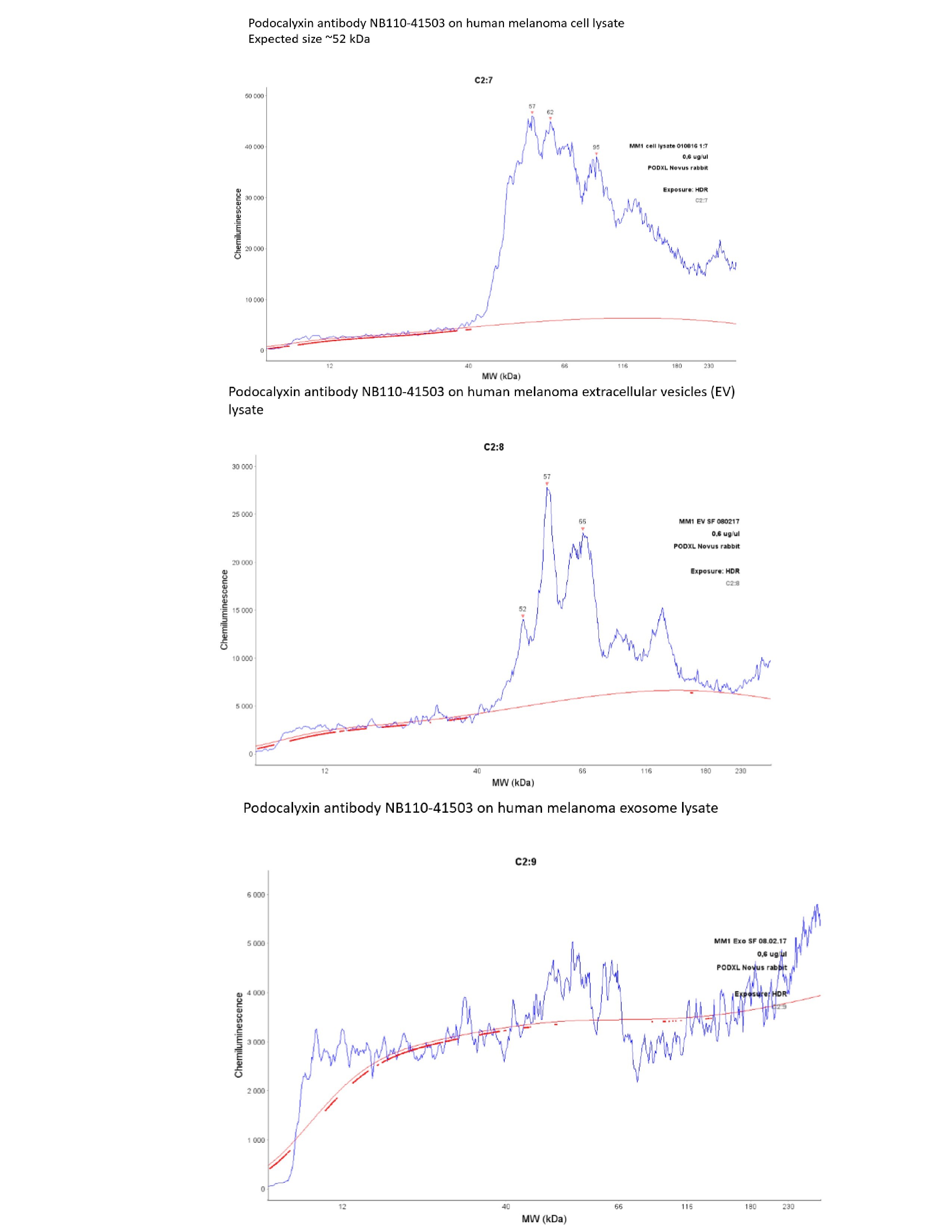
Application: Simple WesternSample Tested: lysates from melanoma cellsSpecies: HumanVerified Customer | Posted 08/21/2017Tested on Peggy Sue. I used lysates from melanoma cells, melanoma extracellular vesicles (EV) and lysates from melanoma exosomes.The polyclonal antibody NB110-41503 yields a very intense signal with several peaks. The peaks are all around the expected size, but I don’t know if the multiple peaks are due to the polyclonal nature of the antibody or different proteins being detected. The signal is absent in the exosome lysate, in line with the observation made using antibody MAB1658.

There are no reviews that match your criteria.
Protocols
View specific protocols for Podocalyxin Like Antibody - BSA Free (NB110-41503):
Podocalyxin Like Antibody:
Culture cells to appropriate density in 35 mm culture dishes or 6-well plates.
1. Remove culture medium and add 10% formalin to the dish. Fix at room temperature for 30 minutes.
2. Remove the formalin and add ice cold methanol. Incubate for 5-10 minutes.
3. Remove methanol and add washing solution (i.e. PBS). Be sure to not let the specimen dry out. Wash three times for 10 minutes.
4. To block nonspecific antibody binding incubate in 10% normal goat serum from 1 hour to overnight at room temperature.
5. Add primary antibody at appropriate dilution and incubate at room temperature from 2 hours to overnight at room temperature.
6. Remove primary antibody and replace with washing solution. Wash three times for 10 minutes.
7. Add secondary antibody at appropriate dilution. Incubate for 1 hour at room temperature.
8. Remove antibody and replace with wash solution, then wash for 10 minutes. Add Hoechst 33258 to wash solution at 1:25,0000 and incubate for 10 minutes. Wash a third time for 10 minutes.
9. Cells can be viewed directly after washing. The plates can also be stored in PBS containing Azide covered in Parafilm (TM). Cells can also be cover-slipped using Fluoromount, with appropriate sealing.
*The above information is only intended as a guide. The researcher should determine what protocol best meets their needs. Please follow safe laboratory procedures.
Podocalyxin Like Antibody:
Western Blot Protocol
1. Perform SDS-PAGE (4-12%) on samples to be analyzed, loading 30 ug of total protein per
lane.
2. Transfer proteins to Nitrocellulose according to the instructions provided by the manufacturer of the transfer
apparatus.
3. Rinse membrane with dH2O and then stain the blot using Ponceau S for 1-2 minutes to access the transfer of proteins onto the nitrocellulose membrane. Rinse the blot in water to remove excess stain and mark the lane locations and locations of molecular weight markers using a pencil.
4. Rinse the blot in TBS for approximately 5 minutes.
5. Block the membrane using 5% non-fat dry milk + 1% BSA in TBS, 1 hour at room temperature.
6. Rinse the membrane in dH2O and then wash the membrane in wash buffer [TBS + 0.1% Tween] 3 times for 10 minutes each.
7. Dilute the rabbit anti-Podolyxin primary antibody (NB 110-41503) in blocking buffer and incubate 2 hours at room temperature.
8. Rinse the membrane in dH2O and then wash the membrane in wash buffer [TBS + 0.1% Tween] 3 times for 10 minutes each.
9. Apply the diluted rabbit-IgG HRP-conjugated secondary antibody in blocking buffer (as per manufacturers
instructions) and incubate 1 hour at room temperature.
10. Wash the blot in wash buffer [TBS + 0.1% Tween] 3 times for 10 minutes each (this step can be repeated as required to reduce background).
11. Apply the detection reagent of choice in accordance with the manufacturers instructions (Pierce, ECL).
Note: Tween-20 can be added to the blocking or antibody diultion buffer at a final concentration of 0.05-0.2%, provided it does not interfere with antibody-antigen binding.
Find general support by application which include: protocols, troubleshooting, illustrated assays, videos and webinars.
- Appropriate Fixation of IHC/ICC Samples
- Cellular Response to Hypoxia Protocols
- ClariTSA™ Fluorophore Kits
- Detection & Visualization of Antibody Binding
- ICC Cell Smear Protocol for Suspension Cells
- ICC Immunocytochemistry Protocol Videos
- ICC for Adherent Cells
- Immunocytochemistry (ICC) Protocol
- Immunocytochemistry Troubleshooting
- Immunofluorescence of Organoids Embedded in Cultrex Basement Membrane Extract
- Immunohistochemistry (IHC) and Immunocytochemistry (ICC) Protocols
- Preparing Samples for IHC/ICC Experiments
- Preventing Non-Specific Staining (Non-Specific Binding)
- Primary Antibody Selection & Optimization
- Protocol for VisUCyte™ HRP Polymer Detection Reagent
- Protocol for the Fluorescent ICC Staining of Cell Smears - Graphic
- Protocol for the Fluorescent ICC Staining of Cultured Cells on Coverslips - Graphic
- Protocol for the Preparation and Fluorescent ICC Staining of Cells on Coverslips
- Protocol for the Preparation and Fluorescent ICC Staining of Non-adherent Cells
- Protocol for the Preparation and Fluorescent ICC Staining of Stem Cells on Coverslips
- Protocol for the Preparation of a Cell Smear for Non-adherent Cell ICC - Graphic
- R&D Systems Quality Control Western Blot Protocol
- TUNEL and Active Caspase-3 Detection by IHC/ICC Protocol
- The Importance of IHC/ICC Controls
- Troubleshooting Guide: Western Blot Figures
- Western Blot Conditions
- Western Blot Protocol
- Western Blot Protocol for Cell Lysates
- Western Blot Troubleshooting
- Western Blot Troubleshooting Guide
- View all Protocols, Troubleshooting, Illustrated assays and Webinars
Loading...