Rad50 Antibody - BSA Free
Novus Biologicals | Catalog # NB100-154
![Simple Western: Rad50 Antibody [NB100-154]](https://resources.rndsystems.com/images/products/Rad50-Antibody-Simple-Western-NB100-154-img0004.jpg "Simple Western: Rad50 Antibody [NB100-154]")
Loading...
Key Product Details
Species Reactivity
Validated:
Human, Mouse, Hamster
Cited:
Human, Mouse, Hamster
Applications
Validated:
Western Blot, Immunocytochemistry/ Immunofluorescence, Simple Western, Immunoprecipitation, ICC/IF (Negative)
Cited:
Western Blot, Immunocytochemistry/ Immunofluorescence, Immunoprecipitation, In vivo assay
Label
Unconjugated
Antibody Source
Polyclonal Rabbit IgG
Format
BSA Free
Loading...
Product Specifications
Immunogen
C-terminal mouse RAD50 (604 amino acids). [UniProt# P70388]
Localization
Upon DNA damage, Rad50 and Mre11 redistribute and are co-localized within the nucleus of damaged cells.
Clonality
Polyclonal
Host
Rabbit
Isotype
IgG
Theoretical MW
153 kDa.
Disclaimer note: The observed molecular weight of the protein may vary from the listed predicted molecular weight due to post translational modifications, post translation cleavages, relative charges, and other experimental factors.
Disclaimer note: The observed molecular weight of the protein may vary from the listed predicted molecular weight due to post translational modifications, post translation cleavages, relative charges, and other experimental factors.
Scientific Data Images for Rad50 Antibody - BSA Free
Simple Western: Rad50 Antibody [NB100-154]
Simple Western: Rad50 Antibody [NB100-154] - Simple Western lane view shows a specific band for Rad50 in 0.5 mg/ml of HeLa lysate. This experiment was performed under reducing conditions using the 12-230 kDa separation system.![Western Blot: Rad50 Antibody [NB100-154]](https://resources.rndsystems.com/images/products/Rad50-Antibody-Western-Blot-NB100-154-img0006.jpg "Western Blot: Rad50 Antibody [NB100-154]")
Western Blot: Rad50 Antibody [NB100-154]
Rad50-Antibody-Western-Blot-NB100-154-img0006.jpg![Western Blot: Rad50 Antibody [NB100-154]](https://resources.rndsystems.com/images/products/Rad50-Antibody-Western-Blot-NB100-154-img0005.jpg "Western Blot: Rad50 Antibody [NB100-154]")
Western Blot: Rad50 Antibody [NB100-154]
Western Blot: Rad50 Antibody [NB100-154] - WB detection of RAD50 protein in 30ug whole cell lysate of 293T cells using Rad50 antibody at 1:2000 dilution (Submitted via Verified Customer Review).![Western Blot: Rad50 Antibody [NB100-154]](https://resources.rndsystems.com/images/products/Rad50-Antibody-Western-Blot-NB100-154-img0002.jpg "Western Blot: Rad50 Antibody [NB100-154]")
Western Blot: Rad50 Antibody [NB100-154]
Western Blot: Rad50 Antibody [NB100-154] - Detection of Rad50 in HeLa nuclear extracts.Applications for Rad50 Antibody - BSA Free
Application
Recommended Usage
Immunocytochemistry/ Immunofluorescence
reported in scientific literature
Immunoprecipitation
1:10-1:500
Simple Western
1:2000
Western Blot
1:2000
Application Notes
This RAD50 antibody is useful for Immunoprecipitation and Western blot, where a band can be seen at 153 kDa. IP has been done with 3-4 X 10(6) cells. ICC/IF does not appear to work. Immunocytochemistry/Immunofluorescence was reported in scientific literature.
In Simple Western only 10 - 15 uL of the recommended dilution is used per data point.
See Simple Western Antibody Database for Simple Western validation: Tested in HeLa lysate 0.5 mg/mL, separated by Size, antibody dilution of 1:2000, apparent MW was 151 kDa. Separated by Size-Wes, Sally Sue/Peggy Sue.
The observed molecular weight of the protein may vary from the listed predicted molecular weight due to post translational modifications, post translation cleavages, relative charges, and other experimental factors.
Reviewed Applications
Read 2 reviews rated 4 using NB100-154 in the following applications:
Formulation, Preparation, and Storage
Purification
Unpurified
Formulation
Whole antisera
Format
BSA Free
Preservative
0.02% Sodium Azide
Concentration
This product is unpurified. The exact concentration of antibody is not quantifiable.
Shipping
The product is shipped with polar packs. Upon receipt, store it immediately at the temperature recommended below.
Stability & Storage
Store at 4C for up to 3 months. For longer storage, aliquot and store at -20C.
Background: Rad50
Alternate Names
DNA repair protein RAD50, EC 3.6, EC 3.6.1.15, EC 3.6.3, EC 3.6.3.27, hRAD50, NBSLD, RAD50 (S. cerevisiae) homolog, RAD50 homolog (S. cerevisiae), RAD502, RAD50-2
Gene Symbol
RAD50
Additional Rad50 Products
Product Documents for Rad50 Antibody - BSA Free
Certificate of Analysis
To download a Certificate of Analysis, please enter a lot or batch number in the search box below.
Product Specific Notices for Rad50 Antibody - BSA Free
This product is for research use only and is not approved for use in humans or in clinical diagnosis. Primary Antibodies are guaranteed for 1 year from date of receipt.
Citations for Rad50 Antibody - BSA Free
 Powered by Bioz
Customer Reviews for Rad50 Antibody - BSA Free (2)
4 out of 5
2 Customer Ratings
Have you used Rad50 Antibody - BSA Free?
Submit a review and receive an Amazon gift card!
$25/€18/£15/$25CAN/¥2500 Yen for a review with an image
$10/€7/£6/$10CAN/¥1110 Yen for a review without an image
Submit a review
Customer Images

Showing
1
-
2 of
2 reviews
Showing All
Filter By:
-
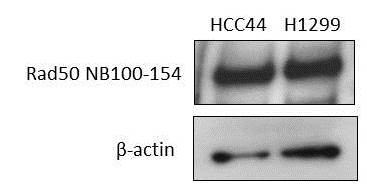
Application: Western BlotSample Tested: human lung cancer cell lineSpecies: HumanVerified Customer | Posted 02/18/2019

-
Application: Western BlotSample Tested: 293T lysateSpecies: HumanVerified Customer | Posted 12/17/2014RAD50 WB in 293T cells
There are no reviews that match your criteria.
Protocols
Find general support by application which include: protocols, troubleshooting, illustrated assays, videos and webinars.
- Appropriate Fixation of IHC/ICC Samples
- Cellular Response to Hypoxia Protocols
- ClariTSA™ Fluorophore Kits
- Detection & Visualization of Antibody Binding
- ICC Cell Smear Protocol for Suspension Cells
- ICC Immunocytochemistry Protocol Videos
- ICC for Adherent Cells
- Immunocytochemistry (ICC) Protocol
- Immunocytochemistry Troubleshooting
- Immunofluorescence of Organoids Embedded in Cultrex Basement Membrane Extract
- Immunohistochemistry (IHC) and Immunocytochemistry (ICC) Protocols
- Immunoprecipitation Protocol
- Preparing Samples for IHC/ICC Experiments
- Preventing Non-Specific Staining (Non-Specific Binding)
- Primary Antibody Selection & Optimization
- Protocol for VisUCyte™ HRP Polymer Detection Reagent
- Protocol for the Fluorescent ICC Staining of Cell Smears - Graphic
- Protocol for the Fluorescent ICC Staining of Cultured Cells on Coverslips - Graphic
- Protocol for the Preparation and Fluorescent ICC Staining of Cells on Coverslips
- Protocol for the Preparation and Fluorescent ICC Staining of Non-adherent Cells
- Protocol for the Preparation and Fluorescent ICC Staining of Stem Cells on Coverslips
- Protocol for the Preparation of a Cell Smear for Non-adherent Cell ICC - Graphic
- R&D Systems Quality Control Western Blot Protocol
- TUNEL and Active Caspase-3 Detection by IHC/ICC Protocol
- The Importance of IHC/ICC Controls
- Troubleshooting Guide: Western Blot Figures
- Western Blot Conditions
- Western Blot Protocol
- Western Blot Protocol for Cell Lysates
- Western Blot Troubleshooting
- Western Blot Troubleshooting Guide
- View all Protocols, Troubleshooting, Illustrated assays and Webinars
Loading...