RAD52 Antibody - BSA Free
Novus Biologicals | Catalog # NBP2-58116
![Western Blot: RAD52 Antibody [NBP2-58116]](https://resources.rndsystems.com/images/products/RAD52-Antibody-Western-Blot-NBP2-58116-img0002.jpg "Western Blot: RAD52 Antibody [NBP2-58116]")
Loading...
Key Product Details
Validated by
Biological Validation
Species Reactivity
Validated:
Human
Cited:
Human
Predicted:
Mouse (90%), Rat (90%). Backed by our 100% Guarantee.
Applications
Validated:
Immunocytochemistry/ Immunofluorescence, Knockdown Validated
Cited:
Western Blot, Immunocytochemistry/ Immunofluorescence, Knockdown Validated
Label
Unconjugated
Antibody Source
Polyclonal Rabbit IgG
Format
BSA Free
Loading...
Product Specifications
Immunogen
This antibody was developed against a recombinant protein corresponding to the following amino acid sequence: ILGGRDSHPAAGGGSVLCFGQCQYTAEEYQAIQKALRQRLGPEYISSRMAGGGQKVCYIEGHRVINLANEMFGYNGWAHSITQQNVDFVDLNNGKFYVGVCAFVRVQLKDGSYHEDVGYGVSE
Clonality
Polyclonal
Host
Rabbit
Isotype
IgG
Scientific Data Images for RAD52 Antibody - BSA Free
Western Blot: RAD52 Antibody [NBP2-58116]
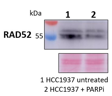
Western Blot: RAD52 Antibody [NBP2-58116] - HCC1937 human breast cancer cells untreated (1) and treated with a PARP inhibitor for 96 h (2). Two RAD52 bands can be appreciated at around 55 kDa. Ponceau red staining is used as total protein loading control (below). Primary antibody dilution 1:500 (overnight, 4C, rotation). Image from verified customer review.![RAD52 Antibody - BSA Free Immunocytochemistry/ Immunofluorescence: RAD52 Antibody [NBP2-58116]](https://resources.rndsystems.com/images/products/nbp2-58116_-immunocytochemistry-immunofluorescence-639174077014331439.jpg "Immunocytochemistry/ Immunofluorescence: RAD52 Antibody [NBP2-58116]")
Immunocytochemistry/ Immunofluorescence: RAD52 Antibody [NBP2-58116]
Staining of human cell line U-2 OS shows localization to nuclear speckles.Applications for RAD52 Antibody - BSA Free
Application
Recommended Usage
Immunocytochemistry/ Immunofluorescence
0.25-2 ug/ml
Knockdown Validated
Reported in scientific publication (PMID: 32690953)
Application Notes
ICC/IF, Fixation Permeabilization: Use PFA/Triton X-100.
Reviewed Applications
Read 1 review rated 4 using NBP2-58116 in the following applications:
Formulation, Preparation, and Storage
Purification
Affinity purified
Formulation
PBS (pH 7.2) and 40% Glycerol
Format
BSA Free
Preservative
0.02% Sodium Azide
Concentration
Concentrations vary lot to lot. See vial label for concentration. If unlisted please contact technical services.
Shipping
The product is shipped with polar packs. Upon receipt, store it immediately at the temperature recommended below.
Stability & Storage
Store at 4C short term. Aliquot and store at -20C long term. Avoid freeze-thaw cycles.
Background: RAD52
Alternate Names
DNA repair protein RAD52 homolog, RAD52 (S. cerevisiae) homolog, RAD52 homolog (S. cerevisiae), recombination protein RAD52, rhabdomyosarcoma antigen MU-RMS-40.23
Gene Symbol
RAD52
Additional RAD52 Products
Product Documents for RAD52 Antibody - BSA Free
Certificate of Analysis
To download a Certificate of Analysis, please enter a lot or batch number in the search box below.
Product Specific Notices for RAD52 Antibody - BSA Free
This product is for research use only and is not approved for use in humans or in clinical diagnosis. Primary Antibodies are guaranteed for 1 year from date of receipt.
Citations for RAD52 Antibody - BSA Free
 Powered by Bioz
Customer Reviews for RAD52 Antibody - BSA Free (1)
4 out of 5
1 Customer Rating
Have you used RAD52 Antibody - BSA Free?
Submit a review and receive an Amazon gift card!
$25/€18/£15/$25CAN/¥2500 Yen for a review with an image
$10/€7/£6/$10CAN/¥1110 Yen for a review without an image
Submit a review
Customer Images
Showing
1
-
1 of
1 review
Showing All
Filter By:
-
Application: Western BlotSample Tested: HCC1937 breast cancer cellsSpecies: HumanVerified Customer | Posted 05/31/2022HCC1937 human breast cancer cells untreated (1) and treated with a PARP inhibitor for 96 h (2). Two RAD52 bands can be appreciated at around 55 kDa. Ponceau red staining is used as total protein loading control (below).Primary antibody dilution 1:500 (overnight, 4ºC, rotation)

There are no reviews that match your criteria.
Protocols
Find general support by application which include: protocols, troubleshooting, illustrated assays, videos and webinars.
- Appropriate Fixation of IHC/ICC Samples
- Cellular Response to Hypoxia Protocols
- ClariTSA™ Fluorophore Kits
- Detection & Visualization of Antibody Binding
- ICC Cell Smear Protocol for Suspension Cells
- ICC Immunocytochemistry Protocol Videos
- ICC for Adherent Cells
- Immunocytochemistry (ICC) Protocol
- Immunocytochemistry Troubleshooting
- Immunofluorescence of Organoids Embedded in Cultrex Basement Membrane Extract
- Immunohistochemistry (IHC) and Immunocytochemistry (ICC) Protocols
- Preparing Samples for IHC/ICC Experiments
- Preventing Non-Specific Staining (Non-Specific Binding)
- Primary Antibody Selection & Optimization
- Protocol for VisUCyte™ HRP Polymer Detection Reagent
- Protocol for the Fluorescent ICC Staining of Cell Smears - Graphic
- Protocol for the Fluorescent ICC Staining of Cultured Cells on Coverslips - Graphic
- Protocol for the Preparation and Fluorescent ICC Staining of Cells on Coverslips
- Protocol for the Preparation and Fluorescent ICC Staining of Non-adherent Cells
- Protocol for the Preparation and Fluorescent ICC Staining of Stem Cells on Coverslips
- Protocol for the Preparation of a Cell Smear for Non-adherent Cell ICC - Graphic
- TUNEL and Active Caspase-3 Detection by IHC/ICC Protocol
- The Importance of IHC/ICC Controls
- View all Protocols, Troubleshooting, Illustrated assays and Webinars
Loading...