RASAL1 Antibody - BSA Free
Novus Biologicals | Catalog # NBP1-32776
![Western Blot: RASAL1 Antibody [NBP1-32776]](https://resources.rndsystems.com/images/products/RASAL1-Antibody-Western-Blot-NBP1-32776-img0006.jpg "Western Blot: RASAL1 Antibody [NBP1-32776]")
Loading...
Key Product Details
Species Reactivity
Validated:
Human, Monkey
Cited:
Mouse
Applications
Validated:
Western Blot, Immunocytochemistry/ Immunofluorescence, Proximity Ligation Assay, Knockdown Validated
Cited:
Western Blot, Proximity Ligation Assay, Knockdown Validated
Label
Unconjugated
Antibody Source
Polyclonal Rabbit IgG
Format
BSA Free
Loading...
Product Specifications
Immunogen
Recombinant protein encompassing a sequence within the C-terminus region of human RASAL1. The exact sequence is proprietary.
Reactivity Notes
Mouse reactivity reported in scientific literature (PMID: 31641113). Monkey reactivity reported from a verified customer review.
Clonality
Polyclonal
Host
Rabbit
Isotype
IgG
Theoretical MW
90 kDa.
Disclaimer note: The observed molecular weight of the protein may vary from the listed predicted molecular weight due to post translational modifications, post translation cleavages, relative charges, and other experimental factors.
Disclaimer note: The observed molecular weight of the protein may vary from the listed predicted molecular weight due to post translational modifications, post translation cleavages, relative charges, and other experimental factors.
Scientific Data Images for RASAL1 Antibody - BSA Free
Western Blot: RASAL1 Antibody [NBP1-32776]
RASAL1-Antibody-Western-Blot-NBP1-32776-img0006.jpg![Immunocytochemistry/ Immunofluorescence: RASAL1 Antibody [NBP1-32776]](https://resources.rndsystems.com/images/products/RASAL1-Antibody-Immunocytochemistry-Immunofluorescence-NBP1-32776-img0003.jpg "Immunocytochemistry/ Immunofluorescence: RASAL1 Antibody [NBP1-32776]")
Immunocytochemistry/ Immunofluorescence: RASAL1 Antibody [NBP1-32776]
Immunocytochemistry/Immunofluorescence: RASAL1 Antibody [NBP1-32776] - Paraformaldehyde-fixed A549, using antibody at 1:200 dilution.![Western Blot: RASAL1 Antibody [NBP1-32776]](https://resources.rndsystems.com/images/products/RASAL1-Antibody-Western-Blot-NBP1-32776-img0004.jpg "Western Blot: RASAL1 Antibody [NBP1-32776]")
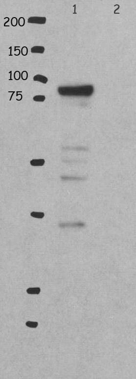
Western Blot: RASAL1 Antibody [NBP1-32776]
Western Blot: RASAL1 Antibody [NBP1-32776] - Sample (30 ug of whole cell lysate) A: A431 7.5% SDS PAGE, antibody diluted at 1:1000.![Western Blot: RASAL1 Antibody [NBP1-32776]](https://resources.rndsystems.com/images/products/RASAL1-Antibody-Western-Blot-NBP1-32776-img0005.jpg "Western Blot: RASAL1 Antibody [NBP1-32776]")
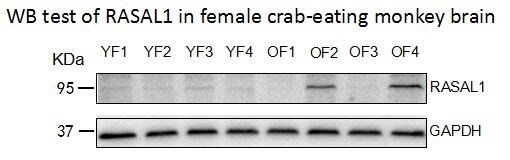
Western Blot: RASAL1 Antibody [NBP1-32776]
Western Blot: RASAL1 Antibody [NBP1-32776] - Protein expression was upregulated in older female crab-eating monkey brain. Image submitted by a verified customer review.
Western Blot: RASAL1 Antibody [NBP1-32776] -
Western Blot: RASAL1 Antibody [NBP1-32776] - Rasal1 inhibits in vitro anti-CD3 & dendritic cell-peptide induced T-cell activation. a Antigen (OVA323-39)-dependent activation & proliferation of DO11.10 CD4 + cells expressing Flag-tagged Rasal1 compared with control cells at different OVA peptide concentrations (5–10 ng). CTLA-Ig was used as a control. Upper inset shows expression of Flag-tagged Rasal using anti-Flag in blotting (n = 3). b Inhibition of T-cell responses by Rasal1 to increasing anti-CD3 concentrations. Upper inset: effects of Rasal1 at 48 & 72 h post-anti-CD3 activation (n = 3). c Anti-CD3-driven NFATc1/AP1 promoter activity in Jurkat T cells was inhibited by the presence of Flag-tagged Rasal1 (n = 3). d siRNA (esiRNA) knockdown of Rasal1 enhances T-cell proliferation. esiRNA were tested for their knockdown efficiency by western blot (top right panel), esiRNA1 & 2 correspond to 500 nM & 1000 nM concentrations (n = 3). e Contact time between D011.10 CD4+T-cells expressing Flag-Rasal1 (red) or empty vector (blue) & OVA peptide on bone marrow dendritic cells (BMDCs). Dot blot of T cell/DC contact duration time in presence of OVA was unaffected by Flag-tagged Rasal1 or vector transfection (n = 3). Representative plots show mean with ± SD. p-value was calculated by unpaired t test. *p < 0.05, **p < 0.01, ***p < 0.001 Image collected & cropped by CiteAb from the following publication (https://pubmed.ncbi.nlm.nih.gov/31641113), licensed under a CC-BY license. Not internally tested by Novus Biologicals.
Western Blot: RASAL1 Antibody [NBP1-32776] -
Western Blot: RASAL1 Antibody [NBP1-32776] - TCR complex purification & Rasal1 association with ZAP-70 in the TCR complex. a Analytic gel from tandem affinity purification (TAP) of TCR plus associated proteins captured from TCR-constituted cells (TCR complex) versus TCR-null negative control cells (control) (left panel). Chart showing the list of TCR-associated proteins identified by MS analysis (right upper panel). The structure of Rasal1 (right lower panel). b Co-precipitation of ZAP-70 with TCR from 3A9 T cells. 3A9 cells were stimulated for 10 min with anti-CD3 followed by immunoprecipitation with anti-TCR zeta & blotting with anti-ZAP-70 or anti-zeta. Lane 1: resting T cells; lane 2: anti-CD3-stimulated T cells. c Rasal1 expression is induced with anti-CD3 activation. Relative fold change expression of Rasal1 versus Rasal3 upon anti-CD3 stimulation of CD4+ & CD8 + peripheral T cells. 18 s rRNA served as a control. d Anti-CD3 induces Rasal1 translocation to the membranes of T cells. Stimulation-dependent association of Rasal1 with the membrane (upper panel) versus cytosolic Rasal1 (lower panel). Anti-actin served as a loading control. LAT was used as a marker of membrane enrichment fractionation (n = 3) Image collected & cropped by CiteAb from the following publication (https://pubmed.ncbi.nlm.nih.gov/31641113), licensed under a CC-BY license. Not internally tested by Novus Biologicals.
Western Blot: RASAL1 Antibody [NBP1-32776] -
Western Blot: RASAL1 Antibody [NBP1-32776] - Rasal1 binds to the immune cell tyrosine kinase ZAP-70. a Proximity ligation assay (PLA) of Rasal1 & ZAP-70 in Jurkat T-cells stained with DAPI, anti-Rasal1, & anti-ZAP-70. Positive PLA signal (fluorescent dots) indicates a distance of 30–40 nm between Rasal1 & ZAP-70 in anti-CD3-activated T cells, indicating their close proximity (n = 2). b Anti-Rasal1 co-precipitates ZAP-70 from primary mouse T cells. Lane 1–2 loading controls, lanes 3-4: anti-Rasal1 IP: lanes 2 & 4. Cells were stimulated with anti-CD3 ligation for 5 min vs IgG control ligation in lanes 1 & 3. c Anti-Rasal1 co-precipitates ZAP-70 from transfected Jurkat T cells. Lanes 1: nonligated (i.e., IgG); lane 2: anti-CD3 ligated T cells for 5 min. Lower panel shows similar level of ZAP-70 expression in lysates of (n = 2) Image collected & cropped by CiteAb from the following publication (https://pubmed.ncbi.nlm.nih.gov/31641113), licensed under a CC-BY license. Not internally tested by Novus Biologicals.
Western Blot: RASAL1 Antibody [NBP1-32776] -
Western Blot: RASAL1 Antibody [NBP1-32776] - Rasal1 binds to the immune cell tyrosine kinase ZAP-70. a Proximity ligation assay (PLA) of Rasal1 & ZAP-70 in Jurkat T-cells stained with DAPI, anti-Rasal1, & anti-ZAP-70. Positive PLA signal (fluorescent dots) indicates a distance of 30–40 nm between Rasal1 & ZAP-70 in anti-CD3-activated T cells, indicating their close proximity (n = 2). b Anti-Rasal1 co-precipitates ZAP-70 from primary mouse T cells. Lane 1–2 loading controls, lanes 3-4: anti-Rasal1 IP: lanes 2 & 4. Cells were stimulated with anti-CD3 ligation for 5 min vs IgG control ligation in lanes 1 & 3. c Anti-Rasal1 co-precipitates ZAP-70 from transfected Jurkat T cells. Lanes 1: nonligated (i.e., IgG); lane 2: anti-CD3 ligated T cells for 5 min. Lower panel shows similar level of ZAP-70 expression in lysates of (n = 2) Image collected & cropped by CiteAb from the following publication (https://pubmed.ncbi.nlm.nih.gov/31641113), licensed under a CC-BY license. Not internally tested by Novus Biologicals.Applications for RASAL1 Antibody - BSA Free
Application
Recommended Usage
Immunocytochemistry/ Immunofluorescence
1:100-1:1000
Western Blot
1:500-1:3000
Application Notes
RASAL1 antibody validated for WB from a verified customer review. Use in Proximity Ligation Assay reported in scientific literature (PMID:31641113).
Reviewed Applications
Read 2 reviews rated 3.5 using NBP1-32776 in the following applications:
Formulation, Preparation, and Storage
Purification
Antigen Affinity-purified
Formulation
0.1M Tris, 0.1M Glycine, 10% Glycerol
Format
BSA Free
Preservative
0.01% Thimerosal
Concentration
Concentrations vary lot to lot. See vial label for concentration. If unlisted please contact technical services.
Shipping
The product is shipped with polar packs. Upon receipt, store it immediately at the temperature recommended below.
Stability & Storage
Aliquot and store at -20C or -80C. Avoid freeze-thaw cycles.
Background: RASAL1
Alternate Names
RAS protein activator like 1 (GAP1 like), RASALGAP1 like protein, rasGAP-activating-like protein 1
Entrez Gene IDs
8437 (Human)
Gene Symbol
RASAL1
UniProt
Additional RASAL1 Products
Product Documents for RASAL1 Antibody - BSA Free
Certificate of Analysis
To download a Certificate of Analysis, please enter a lot or batch number in the search box below.
Product Specific Notices for RASAL1 Antibody - BSA Free
This product is for research use only and is not approved for use in humans or in clinical diagnosis. Primary Antibodies are guaranteed for 1 year from date of receipt.
⚠ WARNING: This product can expose you to chemicals including mercury, which is known to the State of California to cause reproductive toxicity with developmental effects. For more information go to www.P65Warnings.ca.gov.Citations for RASAL1 Antibody - BSA Free
 Powered by Bioz
Customer Reviews for RASAL1 Antibody - BSA Free (2)
3.5 out of 5
2 Customer Ratings
Have you used RASAL1 Antibody - BSA Free?
Submit a review and receive an Amazon gift card!
$25/€18/£15/$25CAN/¥2500 Yen for a review with an image
$10/€7/£6/$10CAN/¥1110 Yen for a review without an image
Submit a review
Customer Images
Showing
1
-
2 of
2 reviews
Showing All
Filter By:
-
Application: Western BlotSample Tested: brain and spinal cordSpecies: MonkeyVerified Customer | Posted 02/13/2019Protein expression was upregulated in older female crab-eating monkey brain.Antibody dilution 1:1000

-
Application: Western BlotSample Tested: 293T lysate transfected with hRasal1Species: HumanVerified Customer | Posted 02/10/2014Western Blot
There are no reviews that match your criteria.
Protocols
Find general support by application which include: protocols, troubleshooting, illustrated assays, videos and webinars.
- Appropriate Fixation of IHC/ICC Samples
- Cellular Response to Hypoxia Protocols
- ClariTSA™ Fluorophore Kits
- Detection & Visualization of Antibody Binding
- ICC Cell Smear Protocol for Suspension Cells
- ICC Immunocytochemistry Protocol Videos
- ICC for Adherent Cells
- Immunocytochemistry (ICC) Protocol
- Immunocytochemistry Troubleshooting
- Immunofluorescence of Organoids Embedded in Cultrex Basement Membrane Extract
- Immunohistochemistry (IHC) and Immunocytochemistry (ICC) Protocols
- Preparing Samples for IHC/ICC Experiments
- Preventing Non-Specific Staining (Non-Specific Binding)
- Primary Antibody Selection & Optimization
- Protocol for VisUCyte™ HRP Polymer Detection Reagent
- Protocol for the Fluorescent ICC Staining of Cell Smears - Graphic
- Protocol for the Fluorescent ICC Staining of Cultured Cells on Coverslips - Graphic
- Protocol for the Preparation and Fluorescent ICC Staining of Cells on Coverslips
- Protocol for the Preparation and Fluorescent ICC Staining of Non-adherent Cells
- Protocol for the Preparation and Fluorescent ICC Staining of Stem Cells on Coverslips
- Protocol for the Preparation of a Cell Smear for Non-adherent Cell ICC - Graphic
- R&D Systems Quality Control Western Blot Protocol
- TUNEL and Active Caspase-3 Detection by IHC/ICC Protocol
- The Importance of IHC/ICC Controls
- Troubleshooting Guide: Western Blot Figures
- Western Blot Conditions
- Western Blot Protocol
- Western Blot Protocol for Cell Lysates
- Western Blot Troubleshooting
- Western Blot Troubleshooting Guide
- View all Protocols, Troubleshooting, Illustrated assays and Webinars
Loading...