![Western Blot: RCC2 Antibody [NB110-40619]](https://resources.rndsystems.com/images/products/RCC2-Antibody-Western-Blot-NB110-40619-img0006.jpg "Western Blot: RCC2 Antibody [NB110-40619]")
Loading...
Key Product Details
Species Reactivity
Validated:
Human, Mouse
Cited:
Human
Applications
Validated:
Immunohistochemistry, Immunohistochemistry-Paraffin, Western Blot, Immunoprecipitation (Negative)
Cited:
Immunocytochemistry/ Immunofluorescence, IF/IHC
Label
Unconjugated
Antibody Source
Polyclonal Rabbit IgG
Loading...
Product Specifications
Immunogen
The immunogen recognized by this antibody maps to a region between residue 475 and the C-terminus (residue 522) of human Regulator of Chromosome Condensation 2 using the numbering given in entry NP_061185.1 (GeneID 55920).
Clonality
Polyclonal
Host
Rabbit
Isotype
IgG
Scientific Data Images for RCC2 Antibody
Western Blot: RCC2 Antibody [NB110-40619]
Western Blot: RCC2 Antibody [NB110-40619] - Detection of both input and immunoprecipitated RCC2 on KYSE-30 and KYSE-450 cell lines. Western blot image submitted by a verified customer review.![Immunohistochemistry-Paraffin: RCC2 Antibody [NB110-40619]](https://resources.rndsystems.com/images/products/RCC2-Antibody-Immunohistochemistry-Paraffin-NB110-40619-img0007.jpg "Immunohistochemistry-Paraffin: RCC2 Antibody [NB110-40619]")
Immunohistochemistry-Paraffin: RCC2 Antibody [NB110-40619]
Immunohistochemistry-Paraffin: RCC2 Antibody [NB110-40619] - RCC2 expression from esophageal cancer tissue from mice. IHC-P image submitted by a verified customer review.![Western Blot: RCC2 Antibody [NB110-40619]](https://resources.rndsystems.com/images/products/RCC2-Antibody-Western-Blot-NB110-40619-img0003.jpg "Western Blot: RCC2 Antibody [NB110-40619]")
Western Blot: RCC2 Antibody [NB110-40619]
Western Blot: RCC2 Antibody [NB110-40619] - Detection of Human and Mouse RCC2 on HeLa whole cell lysate using NB110-40619. RCC2 was immunoprecipitated by rabbit anti-RCC2 antibody NB110-40618.![Immunohistochemistry-Paraffin: RCC2 Antibody [NB110-40619]](https://resources.rndsystems.com/images/products/RCC2-Antibody-Immunohistochemistry-NB110-40619-img0005.jpg "Immunohistochemistry-Paraffin: RCC2 Antibody [NB110-40619]")
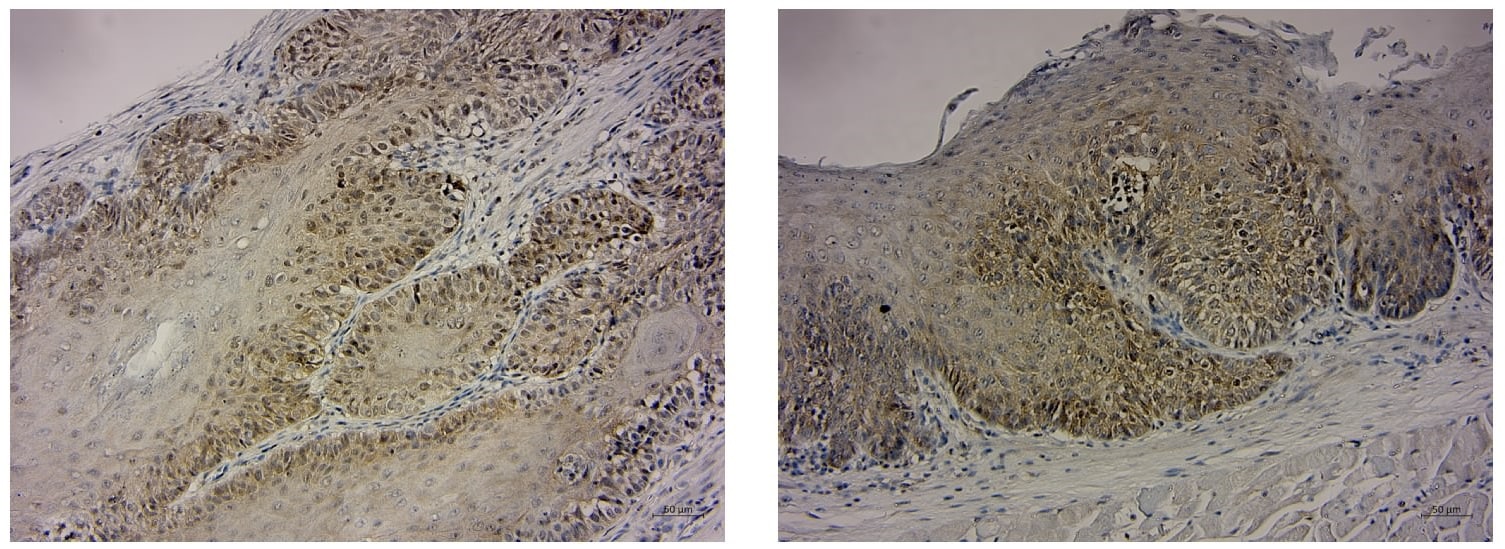
Immunohistochemistry-Paraffin: RCC2 Antibody [NB110-40619]
Immunohistochemistry-Paraffin: RCC2 Antibody [NB110-40619] - Sections of human skin basal cell carcinoma (left) and mouse CT26 colon carcinoma (right). Affinity purified rabbit anti- RCC2 used at a dilution of 1:1000 (0.2 ug/mL) and 1:200 (1 ug/mL). Detection: DABApplications for RCC2 Antibody
Application
Recommended Usage
Immunohistochemistry
1:2000 - 1:10000
Immunohistochemistry-Paraffin
1:2000 - 1:10000
Western Blot
1:2000 - 1:10000
Application Notes
Epitope retrieval with citrate buffer pH 6.0 is recommended for FFPE tissue sections. RCC2 antibody validated for IHC-P, WB from verified customer reviews.
Reviewed Applications
Read 2 reviews rated 5 using NB110-40619 in the following applications:
Formulation, Preparation, and Storage
Purification
Immunogen affinity purified
Formulation
TBS, 0.1% BSA
Preservative
0.09% Sodium Azide
Concentration
0.2 mg/ml
Shipping
The product is shipped with polar packs. Upon receipt, store it immediately at the temperature recommended below.
Stability & Storage
Store at 4C. Do not freeze.
Background: RCC2
Alternate Names
DKFZp762N0610, KIAA1470, protein RCC2, RCC1-like protein TD-60, regulator of chromosome condensation 2, TD60, TD-60, Telophase disk protein of 60 kDa
Gene Symbol
RCC2
UniProt
Additional RCC2 Products
Product Documents for RCC2 Antibody
Certificate of Analysis
To download a Certificate of Analysis, please enter a lot or batch number in the search box below.
Product Specific Notices for RCC2 Antibody
This product is for research use only and is not approved for use in humans or in clinical diagnosis. Primary Antibodies are guaranteed for 1 year from date of receipt.
Citations for RCC2 Antibody
 Powered by Bioz
Customer Reviews for RCC2 Antibody (2)
5 out of 5
2 Customer Ratings
Have you used RCC2 Antibody?
Submit a review and receive an Amazon gift card!
$25/€18/£15/$25CAN/¥2500 Yen for a review with an image
$10/€7/£6/$10CAN/¥1110 Yen for a review without an image
Submit a review
Customer Images

Showing
1
-
2 of
2 reviews
Showing All
Filter By:
-
Application: Immunohistochemistry-ParaffinSample Tested: Human Esophagus tissueSpecies: MouseVerified Customer | Posted 11/04/2020Figure legend: RCC2 expression from esophageal cancer tissue from mice.Antigen retrieval with citrate buffer pH 6

-
Application: Western BlotSample Tested: 293T cell line and Cancer Cell lysateSpecies: HumanVerified Customer | Posted 03/19/2019Detection of RCC2 (IP RCC2) and Input.
There are no reviews that match your criteria.
Protocols
Find general support by application which include: protocols, troubleshooting, illustrated assays, videos and webinars.
- Antigen Retrieval Protocol (PIER)
- Antigen Retrieval for Frozen Sections Protocol
- Appropriate Fixation of IHC/ICC Samples
- Cellular Response to Hypoxia Protocols
- Chromogenic IHC Staining of Formalin-Fixed Paraffin-Embedded (FFPE) Tissue Protocol
- Chromogenic Immunohistochemistry Staining of Frozen Tissue
- ClariTSA™ Fluorophore Kits
- Detection & Visualization of Antibody Binding
- Fluorescent IHC Staining of Frozen Tissue Protocol
- Graphic Protocol for Heat-induced Epitope Retrieval
- Graphic Protocol for the Preparation and Fluorescent IHC Staining of Frozen Tissue Sections
- Graphic Protocol for the Preparation and Fluorescent IHC Staining of Paraffin-embedded Tissue Sections
- Graphic Protocol for the Preparation of Gelatin-coated Slides for Histological Tissue Sections
- IHC Sample Preparation (Frozen sections vs Paraffin)
- Immunofluorescent IHC Staining of Formalin-Fixed Paraffin-Embedded (FFPE) Tissue Protocol
- Immunohistochemistry (IHC) and Immunocytochemistry (ICC) Protocols
- Immunohistochemistry Frozen Troubleshooting
- Immunohistochemistry Paraffin Troubleshooting
- Preparing Samples for IHC/ICC Experiments
- Preventing Non-Specific Staining (Non-Specific Binding)
- Primary Antibody Selection & Optimization
- Protocol for Heat-Induced Epitope Retrieval (HIER)
- Protocol for Making a 4% Formaldehyde Solution in PBS
- Protocol for VisUCyte™ HRP Polymer Detection Reagent
- Protocol for the Preparation & Fixation of Cells on Coverslips
- Protocol for the Preparation and Chromogenic IHC Staining of Frozen Tissue Sections
- Protocol for the Preparation and Chromogenic IHC Staining of Frozen Tissue Sections - Graphic
- Protocol for the Preparation and Chromogenic IHC Staining of Paraffin-embedded Tissue Sections
- Protocol for the Preparation and Chromogenic IHC Staining of Paraffin-embedded Tissue Sections - Graphic
- Protocol for the Preparation and Fluorescent IHC Staining of Frozen Tissue Sections
- Protocol for the Preparation and Fluorescent IHC Staining of Paraffin-embedded Tissue Sections
- Protocol for the Preparation of Gelatin-coated Slides for Histological Tissue Sections
- R&D Systems Quality Control Western Blot Protocol
- TUNEL and Active Caspase-3 Detection by IHC/ICC Protocol
- The Importance of IHC/ICC Controls
- Troubleshooting Guide: Immunohistochemistry
- Troubleshooting Guide: Western Blot Figures
- Western Blot Conditions
- Western Blot Protocol
- Western Blot Protocol for Cell Lysates
- Western Blot Troubleshooting
- Western Blot Troubleshooting Guide
- View all Protocols, Troubleshooting, Illustrated assays and Webinars
Loading...