RUNX2/CBFA1 Antibody - BSA Free
Novus Biologicals | Catalog # NBP2-24755
![Western Blot: RUNX2/CBFA1 Antibody [NBP2-24755]](https://resources.rndsystems.com/images/products/RUNX2-CBFA1-Antibody-Western-Blot-NBP2-24755-img0002.jpg "Western Blot: RUNX2/CBFA1 Antibody [NBP2-24755]")
Loading...
Key Product Details
Species Reactivity
Validated:
Human, Mouse, Chicken, Equine, Goat, Primate
Cited:
Human, Mouse
Predicted:
Canine (90%), Rat (90%). Backed by our 100% Guarantee.
Applications
Validated:
Immunohistochemistry, Immunohistochemistry-Paraffin, Western Blot
Cited:
Western Blot, Knockdown Validated
Label
Unconjugated
Antibody Source
Polyclonal Rabbit IgG
Format
BSA Free
Loading...
Product Specifications
Immunogen
This RUNX2/CBFA1 Antibody was developed against a synthetic peptide corresponding to somewhere between amino acids 250-300 of human RUNX2 was used as immunogen for RUNX2/CBFA1 Antibody. RUNX2 and RUNX1 share an approximate 66% homology in peptide sequence used as immunogen.
Reactivity Notes
Goat reactivity reported from a verified customer review.
Clonality
Polyclonal
Host
Rabbit
Isotype
IgG
Theoretical MW
56.6 kDa.
Disclaimer note: The observed molecular weight of the protein may vary from the listed predicted molecular weight due to post translational modifications, post translation cleavages, relative charges, and other experimental factors.
Disclaimer note: The observed molecular weight of the protein may vary from the listed predicted molecular weight due to post translational modifications, post translation cleavages, relative charges, and other experimental factors.
Scientific Data Images for RUNX2/CBFA1 Antibody - BSA Free
Western Blot: RUNX2/CBFA1 Antibody [NBP2-24755]
Western Blot: RUNX2/CBFA1 Antibody [NBP2-24755] - Analysis of human RUNX2 in Saos-2 cell lysate in the 1) absence and 2) presence of immunizing peptide using RUNX2 antibody at 2 ug/mL.
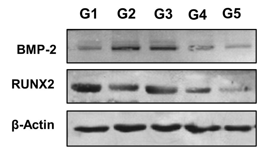
Western Blot: Rabbit Polyclonal RUNX2/CBFA1 Antibody [IMGENEX: IMG-5985A] [NBP2-24755]
Western Blot: Rabbit Polyclonal RUNX2/CBFA1 Antibody [IMGENEX: IMG-5985A] [NBP2-24755] - Bone lysate homogenate from goats. SDS-PAGE with 0.3 mg of total protein from the homogenate per well. RUNX/CBFA1, NBP2-24755 was used at a dilution of 1:1000 and BMP-2, NBP1-32218 was used at a dilution of 1:1000 in TBS Tween and 5% milk powder. The protein was labeled at 70 kDa. Quantification of RUNX2 in bone graft in the tibia of intrailiac cellularized goats at different times compared with tibial autograft and untreated bone defect. Evaluation of pre-cellularized graft in the iliac crest in the treatment of bone defects in the tibia of goats. G1: cellularized graft for 4 weeks applied to the tibial bone defect, G2: cellularized graft for 6 weeks applied to the tibial bone defect, G3: cellularized graft for 8 weeks applied to the tibial bone defect, G4: tibial autograft and G5: untreated bone defect. All groups were evaluated 120 days after graft implantation. Image from a verified customer review.
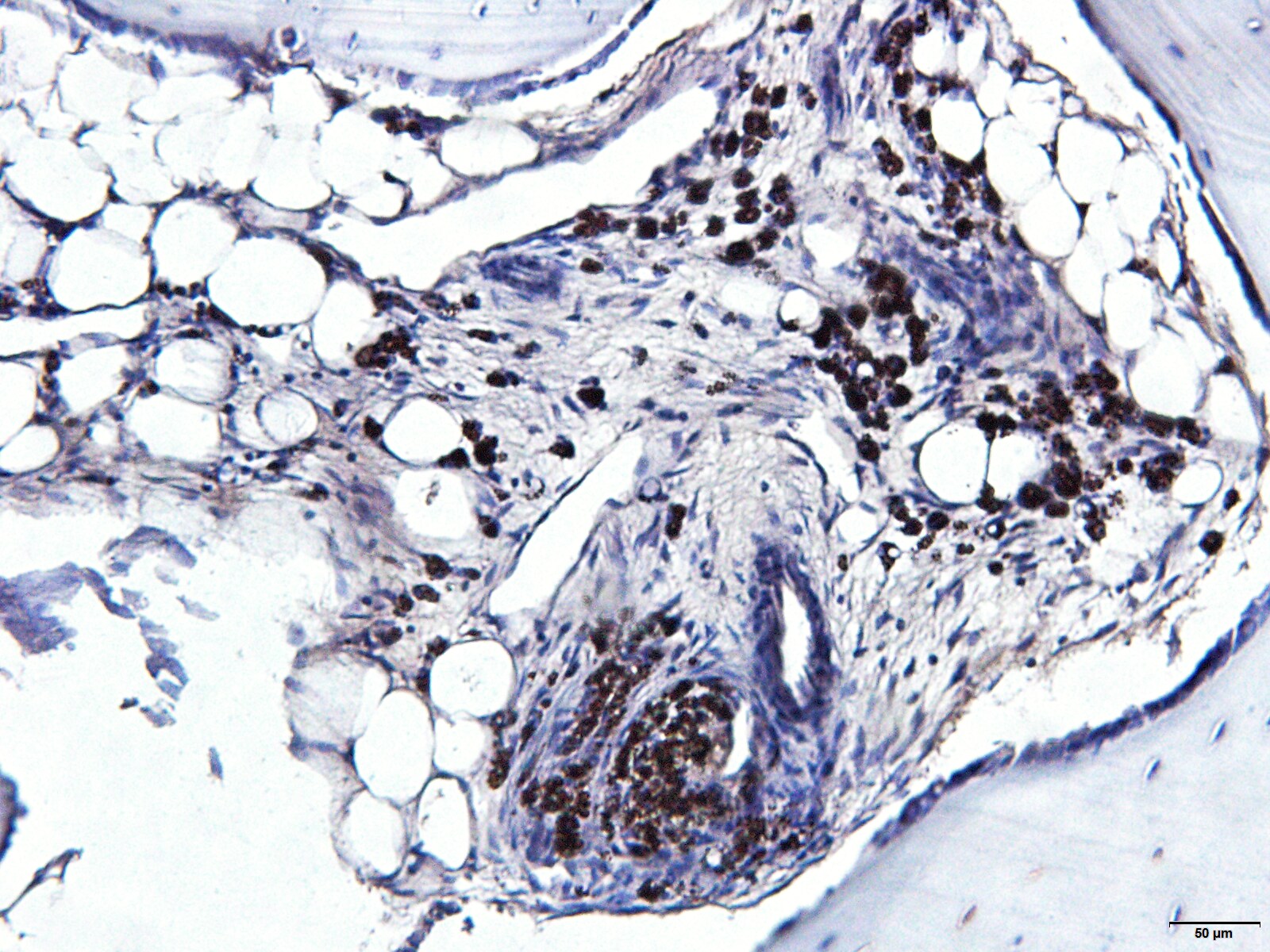
Immunohistochemistry-Paraffin: Rabbit Polyclonal RUNX2/CBFA1 Antibody [IMGENEX: IMG-5985A] [NBP2-24755]
The histological sections were deparaffinized, peroxidase blocked in methanol and 30% hydrogen peroxide (9:1), and antigen retrieval was performed with porcine gastric pepsin and HCl at pH 3.0 in an oven at 37°C for 30 minutes. Then, blocking with BSA was performed for 1 hour followed by incubation with the primary antibody RUNX2/CBA1 Antibody NBP2-24755 (1:100) overnight. Subsequently, excess primary antibody was removed with PBS pH 7.6 and incubated with the secondary antibody (goat anti-rabbit IgG - NB7160 (1:200) for 1 hour and polymer was applied for 30 minutes. Finally, 150 μL of DAB was applied for five minutes and counterstained with hematoxylin. Image from a verified customer review.Applications for RUNX2/CBFA1 Antibody - BSA Free
Application
Recommended Usage
Immunohistochemistry
Validated for IHC from a verified customer review.
Immunohistochemistry-Paraffin
Validated from IHC-P from a verified customer review.
Western Blot
1 - 3 ug/mL
Reviewed Applications
Read 2 reviews rated 5 using NBP2-24755 in the following applications:
Formulation, Preparation, and Storage
Purification
Protein G purified
Formulation
PBS
Format
BSA Free
Preservative
0.05% Sodium Azide
Concentration
1.0 mg/ml
Shipping
The product is shipped with polar packs. Upon receipt, store it immediately at the temperature recommended below.
Stability & Storage
Store at -20C. Avoid freeze-thaw cycles.
Background: RUNX2/CBFA1
Functionally, RUNX2 promotes the expression of osteoblast-specific genes vital for the osteoblast differentiation and proliferation process including type I collagen, osteocalcin (OCN), and alkaline phosphatase (APC) (1, 3). Further evidence for the role of RUNX2 is highlighted by a study of Runx2-/-mice which completely lack osteoblasts (4). Additionally, RUNX2 is also required for chondrocyte maturation, which are the cells responsible for cartilage formation (1, 3, 5). Given the role of RUNX2 in bone and cartilage maturation and formation, it is clear that defects or mutations in RUNX2 cause various bone and bone-related diseases (3, 6, 7). For instance, cleidocranial dysplasia (CCD), which presents with delayed cranial suture closure phenotypes, hypoplastic clavicles, extra teeth, and short stature, is caused by haploinsufficiency in RUNX2 (2, 3, 6). Furthermore, metaphyseal dysplasia with maxillary hypoplasia and brachydactyly (MDMHB) is a bone dysplasia disorder with a phenotype of abnormalities in the long bones, an underdeveloped jawbone, and short fingers that is caused by a duplication in RUNX2 (6). Finally, RUNX2 has been shown to be upregulated in mouse models of the joint disorder osteoarthritis (OA) and may be a potential molecular target for disease treatment (7).
Alternative names for RUNX2 include Acute myeloid leukemia 3 protein CBFA1, CBF-alpha-1, CCD1, CCDAML3, CLCD, Core-binding factor subunit alpha-1, MGC120023, ML3, oncogene AML-3, OSF2, osteoblast-specific transcription factor 2, PEA2aA, PEA2-alpha A, PEBP2A, polyomavirus enhancer-binding protein 2 alpha A subunit, runt related transcription factor 2, SL3/AKV core-binding factor alpha A subunit, and SL3-3 enhancer factor 1 alpha A subunit.
References
1. Ferreira, L. B., Gimba, E., Vinagre, J., Sobrinho-Simoes, M., & Soares, P. (2020). Molecular Aspects of Thyroid Calcification. International journal of molecular sciences. https://doi.org/10.3390/ijms21207718
2. Kim, W. J., Shin, H. L., Kim, B. S., Kim, H. J., & Ryoo, H. M. (2020). RUNX2-modifying enzymes: therapeutic targets for bone diseases. Experimental & molecular medicine. https://doi.org/10.1038/s12276-020-0471-4
3. Vimalraj, S., Arumugam, B., Miranda, P. J., & Selvamurugan, N. (2015). Runx2: Structure, function, and phosphorylation in osteoblast differentiation. International journal of biological macromolecules. https://doi.org/10.1016/j.ijbiomac.2015.04.008
4. Uniprot (Q13950)
5. Komori T. (2017). Roles of Runx2 in Skeletal Development. Advances in experimental medicine and biology. https://doi.org/10.1007/978-981-10-3233-2_6
6. Moffatt, P., Ben Amor, M., Glorieux, F. H., Roschger, P., Klaushofer, K., Schwartzentruber, J. A., Paterson, A. D., Hu, P., Marshall, C., FORGE Canada Consortium, Fahiminiya, S., Majewski, J., Beaulieu, C. L., Boycott, K. M., & Rauch, F. (2013). Metaphyseal dysplasia with maxillary hypoplasia and brachydactyly is caused by a duplication in RUNX2. American journal of human genetics. https://doi.org/10.1016/j.ajhg.2012.12.001
7. Chen, D., Kim, D. J., Shen, J., Zou, Z., & O'Keefe, R. J. (2019). Runx2 plays a central role in Osteoarthritis development. Journal of orthopaedic translation. https://doi.org/10.1016/j.jot.2019.11.008
Long Name
Runt-related Transcription Factor 2
Alternate Names
CBFA1
Gene Symbol
RUNX2
UniProt
Additional RUNX2/CBFA1 Products
Product Documents for RUNX2/CBFA1 Antibody - BSA Free
Certificate of Analysis
To download a Certificate of Analysis, please enter a lot or batch number in the search box below.
Product Specific Notices for RUNX2/CBFA1 Antibody - BSA Free
This product is for research use only and is not approved for use in humans or in clinical diagnosis. Primary Antibodies are guaranteed for 1 year from date of receipt.
Citations for RUNX2/CBFA1 Antibody - BSA Free
 Powered by Bioz
Customer Reviews for RUNX2/CBFA1 Antibody - BSA Free (2)
5 out of 5
2 Customer Ratings
Have you used RUNX2/CBFA1 Antibody - BSA Free?
Submit a review and receive an Amazon gift card!
$25/€18/£15/$25CAN/¥2500 Yen for a review with an image
$10/€7/£6/$10CAN/¥1110 Yen for a review without an image
Submit a review
Customer Images
Showing
1
-
2 of
2 reviews
Showing All
Filter By:
-
Application: Immunohistochemistry-ParaffinSample Tested: Bone and Bone ExtractsSpecies: GoatVerified Customer | Posted 07/29/2025The f histological goat bone fragments were incubated with the primary antibody RUNX2 NBP2-24755 overnight. Subsequently, the secondary antibody goat anti-rabbit IgG - NB7160 was applied and incubated for 1 hour.Validation of an immunohistochemistry technique for goats
 Bio-Techne ResponseThis review reflects a new species or application tested on a primary antibody.
 -
Application: Western BlotSample Tested: Bone and Bone ExtractsSpecies: GoatVerified Customer | Posted 05/13/2025Bone lysate homogenate from goats. SDS-PAGE with 0.3 mg of total protein from the homogenate per well. RUNX/CBFA1, NBP2-24755 was used at a dilution of 1:1000 in TBS Tween and 5% milk powder. The protein was labeled at 70 kDa.Quantification of RUNX2 in bone graft in the tibia of intrailiac cellularized goats at different times compared with tibial autograft and untreated bone defect. Evaluation of pre-cellularized graft in the iliac crest in the treatment of bone defects in the tibia of goats. G1: cellularized graft for 4 weeks applied to the tibial bone defect, G2: cellularized graft for 6 weeks applied to the tibial bone defect, G3: cellularized graft for 8 weeks applied to the tibial bone defect, G4: tibial autograft and G5: untreated bone defect. All groups were evaluated 120 days after graft implantation.
Bio-Techne ResponseThis review reflects a new species or application tested on a primary antibody.
There are no reviews that match your criteria.
Protocols
Find general support by application which include: protocols, troubleshooting, illustrated assays, videos and webinars.
- Antigen Retrieval Protocol (PIER)
- Antigen Retrieval for Frozen Sections Protocol
- Appropriate Fixation of IHC/ICC Samples
- Cellular Response to Hypoxia Protocols
- Chromogenic IHC Staining of Formalin-Fixed Paraffin-Embedded (FFPE) Tissue Protocol
- Chromogenic Immunohistochemistry Staining of Frozen Tissue
- ClariTSA™ Fluorophore Kits
- Detection & Visualization of Antibody Binding
- Fluorescent IHC Staining of Frozen Tissue Protocol
- Graphic Protocol for Heat-induced Epitope Retrieval
- Graphic Protocol for the Preparation and Fluorescent IHC Staining of Frozen Tissue Sections
- Graphic Protocol for the Preparation and Fluorescent IHC Staining of Paraffin-embedded Tissue Sections
- Graphic Protocol for the Preparation of Gelatin-coated Slides for Histological Tissue Sections
- IHC Sample Preparation (Frozen sections vs Paraffin)
- Immunofluorescent IHC Staining of Formalin-Fixed Paraffin-Embedded (FFPE) Tissue Protocol
- Immunohistochemistry (IHC) and Immunocytochemistry (ICC) Protocols
- Immunohistochemistry Frozen Troubleshooting
- Immunohistochemistry Paraffin Troubleshooting
- Preparing Samples for IHC/ICC Experiments
- Preventing Non-Specific Staining (Non-Specific Binding)
- Primary Antibody Selection & Optimization
- Protocol for Heat-Induced Epitope Retrieval (HIER)
- Protocol for Making a 4% Formaldehyde Solution in PBS
- Protocol for VisUCyte™ HRP Polymer Detection Reagent
- Protocol for the Preparation & Fixation of Cells on Coverslips
- Protocol for the Preparation and Chromogenic IHC Staining of Frozen Tissue Sections
- Protocol for the Preparation and Chromogenic IHC Staining of Frozen Tissue Sections - Graphic
- Protocol for the Preparation and Chromogenic IHC Staining of Paraffin-embedded Tissue Sections
- Protocol for the Preparation and Chromogenic IHC Staining of Paraffin-embedded Tissue Sections - Graphic
- Protocol for the Preparation and Fluorescent IHC Staining of Frozen Tissue Sections
- Protocol for the Preparation and Fluorescent IHC Staining of Paraffin-embedded Tissue Sections
- Protocol for the Preparation of Gelatin-coated Slides for Histological Tissue Sections
- R&D Systems Quality Control Western Blot Protocol
- TUNEL and Active Caspase-3 Detection by IHC/ICC Protocol
- The Importance of IHC/ICC Controls
- Troubleshooting Guide: Immunohistochemistry
- Troubleshooting Guide: Western Blot Figures
- Western Blot Conditions
- Western Blot Protocol
- Western Blot Protocol for Cell Lysates
- Western Blot Troubleshooting
- Western Blot Troubleshooting Guide
- View all Protocols, Troubleshooting, Illustrated assays and Webinars
FAQs for RUNX2/CBFA1 Antibody - BSA Free
Showing
1
-
1 of
1 FAQ
Showing All
-
Q: We would like an anti-RUNX2 for IHC-P which share cross reactivity with Rat, but not with Human.
A: We don't have any data for our RUNX2 antibodies that confirms they will NOT detect the human protein. When we can confirm that an antibody will not react with a certain species, we display a (-) sign on the datasheet. Otherwise, if the species is not listed it means that it has not been tested.


