![Western Blot: BMPR-II Antibody [NBP1-32218]](https://resources.rndsystems.com/images/products/BMPR-II-Antibody-Western-Blot-NBP1-32218-img0003.jpg "Western Blot: BMPR-II Antibody [NBP1-32218]")
Loading...
Key Product Details
Validated by
Knockout/Knockdown
Species Reactivity
Validated:
Human, Goat
Predicted:
Bovine (93%), Mouse (92%), Porcine (92%), Rat (92%), Rhesus Macaque (98%), Sheep (93%). Backed by our 100% Guarantee.
Applications
Knockout Validated, Immunohistochemistry, Immunohistochemistry-Paraffin, Western Blot
Label
Unconjugated
Antibody Source
Polyclonal Rabbit IgG
Loading...
Product Specifications
Immunogen
Recombinant protein encompassing a sequence within the C-terminus region of human BMPR-II. The exact sequence is proprietary.
Reactivity Notes
Chicken (86%). Goat reactivity reported from a verified customer review.
Localization
Membrane
Clonality
Polyclonal
Host
Rabbit
Isotype
IgG
Theoretical MW
115 kDa.
Disclaimer note: The observed molecular weight of the protein may vary from the listed predicted molecular weight due to post translational modifications, post translation cleavages, relative charges, and other experimental factors.
Disclaimer note: The observed molecular weight of the protein may vary from the listed predicted molecular weight due to post translational modifications, post translation cleavages, relative charges, and other experimental factors.
Scientific Data Images for BMPR-II Antibody
Western Blot: BMPR-II Antibody [NBP1-32218]
Western Blot: BMPR-II Antibody [NBP1-32218] - Wild-type (WT) and BMPR2 knockout (KO) HeLa cell extracts (30 ug) were separated by 5% SDS-PAGE, and the membrane was blotted with BMPR2 antibody diluted at 1:500. HRP-conjugated anti-rabbit IgG antibody was used to detect the primary antibody.![Western Blot: BMPR-II Antibody [NBP1-32218]](https://resources.rndsystems.com/images/products/BMPR-II-Antibody-Western-Blot-NBP1-32218-img0002.jpg "Western Blot: BMPR-II Antibody [NBP1-32218]")
Western Blot: BMPR-II Antibody [NBP1-32218]
Western Blot: BMPR-II Antibody [NBP1-32218] - Various whole cell extracts (30 ug) were separated by 5% SDS-PAGE, and the membrane was blotted with BMPR2 antibody [C1C3] diluted at 1:500. The HRP-conjugated anti-rabbit IgG antibody (NBP2-19301) was used to detect the primary antibody.
Western Blot: Rabbit Polyclonal BMPR-II Antibody [NBP1-32218]
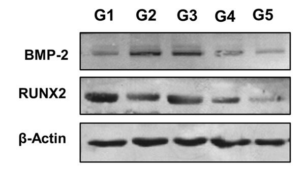
Western Blot: Rabbit Polyclonal BMPR-II Antibody [NBP1-32218] - Bone lysate homogenate from goats. SDS-PAGE with 0.3 mg of total protein from the homogenate per well. BMP-2, NBP1-32218, was used at a dilution of 1:1000 and RUNX/CBFA1, NBP2-24755 was used at a dilution of 1:1000 in TBS Tween and 5% powdered milk. The protein was labeled at 150 kDa. Quantification of BMP-2 in bone graft in the tibia of intrailiac cellularized goats at different times compared with tibial autograft and untreated bone defect. Evaluation of pre-cellularized graft in the iliac crest in the treatment of bone defects in the tibia of goats. G1: cellularized graft for 4 weeks applied to the tibial bone defect, G2: cellularized graft for 6 weeks applied to the tibial bone defect, G3: cellularized graft for 8 weeks applied to the tibial bone defect, G4: tibial autograft and G5: untreated bone defect. All groups were evaluated 120 days after graft implantation. Image from a verified customer review.
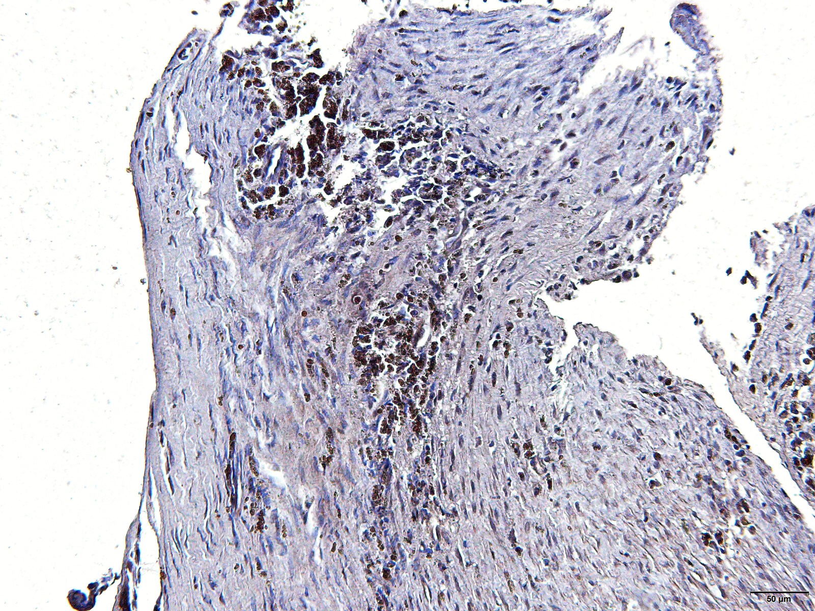
Immunohistochemistry-Paraffin: Rabbit Polyclonal BMPR-II Antibody [NBP1-32218]
The sections of histological sections of goat bone fragments were deparaffinized, peroxidase blocked in methanol and 30% hydrogen peroxide (9:1), and antigen retrieval was performed with porcine gastric pepsin and HCl at pH 3.0 in an oven at 37°C for 30 minutes. Then, blocking with BSA was performed for 1 hour followed by incubation with the primary antibody BMPR-2 antibody NBP1-32218 (1:100) overnight. Subsequently, excess primary antibody was removed with PBS pH 7.6 and incubated with the secondary antibody (goat anti-rabbit IgG - NB7160 (1:200) for 1 hour and polymer was applied for 30 minutes. Finally, 150 μL of DAB was applied for five minutes and counterstained with hematoxylin. Image from a verified customer review.Applications for BMPR-II Antibody
Application
Recommended Usage
Immunohistochemistry
Validated for IHC from a verified customer review.
Immunohistochemistry-Paraffin
Validated for IHC-P from a verified customer review.
Western Blot
1:500-1:3000
Reviewed Applications
Read 2 reviews rated 5 using NBP1-32218 in the following applications:
Formulation, Preparation, and Storage
Purification
Antigen Affinity-purified
Formulation
PBS, 1% BSA, 20% Glycerol
Preservative
0.025% Proclin 300
Concentration
Concentrations vary lot to lot. See vial label for concentration. If unlisted please contact technical services.
Shipping
The product is shipped with polar packs. Upon receipt, store it immediately at the temperature recommended below.
Stability & Storage
Aliquot and store at -20C or -80C. Avoid freeze-thaw cycles.
Background: BMPR-II
Long Name
Bone Morphogenetic Protein Receptor II
Alternate Names
BMPR2, BMPRII
Gene Symbol
BMPR2
Additional BMPR-II Products
Product Documents for BMPR-II Antibody
Certificate of Analysis
To download a Certificate of Analysis, please enter a lot or batch number in the search box below.
Product Specific Notices for BMPR-II Antibody
This product is for research use only and is not approved for use in humans or in clinical diagnosis. Primary Antibodies are guaranteed for 1 year from date of receipt.
Related Research Areas
Customer Reviews for BMPR-II Antibody (2)
5 out of 5
2 Customer Ratings
Have you used BMPR-II Antibody?
Submit a review and receive an Amazon gift card!
$25/€18/£15/$25CAN/¥2500 Yen for a review with an image
$10/€7/£6/$10CAN/¥1110 Yen for a review without an image
Submit a review
Customer Images
Showing
1
-
2 of
2 reviews
Showing All
Filter By:
-
Application: Immunohistochemistry-ParaffinSample Tested: Bone and Bone ExtractsSpecies: GoatVerified Customer | Posted 07/29/2025The f histological goat bone fragments were incubated with the primary antibody BMPR22 NBP1-32218 overnight. Subsequently, the secondary antibody goat anti-rabbit IgG - NB7160 was applied and incubated for 1 hour.Validation of an immunohistochemistry technique for goats
 Bio-Techne ResponseThis review reflects a new species or application tested on a primary antibody.
 -
Application: Western BlotSample Tested: Bone and Bone ExtractsSpecies: GoatVerified Customer | Posted 05/13/2025Bone lysate homogenate from goats. SDS-PAGE with 0.3 mg of total protein from the homogenate per well. BMP-2, NBP1-32218, was used at a dilution of 1:1000 in TBS Tween and 5% powdered milk. The protein was labeled at 150 kDa.Quantification of BMP-2 in bone graft in the tibia of intrailiac cellularized goats at different times compared with tibial autograft and untreated bone defect. Evaluation of pre-cellularized graft in the iliac crest in the treatment of bone defects in the tibia of goats. G1: cellularized graft for 4 weeks applied to the tibial bone defect, G2: cellularized graft for 6 weeks applied to the tibial bone defect, G3: cellularized graft for 8 weeks applied to the tibial bone defect, G4: tibial autograft and G5: untreated bone defect. All groups were evaluated 120 days after graft implantation.
Bio-Techne ResponseThis review reflects a new species or application tested on a primary antibody.
There are no reviews that match your criteria.
Protocols
Find general support by application which include: protocols, troubleshooting, illustrated assays, videos and webinars.
- Antigen Retrieval Protocol (PIER)
- Antigen Retrieval for Frozen Sections Protocol
- Appropriate Fixation of IHC/ICC Samples
- Cellular Response to Hypoxia Protocols
- Chromogenic IHC Staining of Formalin-Fixed Paraffin-Embedded (FFPE) Tissue Protocol
- Chromogenic Immunohistochemistry Staining of Frozen Tissue
- ClariTSA™ Fluorophore Kits
- Detection & Visualization of Antibody Binding
- Fluorescent IHC Staining of Frozen Tissue Protocol
- Graphic Protocol for Heat-induced Epitope Retrieval
- Graphic Protocol for the Preparation and Fluorescent IHC Staining of Frozen Tissue Sections
- Graphic Protocol for the Preparation and Fluorescent IHC Staining of Paraffin-embedded Tissue Sections
- Graphic Protocol for the Preparation of Gelatin-coated Slides for Histological Tissue Sections
- IHC Sample Preparation (Frozen sections vs Paraffin)
- Immunofluorescent IHC Staining of Formalin-Fixed Paraffin-Embedded (FFPE) Tissue Protocol
- Immunohistochemistry (IHC) and Immunocytochemistry (ICC) Protocols
- Immunohistochemistry Frozen Troubleshooting
- Immunohistochemistry Paraffin Troubleshooting
- Preparing Samples for IHC/ICC Experiments
- Preventing Non-Specific Staining (Non-Specific Binding)
- Primary Antibody Selection & Optimization
- Protocol for Heat-Induced Epitope Retrieval (HIER)
- Protocol for Making a 4% Formaldehyde Solution in PBS
- Protocol for VisUCyte™ HRP Polymer Detection Reagent
- Protocol for the Preparation & Fixation of Cells on Coverslips
- Protocol for the Preparation and Chromogenic IHC Staining of Frozen Tissue Sections
- Protocol for the Preparation and Chromogenic IHC Staining of Frozen Tissue Sections - Graphic
- Protocol for the Preparation and Chromogenic IHC Staining of Paraffin-embedded Tissue Sections
- Protocol for the Preparation and Chromogenic IHC Staining of Paraffin-embedded Tissue Sections - Graphic
- Protocol for the Preparation and Fluorescent IHC Staining of Frozen Tissue Sections
- Protocol for the Preparation and Fluorescent IHC Staining of Paraffin-embedded Tissue Sections
- Protocol for the Preparation of Gelatin-coated Slides for Histological Tissue Sections
- R&D Systems Quality Control Western Blot Protocol
- TUNEL and Active Caspase-3 Detection by IHC/ICC Protocol
- The Importance of IHC/ICC Controls
- Troubleshooting Guide: Immunohistochemistry
- Troubleshooting Guide: Western Blot Figures
- Western Blot Conditions
- Western Blot Protocol
- Western Blot Protocol for Cell Lysates
- Western Blot Troubleshooting
- Western Blot Troubleshooting Guide
- View all Protocols, Troubleshooting, Illustrated assays and Webinars
Loading...