S100A9 Antibody (MAC387) [Alexa Fluor® 488]
Novus Biologicals | Catalog # NBP2-47667AF488
Key Product Details
Species Reactivity
Applications
Label
Antibody Source
Product Specifications
Immunogen
Localization
Specificity
Clonality
Host
Isotype
Scientific Data Images for S100A9 Antibody (MAC387) [Alexa Fluor® 488]
Applications for S100A9 Antibody (MAC387) [Alexa Fluor® 488]
CyTOF-ready
Flow Cytometry
Immunocytochemistry/ Immunofluorescence
Immunofluorescence
Immunohistochemistry
Immunohistochemistry-Paraffin
Reviewed Applications
Read 1 review rated 4 using NBP2-47667AF488 in the following applications:
Spectra Viewer
Plan Your Experiments
Use our spectra viewer to interactively plan your experiments, assessing multiplexing options. View the excitation and emission spectra for our fluorescent dye range and other commonly used dyes.
Spectra Viewer
Flow Cytometry Panel Builder
Bio-Techne Knows Flow Cytometry
Save time and reduce costly mistakes by quickly finding compatible reagents using the Panel Builder Tool.
Advanced Features
- Spectra Viewer - Custom analysis of spectra from multiple fluorochromes
- Spillover Popups - Visualize the spectra of individual fluorochromes
- Antigen Density Selector - Match fluorochrome brightness with antigen density

Formulation, Preparation, and Storage
Purification
Formulation
Preservative
Concentration
Shipping
Stability & Storage
Background: S100A9
Long Name
Alternate Names
Gene Symbol
Additional S100A9 Products
Product Documents for S100A9 Antibody (MAC387) [Alexa Fluor® 488]
Certificate of Analysis
To download a Certificate of Analysis, please enter a lot or batch number in the search box below.
Product Specific Notices for S100A9 Antibody (MAC387) [Alexa Fluor® 488]
Alexa Fluor (R) products are provided under an intellectual property license from Life Technologies Corporation. The purchase of this product conveys to the buyer the non-transferable right to use the purchased product and components of the product only in research conducted by the buyer (whether the buyer is an academic or for-profit entity). The sale of this product is expressly conditioned on the buyer not using the product or its components, or any materials made using the product or its components, in any activity to generate revenue, which may include, but is not limited to use of the product or its components: (i) in manufacturing; (ii) to provide a service, information, or data in return for payment; (iii) for therapeutic, diagnostic or prophylactic purposes; or (iv) for resale, regardless of whether they are resold for use in research. For information on purchasing a license to this product for purposes other than as described above, contact Life Technologies Corporation, 5791 Van Allen Way, Carlsbad, CA 92008 USA or outlicensing@lifetech.com. This conjugate is made on demand. Actual recovery may vary from the stated volume of this product. The volume will be greater than or equal to the unit size stated on the datasheet.
This product is for research use only and is not approved for use in humans or in clinical diagnosis. Primary Antibodies are guaranteed for 1 year from date of receipt.
Customer Reviews for S100A9 Antibody (MAC387) [Alexa Fluor® 488] (1)
Have you used S100A9 Antibody (MAC387) [Alexa Fluor® 488]?
Submit a review and receive an Amazon gift card!
$25/€18/£15/$25CAN/¥2500 Yen for a review with an image
$10/€7/£6/$10CAN/¥1110 Yen for a review without an image
Submit a review
Customer Images
![S100A9 Antibody (MAC387) [Alexa Fluor® 488] NBP2-47667AF488](https://resources.rndsystems.com/images/reviews/review_nbp2-47667af488_38951.png)
-
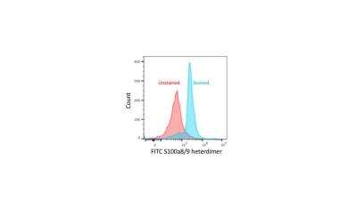
Application: Flow CytometrySample Tested: Murine Splenocytes (C57B6)Species: MouseVerified Customer | Posted 01/28/2018Mouse splenocytes stained with or without AF488 S100a8/a9 antibody.Mouse splenocytes were fixed and permeabilized with Foxp3 Fixation/Permeabilization kit. Splenocytes were incubated with Fc block for 5 mins at RT and stained with 1:100 dilution of this antibody for 25 mins at RT. Data was acquired on a Cytoflex LX and analyzed using FlowJo 10.
![S100A9 Antibody (MAC387) [Alexa Fluor® 488] NBP2-47667AF488](data:image/png;base64,R0lGODlhAQABAAD/ACwAAAAAAQABAAACADs=)
There are no reviews that match your criteria.
Protocols
Find general support by application which include: protocols, troubleshooting, illustrated assays, videos and webinars.
- 7-Amino Actinomycin D (7-AAD) Cell Viability Flow Cytometry Protocol
- Antigen Retrieval Protocol (PIER)
- Antigen Retrieval for Frozen Sections Protocol
- Appropriate Fixation of IHC/ICC Samples
- Cellular Response to Hypoxia Protocols
- Chromogenic IHC Staining of Formalin-Fixed Paraffin-Embedded (FFPE) Tissue Protocol
- Chromogenic Immunohistochemistry Staining of Frozen Tissue
- ClariTSA™ Fluorophore Kits
- Detection & Visualization of Antibody Binding
- Extracellular Membrane Flow Cytometry Protocol
- Flow Cytometry Protocol for Cell Surface Markers
- Flow Cytometry Protocol for Staining Membrane Associated Proteins
- Flow Cytometry Staining Protocols
- Flow Cytometry Troubleshooting Guide
- Fluorescent IHC Staining of Frozen Tissue Protocol
- Graphic Protocol for Heat-induced Epitope Retrieval
- Graphic Protocol for the Preparation and Fluorescent IHC Staining of Frozen Tissue Sections
- Graphic Protocol for the Preparation and Fluorescent IHC Staining of Paraffin-embedded Tissue Sections
- Graphic Protocol for the Preparation of Gelatin-coated Slides for Histological Tissue Sections
- ICC Cell Smear Protocol for Suspension Cells
- ICC Immunocytochemistry Protocol Videos
- ICC for Adherent Cells
- IHC Sample Preparation (Frozen sections vs Paraffin)
- Immunocytochemistry (ICC) Protocol
- Immunocytochemistry Troubleshooting
- Immunofluorescence of Organoids Embedded in Cultrex Basement Membrane Extract
- Immunofluorescent IHC Staining of Formalin-Fixed Paraffin-Embedded (FFPE) Tissue Protocol
- Immunohistochemistry (IHC) and Immunocytochemistry (ICC) Protocols
- Immunohistochemistry Frozen Troubleshooting
- Immunohistochemistry Paraffin Troubleshooting
- Intracellular Flow Cytometry Protocol Using Alcohol (Methanol)
- Intracellular Flow Cytometry Protocol Using Detergents
- Intracellular Nuclear Staining Flow Cytometry Protocol Using Detergents
- Intracellular Staining Flow Cytometry Protocol Using Alcohol Permeabilization
- Intracellular Staining Flow Cytometry Protocol Using Detergents to Permeabilize Cells
- Preparing Samples for IHC/ICC Experiments
- Preventing Non-Specific Staining (Non-Specific Binding)
- Primary Antibody Selection & Optimization
- Propidium Iodide Cell Viability Flow Cytometry Protocol
- Protocol for Heat-Induced Epitope Retrieval (HIER)
- Protocol for Liperfluo
- Protocol for Making a 4% Formaldehyde Solution in PBS
- Protocol for VisUCyte™ HRP Polymer Detection Reagent
- Protocol for the Characterization of Human Th22 Cells
- Protocol for the Characterization of Human Th9 Cells
- Protocol for the Fluorescent ICC Staining of Cell Smears - Graphic
- Protocol for the Fluorescent ICC Staining of Cultured Cells on Coverslips - Graphic
- Protocol for the Preparation & Fixation of Cells on Coverslips
- Protocol for the Preparation and Chromogenic IHC Staining of Frozen Tissue Sections
- Protocol for the Preparation and Chromogenic IHC Staining of Frozen Tissue Sections - Graphic
- Protocol for the Preparation and Chromogenic IHC Staining of Paraffin-embedded Tissue Sections
- Protocol for the Preparation and Chromogenic IHC Staining of Paraffin-embedded Tissue Sections - Graphic
- Protocol for the Preparation and Fluorescent ICC Staining of Cells on Coverslips
- Protocol for the Preparation and Fluorescent ICC Staining of Non-adherent Cells
- Protocol for the Preparation and Fluorescent ICC Staining of Stem Cells on Coverslips
- Protocol for the Preparation and Fluorescent IHC Staining of Frozen Tissue Sections
- Protocol for the Preparation and Fluorescent IHC Staining of Paraffin-embedded Tissue Sections
- Protocol for the Preparation of Gelatin-coated Slides for Histological Tissue Sections
- Protocol for the Preparation of a Cell Smear for Non-adherent Cell ICC - Graphic
- Protocol: Annexin V and PI Staining by Flow Cytometry
- Protocol: Annexin V and PI Staining for Apoptosis by Flow Cytometry
- TUNEL and Active Caspase-3 Detection by IHC/ICC Protocol
- The Importance of IHC/ICC Controls
- Troubleshooting Guide: Fluorokine Flow Cytometry Kits
- Troubleshooting Guide: Immunohistochemistry
- View all Protocols, Troubleshooting, Illustrated assays and Webinars
FAQs for S100A9 Antibody (MAC387) [Alexa Fluor® 488]
-
Q: Have any of your S100A9 antibodies been tested for use in neutralization, or blocking assays, on human samples?
A: Unfortunately at this time we have not tested, or received customer feedback on our S100A9 antibodies for use in blocking or neutralizing assays. If you were planning on testing any of our S100A9 antibodies for neutralizing or blocking capabilities, we would recommend our Innovator’s Reward Program. Under the terms of this program we would be happy to provide a credit for a free vial in exchange for new data on this previously untested or reported application. Please submit this data in the form of an online review. Additional Innovators Reward Program information can be found using this link.
-
Q: We would like to stain paraffin embedded human intestinal tissue for Calprotectin. One question will be which cells express the majority of Calprotectin - invading neutrophils vs epithelium for example. The antibody list you provide is quite extensive. Which antibody would you recommend for our purpose? Which tissue would you recommend as positive control.
A:
Nine of our antibodies to Calprotectin have been validated for IHC-P with human tissue, and you can see all of these products. A number of our IHC images were generated using human spleen samples, and as such this may be a good choice of a positive control tissue. We do sell a human spleen slide product, which is suitable for IHC-P. Unfortunately I do not have an in-depth knowledge of Calprotectin, however the image shown for our antibody with catalogue number NBP1-02826 demonstrates clear staining of neutrophils. The following paper also suggests that the protein is abundant in neutrophils: PMID 11435495.
-
Q: Have any of your S100A9 antibodies been tested for use in neutralization, or blocking assays, on human samples?
A: Unfortunately at this time we have not tested, or received customer feedback on our S100A9 antibodies for use in blocking or neutralizing assays. If you were planning on testing any of our S100A9 antibodies for neutralizing or blocking capabilities, we would recommend our Innovator’s Reward Program. Under the terms of this program we would be happy to provide a credit for a free vial in exchange for new data on this previously untested or reported application. Please submit this data in the form of an online review. Additional Innovators Reward Program information can be found using this link.
-
Q: We would like to stain paraffin embedded human intestinal tissue for Calprotectin. One question will be which cells express the majority of Calprotectin - invading neutrophils vs epithelium for example. The antibody list you provide is quite extensive. Which antibody would you recommend for our purpose? Which tissue would you recommend as positive control.
A:
Nine of our antibodies to Calprotectin have been validated for IHC-P with human tissue, and you can see all of these products. A number of our IHC images were generated using human spleen samples, and as such this may be a good choice of a positive control tissue. We do sell a human spleen slide product, which is suitable for IHC-P. Unfortunately I do not have an in-depth knowledge of Calprotectin, however the image shown for our antibody with catalogue number NBP1-02826 demonstrates clear staining of neutrophils. The following paper also suggests that the protein is abundant in neutrophils: PMID 11435495.