SERPINB13 Antibody (OTI2B6)
Novus Biologicals | Catalog # NBP2-01312
Key Product Details
Species Reactivity
Validated:
Human
Cited:
Human
Applications
Validated:
Immunohistochemistry, Immunohistochemistry-Paraffin, Western Blot
Cited:
IF/IHC
Label
Unconjugated
Antibody Source
Monoclonal Mouse IgG1 Clone # OTI2B6
Loading...
Product Specifications
Immunogen
Full length human recombinant protein of human SERPINB13(NP_036529) produced in HEK293T cell.
Clonality
Monoclonal
Host
Mouse
Isotype
IgG1
Theoretical MW
44.1 kDa.
Disclaimer note: The observed molecular weight of the protein may vary from the listed predicted molecular weight due to post translational modifications, post translation cleavages, relative charges, and other experimental factors.
Disclaimer note: The observed molecular weight of the protein may vary from the listed predicted molecular weight due to post translational modifications, post translation cleavages, relative charges, and other experimental factors.
Scientific Data Images for SERPINB13 Antibody (OTI2B6)
![Western Blot: SERPINB13 Antibody (OTI2B6) [NBP2-01312]](https://resources.rndsystems.com/images/products/SERPINB13-Antibody-2B6-Western-Blot-NBP2-01312-img0001.jpg "Western Blot: SERPINB13 Antibody (OTI2B6) [NBP2-01312]")
Western Blot: SERPINB13 Antibody (OTI2B6) [NBP2-01312]
Western Blot: SERPINB13 Antibody (2B6) [NBP2-01312] - HEK293T cells were transfected with the pCMV6-ENTRY control (Left lane) or pCMV6-ENTRY SERPINB13 (Right lane) cDNA for 48 hrs and lysed. Equivalent amounts of cell lysates (5 ug per lane) were separated by SDS-PAGE and immunoblotted with anti-SERPINB13.![Immunohistochemistry: SERPINB13 Antibody (OTI2B6) [NBP2-01312]](https://resources.rndsystems.com/images/products/SERPINB13-Antibody-OTI2B6-Immunohistochemistry-NBP2-01312-img0006.jpg "Immunohistochemistry: SERPINB13 Antibody (OTI2B6) [NBP2-01312]")
Immunohistochemistry: SERPINB13 Antibody (OTI2B6) [NBP2-01312]
SERPINB13-Antibody-OTI2B6-Immunohistochemistry-NBP2-01312-img0006.jpg![Immunohistochemistry-Paraffin: SERPINB13 Antibody (OTI2B6) [NBP2-01312]](https://resources.rndsystems.com/images/products/SERPINB13-Antibody-2B6-Immunohistochemistry-Paraffin-NBP2-01312-img0002.jpg "Immunohistochemistry-Paraffin: SERPINB13 Antibody (OTI2B6) [NBP2-01312]")
Immunohistochemistry-Paraffin: SERPINB13 Antibody (OTI2B6) [NBP2-01312]
Immunohistochemistry-Paraffin: SERPINB13 Antibody (2B6) [NBP2-01312] - Staining of paraffin-embedded Adenocarcinoma of Human breast tissue using anti-SERPINB13 mouse monoclonal antibody.![Immunohistochemistry-Paraffin: SERPINB13 Antibody (OTI2B6) [NBP2-01312]](https://resources.rndsystems.com/images/products/SERPINB13-Antibody-2B6-Immunohistochemistry-Paraffin-NBP2-01312-img0003.jpg "Immunohistochemistry-Paraffin: SERPINB13 Antibody (OTI2B6) [NBP2-01312]")
Immunohistochemistry-Paraffin: SERPINB13 Antibody (OTI2B6) [NBP2-01312]
Immunohistochemistry-Paraffin: SERPINB13 Antibody (2B6) [NBP2-01312] - Staining of paraffin-embedded Carcinoma of Human bladder tissue using anti-SERPINB13 mouse monoclonal antibody.![Immunohistochemistry-Paraffin: SERPINB13 Antibody (OTI2B6) [NBP2-01312]](https://resources.rndsystems.com/images/products/SERPINB13-Antibody-2B6-Immunohistochemistry-Paraffin-NBP2-01312-img0004.jpg "Immunohistochemistry-Paraffin: SERPINB13 Antibody (OTI2B6) [NBP2-01312]")
Immunohistochemistry-Paraffin: SERPINB13 Antibody (OTI2B6) [NBP2-01312]
Immunohistochemistry-Paraffin: SERPINB13 Antibody (2B6) [NBP2-01312] - Staining of paraffin-embedded Human Kidney tissue using anti-SERPINB13 mouse monoclonal antibody.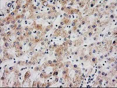
Immunohistochemistry-Paraffin: SERPINB13 Antibody (2B6) [NBP2-01312] Staining of paraffin-embedded Human liver tissue using anti-SERPINB13 mouse monoclonal antibody.
 [NBP2-01312] -")
Immunohistochemistry: SERPINB13 Antibody (OTI2B6) [NBP2-01312] -
Immunohistochemistry: SERPINB13 Antibody (OTI2B6) [NBP2-01312] - Cytoplasmic staining was detected for (a) MTSS1, (b) SCAMP1 & (c) Serpin B13 protein in the malignant epithelium of breast cancer. Alternatively, the majority of ‘pure’ HER2+ (HER2+/ER−/PR−) tumours showed loss or in (d) MTSS1, (e) SCAMP1 & (f) SERPIN B13 staining. g Distribution of MTSS1, SCAMP1 & SERPIN B13 in the ‘pure’ HER2 patient class versus other tumour classes Image collected & cropped by CiteAb from the following publication (https://www.nature.com/articles/s41419-018-0364-9), licensed under a CC-BY license. Not internally tested by Novus Biologicals. [NBP2-01312] -")
Immunohistochemistry: SERPINB13 Antibody (OTI2B6) [NBP2-01312] -
Immunohistochemistry: SERPINB13 Antibody (OTI2B6) [NBP2-01312] - Cytoplasmic staining was detected for (a) MTSS1, (b) SCAMP1 & (c) Serpin B13 protein in the malignant epithelium of breast cancer. Alternatively, the majority of ‘pure’ HER2+ (HER2+/ER−/PR−) tumours showed loss or in (d) MTSS1, (e) SCAMP1 & (f) SERPIN B13 staining. g Distribution of MTSS1, SCAMP1 & SERPIN B13 in the ‘pure’ HER2 patient class versus other tumour classes Image collected & cropped by CiteAb from the following publication (https://www.nature.com/articles/s41419-018-0364-9), licensed under a CC-BY license. Not internally tested by Novus Biologicals.Applications for SERPINB13 Antibody (OTI2B6)
Application
Recommended Usage
Immunohistochemistry
1:150
Immunohistochemistry-Paraffin
1:150
Western Blot
1:2000
Formulation, Preparation, and Storage
Purification
Immunogen affinity purified
Formulation
PBS (pH 7.3), 1.0% BSA and 50% Glycerol
Preservative
0.02% Sodium Azide
Concentration
1 mg/ml
Shipping
The product is shipped with polar packs. Upon receipt, store it immediately at the temperature recommended below.
Stability & Storage
Store at -20C. Avoid freeze-thaw cycles.
Background: SERPINB13
Alternate Names
HaCaT UV-repressible serpin, headpin, HSHUR7SEQ, HUR7, HURPIN, MGC126870, Peptidase inhibitor 13, PI-13, PI13hurpin, protease inhibitor 13 (hurpin, headpin), Proteinase inhibitor 13, serine (or cysteine) proteinase inhibitor, clade B (ovalbumin), member 13, serpin B13, serpin peptidase inhibitor, clade B (ovalbumin), member 13, UV-B repressed sequence, HUR 7
Entrez Gene IDs
5275 (Human)
Gene Symbol
SERPINB13
Additional SERPINB13 Products
Product Documents for SERPINB13 Antibody (OTI2B6)
Certificate of Analysis
To download a Certificate of Analysis, please enter a lot or batch number in the search box below.
Product Specific Notices for SERPINB13 Antibody (OTI2B6)
This product is for research use only and is not approved for use in humans or in clinical diagnosis. Primary Antibodies are guaranteed for 1 year from date of receipt.
Citations for SERPINB13 Antibody (OTI2B6)
 Powered by Bioz
Customer Reviews for SERPINB13 Antibody (OTI2B6)
There are currently no reviews for this product. Be the first to review SERPINB13 Antibody (OTI2B6) and earn rewards!
Have you used SERPINB13 Antibody (OTI2B6)?
Submit a review and receive an Amazon gift card!
$25/€18/£15/$25CAN/¥2500 Yen for a review with an image
$10/€7/£6/$10CAN/¥1110 Yen for a review without an image
Submit a review
Protocols
Find general support by application which include: protocols, troubleshooting, illustrated assays, videos and webinars.
- Antigen Retrieval Protocol (PIER)
- Antigen Retrieval for Frozen Sections Protocol
- Appropriate Fixation of IHC/ICC Samples
- Cellular Response to Hypoxia Protocols
- Chromogenic IHC Staining of Formalin-Fixed Paraffin-Embedded (FFPE) Tissue Protocol
- Chromogenic Immunohistochemistry Staining of Frozen Tissue
- ClariTSA™ Fluorophore Kits
- Detection & Visualization of Antibody Binding
- Fluorescent IHC Staining of Frozen Tissue Protocol
- Graphic Protocol for Heat-induced Epitope Retrieval
- Graphic Protocol for the Preparation and Fluorescent IHC Staining of Frozen Tissue Sections
- Graphic Protocol for the Preparation and Fluorescent IHC Staining of Paraffin-embedded Tissue Sections
- Graphic Protocol for the Preparation of Gelatin-coated Slides for Histological Tissue Sections
- IHC Sample Preparation (Frozen sections vs Paraffin)
- Immunofluorescent IHC Staining of Formalin-Fixed Paraffin-Embedded (FFPE) Tissue Protocol
- Immunohistochemistry (IHC) and Immunocytochemistry (ICC) Protocols
- Immunohistochemistry Frozen Troubleshooting
- Immunohistochemistry Paraffin Troubleshooting
- Preparing Samples for IHC/ICC Experiments
- Preventing Non-Specific Staining (Non-Specific Binding)
- Primary Antibody Selection & Optimization
- Protocol for Heat-Induced Epitope Retrieval (HIER)
- Protocol for Making a 4% Formaldehyde Solution in PBS
- Protocol for VisUCyte™ HRP Polymer Detection Reagent
- Protocol for the Preparation & Fixation of Cells on Coverslips
- Protocol for the Preparation and Chromogenic IHC Staining of Frozen Tissue Sections
- Protocol for the Preparation and Chromogenic IHC Staining of Frozen Tissue Sections - Graphic
- Protocol for the Preparation and Chromogenic IHC Staining of Paraffin-embedded Tissue Sections
- Protocol for the Preparation and Chromogenic IHC Staining of Paraffin-embedded Tissue Sections - Graphic
- Protocol for the Preparation and Fluorescent IHC Staining of Frozen Tissue Sections
- Protocol for the Preparation and Fluorescent IHC Staining of Paraffin-embedded Tissue Sections
- Protocol for the Preparation of Gelatin-coated Slides for Histological Tissue Sections
- R&D Systems Quality Control Western Blot Protocol
- TUNEL and Active Caspase-3 Detection by IHC/ICC Protocol
- The Importance of IHC/ICC Controls
- Troubleshooting Guide: Immunohistochemistry
- Troubleshooting Guide: Western Blot Figures
- Western Blot Conditions
- Western Blot Protocol
- Western Blot Protocol for Cell Lysates
- Western Blot Troubleshooting
- Western Blot Troubleshooting Guide
- View all Protocols, Troubleshooting, Illustrated assays and Webinars
Loading...