SLC16A3 Antibody - BSA Free
Novus Biologicals | Catalog # NBP1-81251
![Immunohistochemistry-Paraffin: SLC16A3 Antibody [NBP1-81251]](https://resources.rndsystems.com/images/products/SLC16A3-Antibody-Immunohistochemistry-Paraffin-NBP1-81251-img0020.jpg "Immunohistochemistry-Paraffin: SLC16A3 Antibody [NBP1-81251]")
Key Product Details
Species Reactivity
Validated:
Cited:
Predicted:
Applications
Validated:
Cited:
Label
Antibody Source
Format
Product Specifications
Immunogen
Reactivity Notes
Clonality
Host
Isotype
Scientific Data Images for SLC16A3 Antibody - BSA Free
Immunohistochemistry-Paraffin: SLC16A3 Antibody [NBP1-81251]
Immunohistochemistry-Paraffin: SLC16A3 Antibody [NBP1-81251] - Staining of human skeletal muscle shows strong cytoplasmic positivity in myocytes.![Immunohistochemistry-Paraffin: SLC16A3 Antibody [NBP1-81251]](https://resources.rndsystems.com/images/products/SLC16A3-Antibody-Immunohistochemistry-Paraffin-NBP1-81251-img0018.jpg "Immunohistochemistry-Paraffin: SLC16A3 Antibody [NBP1-81251]")
Immunohistochemistry-Paraffin: SLC16A3 Antibody [NBP1-81251]
Immunohistochemistry-Paraffin: SLC16A3 Antibody [NBP1-81251] - Staining of human lung shows strong membranous positivity in macrophages.![Immunohistochemistry-Paraffin: SLC16A3 Antibody [NBP1-81251]](https://resources.rndsystems.com/images/products/SLC16A3-Antibody-Immunohistochemistry-Paraffin-NBP1-81251-img0019.jpg "Immunohistochemistry-Paraffin: SLC16A3 Antibody [NBP1-81251]")
Immunohistochemistry-Paraffin: SLC16A3 Antibody [NBP1-81251]
Immunohistochemistry-Paraffin: SLC16A3 Antibody [NBP1-81251] - Staining of human testis shows strong membranous positivity in cells in seminiferous ducts.![Immunohistochemistry-Paraffin: SLC16A3 Antibody [NBP1-81251]](https://resources.rndsystems.com/images/products/SLC16A3-Antibody-Immunohistochemistry-Paraffin-NBP1-81251-img0017.jpg "Immunohistochemistry-Paraffin: SLC16A3 Antibody [NBP1-81251]")
Immunohistochemistry-Paraffin: SLC16A3 Antibody [NBP1-81251]
Immunohistochemistry-Paraffin: SLC16A3 Antibody [NBP1-81251] - Staining of human placenta shows strong membranous positivity in trophoblastic cells.![Simple Western: SLC16A3 Antibody [NBP1-81251]](https://resources.rndsystems.com/images/products/SLC16A3-Antibody-Simple-Western-NBP1-81251-img0015.jpg "Simple Western: SLC16A3 Antibody [NBP1-81251]")
Simple Western: SLC16A3 Antibody [NBP1-81251]
Simple Western: SLC16A3 Antibody [NBP1-81251] - Simple Western: SLC16A3 Antibody [NBP1-81251] - Electropherogram image(s) of corresponding Simple Western lane view. SLC16A3 antibody was used at 1:250 dilution on U-251MG lysates(s)![Simple Western: SLC16A3 Antibody [NBP1-81251]](https://resources.rndsystems.com/images/products/SLC16A3-Antibody-Simple-Western-NBP1-81251-img0016.jpg "Simple Western: SLC16A3 Antibody [NBP1-81251]")
Simple Western: SLC16A3 Antibody [NBP1-81251]
Simple Western: SLC16A3 Antibody [NBP1-81251] - Simple Western: SLC16A3 Antibody [NBP1-81251] - Electropherogram image(s) of corresponding Simple Western lane view. SLC16A3 antibody was used at 1:250 dilution on HeLa lysates(s).![SLC16A3 Antibody - BSA Free Immunocytochemistry/ Immunofluorescence: SLC16A3 Antibody [NBP1-81251]](https://resources.rndsystems.com/images/products/nbp1-81251_-immunocytochemistry-immunofluorescence-639174076361144394.jpg "Immunocytochemistry/ Immunofluorescence: SLC16A3 Antibody [NBP1-81251]")
Immunocytochemistry/ Immunofluorescence: SLC16A3 Antibody [NBP1-81251]
Staining of human cell line U-251 MG shows localization to plasma membrane.Applications for SLC16A3 Antibody - BSA Free
Immunocytochemistry/ Immunofluorescence
Immunohistochemistry
Immunohistochemistry-Paraffin
Simple Western
See Simple Western Antibody Database for Simple Western validation: Tested in RT-4 and U-251MG, separated by Size, antibody dilution of 1:250, apparent MW was 53 kDa. Separated by Size-Wes, Sally Sue/Peggy Sue.
Reviewed Applications
Read 1 review rated 5 using NBP1-81251 in the following applications:
Formulation, Preparation, and Storage
Purification
Formulation
Format
Preservative
Concentration
Shipping
Stability & Storage
Background: SLC16A3
Long Name
Alternate Names
Gene Symbol
UniProt
Additional SLC16A3 Products
Product Documents for SLC16A3 Antibody - BSA Free
Certificate of Analysis
To download a Certificate of Analysis, please enter a lot or batch number in the search box below.
Product Specific Notices for SLC16A3 Antibody - BSA Free
This product is for research use only and is not approved for use in humans or in clinical diagnosis. Primary Antibodies are guaranteed for 1 year from date of receipt.
Related Research Areas
Citations for SLC16A3 Antibody - BSA Free
 Powered by Bioz
Customer Reviews for SLC16A3 Antibody - BSA Free (1)
Have you used SLC16A3 Antibody - BSA Free?
Submit a review and receive an Amazon gift card!
$25/€18/£15/$25CAN/¥2500 Yen for a review with an image
$10/€7/£6/$10CAN/¥1110 Yen for a review without an image
Submit a review
Customer Images
-
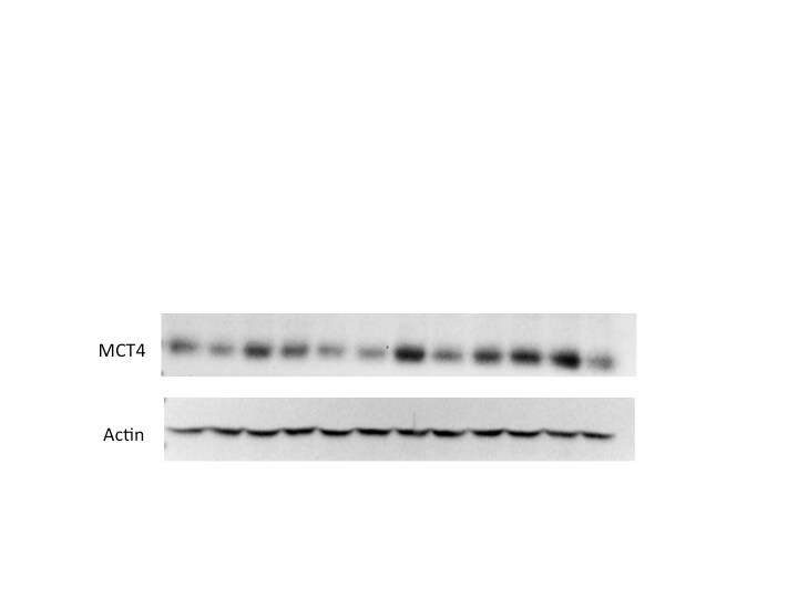
Application: Western BlotSample Tested:Species: HumanVerified Customer | Posted 06/04/2015Immunoblot assay of MCT4 expression in human breast cancer cell lines

There are no reviews that match your criteria.
Protocols
Find general support by application which include: protocols, troubleshooting, illustrated assays, videos and webinars.
- Antigen Retrieval Protocol (PIER)
- Antigen Retrieval for Frozen Sections Protocol
- Appropriate Fixation of IHC/ICC Samples
- Cellular Response to Hypoxia Protocols
- Chromogenic IHC Staining of Formalin-Fixed Paraffin-Embedded (FFPE) Tissue Protocol
- Chromogenic Immunohistochemistry Staining of Frozen Tissue
- ClariTSA™ Fluorophore Kits
- Detection & Visualization of Antibody Binding
- Fluorescent IHC Staining of Frozen Tissue Protocol
- Graphic Protocol for Heat-induced Epitope Retrieval
- Graphic Protocol for the Preparation and Fluorescent IHC Staining of Frozen Tissue Sections
- Graphic Protocol for the Preparation and Fluorescent IHC Staining of Paraffin-embedded Tissue Sections
- Graphic Protocol for the Preparation of Gelatin-coated Slides for Histological Tissue Sections
- ICC Cell Smear Protocol for Suspension Cells
- ICC Immunocytochemistry Protocol Videos
- ICC for Adherent Cells
- IHC Sample Preparation (Frozen sections vs Paraffin)
- Immunocytochemistry (ICC) Protocol
- Immunocytochemistry Troubleshooting
- Immunofluorescence of Organoids Embedded in Cultrex Basement Membrane Extract
- Immunofluorescent IHC Staining of Formalin-Fixed Paraffin-Embedded (FFPE) Tissue Protocol
- Immunohistochemistry (IHC) and Immunocytochemistry (ICC) Protocols
- Immunohistochemistry Frozen Troubleshooting
- Immunohistochemistry Paraffin Troubleshooting
- Preparing Samples for IHC/ICC Experiments
- Preventing Non-Specific Staining (Non-Specific Binding)
- Primary Antibody Selection & Optimization
- Protocol for Heat-Induced Epitope Retrieval (HIER)
- Protocol for Making a 4% Formaldehyde Solution in PBS
- Protocol for VisUCyte™ HRP Polymer Detection Reagent
- Protocol for the Fluorescent ICC Staining of Cell Smears - Graphic
- Protocol for the Fluorescent ICC Staining of Cultured Cells on Coverslips - Graphic
- Protocol for the Preparation & Fixation of Cells on Coverslips
- Protocol for the Preparation and Chromogenic IHC Staining of Frozen Tissue Sections
- Protocol for the Preparation and Chromogenic IHC Staining of Frozen Tissue Sections - Graphic
- Protocol for the Preparation and Chromogenic IHC Staining of Paraffin-embedded Tissue Sections
- Protocol for the Preparation and Chromogenic IHC Staining of Paraffin-embedded Tissue Sections - Graphic
- Protocol for the Preparation and Fluorescent ICC Staining of Cells on Coverslips
- Protocol for the Preparation and Fluorescent ICC Staining of Non-adherent Cells
- Protocol for the Preparation and Fluorescent ICC Staining of Stem Cells on Coverslips
- Protocol for the Preparation and Fluorescent IHC Staining of Frozen Tissue Sections
- Protocol for the Preparation and Fluorescent IHC Staining of Paraffin-embedded Tissue Sections
- Protocol for the Preparation of Gelatin-coated Slides for Histological Tissue Sections
- Protocol for the Preparation of a Cell Smear for Non-adherent Cell ICC - Graphic
- TUNEL and Active Caspase-3 Detection by IHC/ICC Protocol
- The Importance of IHC/ICC Controls
- Troubleshooting Guide: Immunohistochemistry
- View all Protocols, Troubleshooting, Illustrated assays and Webinars
FAQs for SLC16A3 Antibody - BSA Free
-
Q: Does the SLC16A3 Antibody (NBP1-81251) also work on frozen sections?
A: Thank you for contacting Novus Biologicals Technical support. NBP1-81251 has not yet been tested or validated for the use on frozen sections. It is validated and guaranteed to work in IHC on paraffin embedded tissues.