
SLC45A4 Antibody
Novus Biologicals | Catalog # NBP1-70394

Loading...
Key Product Details
Species Reactivity
Human
Applications
Immunohistochemistry, Immunohistochemistry-Paraffin, Western Blot
Label
Unconjugated
Antibody Source
Polyclonal Rabbit IgG
Loading...
Product Specifications
Immunogen
A synthetic peptide from internal region of human SLC45A4 conjugated to an blue carrier protein was used as the antigen.
Clonality
Polyclonal
Host
Rabbit
Isotype
IgG
Description
This peptide is soluble in PBS and water.
Scientific Data Images for SLC45A4 Antibody
Western Blot: SLC45A4 Antibody [NBP1-70394] -
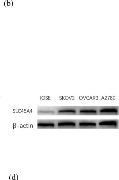
SLC45A4 is overexpressed in EOC (a) Comparison of SLC45A4 RNA expression between normal ovarian tissue and OC tissue was analyzed using data from TCGA and GEPIA databases. (b) Expression of SLC45A4 in OC tissue in HPA database. (c) IHC analysis showed that SLC45A4 protein expression in EOC was higher than that in normal fallopian tubes and ovaries. Scale = 250 μm.(d) The mRNA and protein expression levels of SLC45A4 in IOSE80, and EOC cells were assessed using qRT-PCR and WB analysis. ∗P < 0.05; ∗∗P < 0.01; ∗∗∗P < 0.001. Image collected and cropped by CiteAb from the following open publication (https://pubmed.ncbi.nlm.nih.gov/39363015), licensed under a CC-BY license. Not internally tested by Novus Biologicals.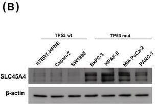
Western Blot: SLC45A4 Antibody [NBP1-70394] -
Knockdown of SLC45A4 inhibits proliferation in TP53 mutant PDA cells in vitro. qRT‐PCR (A) and western blotting (B) showed a higher expression of SLC45A4 in TP53 mutant cell lines compared to TP53 wild‐type cell lines. Wt, wild‐type; Mut, mutation. Knockdown of SLC45A4 was confirmed by qRT‐PCR (C) and western blotting (D). MTS (E), colony formation (F) and EdU incorporation (G) assays were performed with si‐SLC45A4 or control cells in hTERT‐HPNE, HPAF‐II and MIA PaCa‐2 cell lines. Scale bar = 200 μm. The data are presented as the mean +/- SD. *p < 0.05, **p < 0.01 and ***p < 0.001 Image collected and cropped by CiteAb from the following open publication (https://pubmed.ncbi.nlm.nih.gov/34010493), licensed under a CC-BY license. Not internally tested by Novus Biologicals.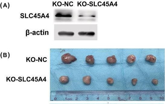
Western Blot: SLC45A4 Antibody [NBP1-70394] -
Knockout of SLC45A4 suppresses TP53 mutant PDA cell growth in vivo. (A) Knockout of SLC45A4 was detected by western blotting. (B) Knockout of SLC45A4 in HPAF‐II cell inhibited tumor growth in vivo (n = 5 per group). (C) Tumor weights and (D) growth curves of xenograft tumors after subcutaneous injection with KO‐SLC45A4 HPAF‐II cell or the control cell. (E) Representative images are shown of immunohistochemical staining of SLC45A4 and Ki67 with statistical analysis. Data are presented as the mean +/- SD (n = 5). *p < 0.05, **p < 0.01 and ***p < 0.001 Image collected and cropped by CiteAb from the following open publication (https://pubmed.ncbi.nlm.nih.gov/34010493), licensed under a CC-BY license. Not internally tested by Novus Biologicals.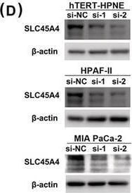
Western Blot: SLC45A4 Antibody [NBP1-70394] -
Knockdown of SLC45A4 inhibits proliferation in TP53 mutant PDA cells in vitro. qRT‐PCR (A) and western blotting (B) showed a higher expression of SLC45A4 in TP53 mutant cell lines compared to TP53 wild‐type cell lines. Wt, wild‐type; Mut, mutation. Knockdown of SLC45A4 was confirmed by qRT‐PCR (C) and western blotting (D). MTS (E), colony formation (F) and EdU incorporation (G) assays were performed with si‐SLC45A4 or control cells in hTERT‐HPNE, HPAF‐II and MIA PaCa‐2 cell lines. Scale bar = 200 μm. The data are presented as the mean +/- SD. *p < 0.05, **p < 0.01 and ***p < 0.001 Image collected and cropped by CiteAb from the following open publication (https://pubmed.ncbi.nlm.nih.gov/34010493), licensed under a CC-BY license. Not internally tested by Novus Biologicals.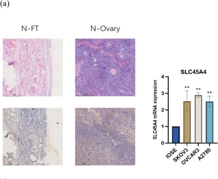
Immunohistochemistry: SLC45A4 Antibody [NBP1-70394] -
SLC45A4 is overexpressed in EOC (a) Comparison of SLC45A4 RNA expression between normal ovarian tissue and OC tissue was analyzed using data from TCGA and GEPIA databases. (b) Expression of SLC45A4 in OC tissue in HPA database. (c) IHC analysis showed that SLC45A4 protein expression in EOC was higher than that in normal fallopian tubes and ovaries. Scale = 250 μm.(d) The mRNA and protein expression levels of SLC45A4 in IOSE80, and EOC cells were assessed using qRT-PCR and WB analysis. ∗P < 0.05; ∗∗P < 0.01; ∗∗∗P < 0.001. Image collected and cropped by CiteAb from the following open publication (https://pubmed.ncbi.nlm.nih.gov/39363015), licensed under a CC-BY license. Not internally tested by Novus Biologicals.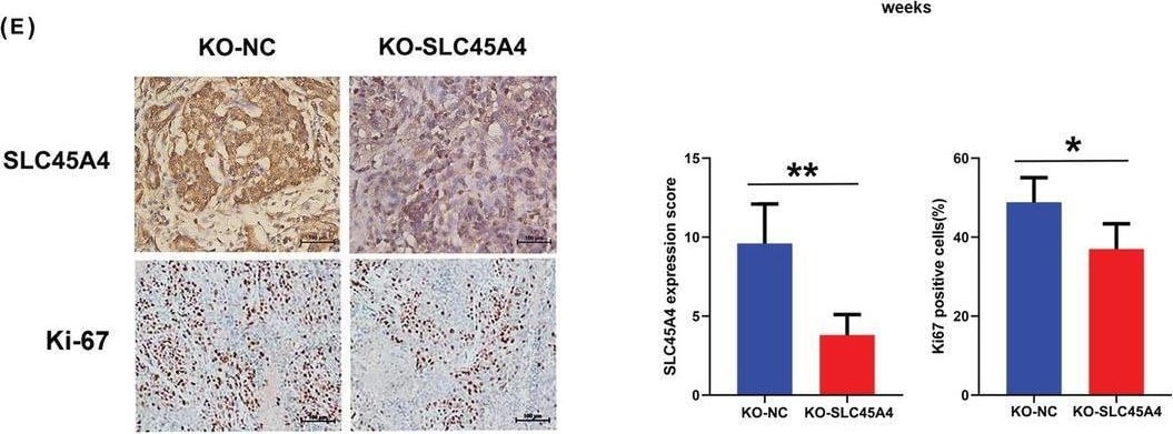
Immunohistochemistry: SLC45A4 Antibody [NBP1-70394] -
Knockout of SLC45A4 suppresses TP53 mutant PDA cell growth in vivo. (A) Knockout of SLC45A4 was detected by western blotting. (B) Knockout of SLC45A4 in HPAF‐II cell inhibited tumor growth in vivo (n = 5 per group). (C) Tumor weights and (D) growth curves of xenograft tumors after subcutaneous injection with KO‐SLC45A4 HPAF‐II cell or the control cell. (E) Representative images are shown of immunohistochemical staining of SLC45A4 and Ki67 with statistical analysis. Data are presented as the mean +/- SD (n = 5). *p < 0.05, **p < 0.01 and ***p < 0.001 Image collected and cropped by CiteAb from the following open publication (https://pubmed.ncbi.nlm.nih.gov/34010493), licensed under a CC-BY license. Not internally tested by Novus Biologicals.Applications for SLC45A4 Antibody
Application
Recommended Usage
Immunohistochemistry
1:300-1:2000
Immunohistochemistry-Paraffin
1:300-1:2000
Western Blot
1:300-1:2000
Formulation, Preparation, and Storage
Purification
Unpurified
Reconstitution
Reconstitute in 0.1 ml of sterile water. Centrifuge to remove any insoluble material. Glycerol may be added (1:1) for additional stability. Please note the sample size is provided in reconstituted format.
Formulation
Lyophilized from whole antisera
Preservative
No Preservative
Concentration
This product is unpurified. The exact concentration of antibody is not quantifiable.
Shipping
The product is shipped with polar packs. Upon receipt, store it immediately at the temperature recommended below.
Stability & Storage
Store at 4C short term. Aliquot and store at -20C long term. Avoid freeze-thaw cycles.
Calculators
Background: SLC45A4
Alternate Names
KIAA1126, solute carrier family 45, member 4
Gene Symbol
SLC45A4
Additional SLC45A4 Products
Product Documents for SLC45A4 Antibody
Certificate of Analysis
To download a Certificate of Analysis, please enter a lot or batch number in the search box below.
Product Specific Notices for SLC45A4 Antibody
This peptide is soluble in PBS and water.
This product is for research use only and is not approved for use in humans or in clinical diagnosis. Primary Antibodies are guaranteed for 1 year from date of receipt.
Citations for SLC45A4 Antibody
 Powered by Bioz
Customer Reviews for SLC45A4 Antibody
There are currently no reviews for this product. Be the first to review SLC45A4 Antibody and earn rewards!
Have you used SLC45A4 Antibody?
Submit a review and receive an Amazon gift card!
$25/€18/£15/$25CAN/¥2500 for a review with an image
$10/€7/£6/$10CAN/¥1110 for a review without an image
Submit a review
Protocols
Find general support by application which include: protocols, troubleshooting, illustrated assays, videos and webinars.
- Antigen Retrieval Protocol (PIER)
- Antigen Retrieval for Frozen Sections Protocol
- Appropriate Fixation of IHC/ICC Samples
- Cellular Response to Hypoxia Protocols
- Chromogenic IHC Staining of Formalin-Fixed Paraffin-Embedded (FFPE) Tissue Protocol
- Chromogenic Immunohistochemistry Staining of Frozen Tissue
- ClariTSA™ Fluorophore Kits
- Detection & Visualization of Antibody Binding
- Fluorescent IHC Staining of Frozen Tissue Protocol
- Graphic Protocol for Heat-induced Epitope Retrieval
- Graphic Protocol for the Preparation and Fluorescent IHC Staining of Frozen Tissue Sections
- Graphic Protocol for the Preparation and Fluorescent IHC Staining of Paraffin-embedded Tissue Sections
- Graphic Protocol for the Preparation of Gelatin-coated Slides for Histological Tissue Sections
- IHC Sample Preparation (Frozen sections vs Paraffin)
- Immunofluorescent IHC Staining of Formalin-Fixed Paraffin-Embedded (FFPE) Tissue Protocol
- Immunohistochemistry (IHC) and Immunocytochemistry (ICC) Protocols
- Immunohistochemistry Frozen Troubleshooting
- Immunohistochemistry Paraffin Troubleshooting
- Preparing Samples for IHC/ICC Experiments
- Preventing Non-Specific Staining (Non-Specific Binding)
- Primary Antibody Selection & Optimization
- Protocol for Heat-Induced Epitope Retrieval (HIER)
- Protocol for Making a 4% Formaldehyde Solution in PBS
- Protocol for VisUCyte™ HRP Polymer Detection Reagent
- Protocol for the Preparation & Fixation of Cells on Coverslips
- Protocol for the Preparation and Chromogenic IHC Staining of Frozen Tissue Sections
- Protocol for the Preparation and Chromogenic IHC Staining of Frozen Tissue Sections - Graphic
- Protocol for the Preparation and Chromogenic IHC Staining of Paraffin-embedded Tissue Sections
- Protocol for the Preparation and Chromogenic IHC Staining of Paraffin-embedded Tissue Sections - Graphic
- Protocol for the Preparation and Fluorescent IHC Staining of Frozen Tissue Sections
- Protocol for the Preparation and Fluorescent IHC Staining of Paraffin-embedded Tissue Sections
- Protocol for the Preparation of Gelatin-coated Slides for Histological Tissue Sections
- R&D Systems Quality Control Western Blot Protocol
- TUNEL and Active Caspase-3 Detection by IHC/ICC Protocol
- The Importance of IHC/ICC Controls
- Troubleshooting Guide: Immunohistochemistry
- Troubleshooting Guide: Western Blot Figures
- Western Blot Conditions
- Western Blot Protocol
- Western Blot Protocol for Cell Lysates
- Western Blot Troubleshooting
- Western Blot Troubleshooting Guide
- View all Protocols, Troubleshooting, Illustrated assays and Webinars
Loading...