SOD1/Cu-Zn SOD Antibody - BSA Free
Novus Biologicals | Catalog # NBP2-24915
![Western Blot: SOD1/Cu-Zn SOD Antibody [NBP2-24915]](https://resources.rndsystems.com/images/products/SOD1-Cu-Zn-SOD-Antibody-Western-Blot-NBP2-24915-img0006.jpg "Western Blot: SOD1/Cu-Zn SOD Antibody [NBP2-24915]")
Key Product Details
Species Reactivity
Validated:
Human, Mouse, Rat, Bovine
Cited:
Human, Mouse, Rat
Applications
Validated:
Immunohistochemistry, Immunohistochemistry-Paraffin, Western Blot
Cited:
Immunohistochemistry-Paraffin, Western Blot
Label
Unconjugated
Antibody Source
Polyclonal Rabbit IgG
Format
BSA Free
Loading...
Product Specifications
Immunogen
The superoxide dismutase enzyme from bovine erythrocytes (CAS # 9054-89-1) was used as the immunogen.
Clonality
Polyclonal
Host
Rabbit
Isotype
IgG
Scientific Data Images for SOD1/Cu-Zn SOD Antibody - BSA Free
Western Blot: SOD1/Cu-Zn SOD Antibody [NBP2-24915]
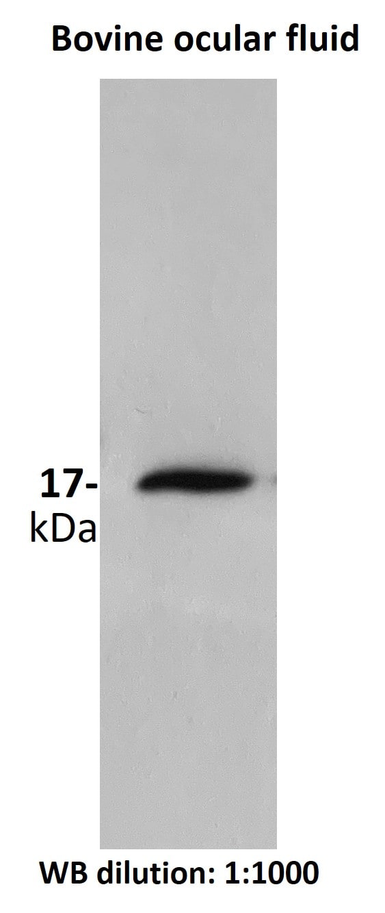
Western Blot: SOD1/Cu-Zn SOD Antibody [NBP2-24915] - Bovine ocular fluid. Antibody at 1:1000. Western blot image submitted by a verified customer review.![Immunohistochemistry-Paraffin: SOD1/Cu-Zn SOD Antibody [NBP2-24915]](https://resources.rndsystems.com/images/products/SOD1-Cu-Zn-SOD-Antibody-Immunohistochemistry-Paraffin-NBP2-24915-img0003.jpg "Immunohistochemistry-Paraffin: SOD1/Cu-Zn SOD Antibody [NBP2-24915]")
Immunohistochemistry-Paraffin: SOD1/Cu-Zn SOD Antibody [NBP2-24915]
Immunohistochemistry-Paraffin: SOD1/Cu-Zn SOD Antibody [NBP2-24915] - Analysis of Superoxide Dismutase 1 in FFPE human liver tissue using an isotype control (top) and NBP2-24915 (bottom) at 5 ug/ml.![Western Blot: SOD1/Cu-Zn SOD Antibody [NBP2-24915]](https://resources.rndsystems.com/images/products/SOD1-Cu-Zn-SOD-Antibody-Western-Blot-NBP2-24915-img0004.jpg "Western Blot: SOD1/Cu-Zn SOD Antibody [NBP2-24915]")
Western Blot: SOD1/Cu-Zn SOD Antibody [NBP2-24915]
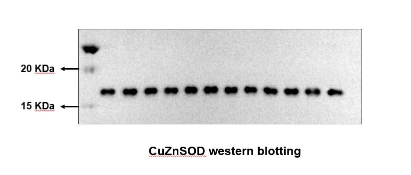
Western Blot: SOD1/Cu-Zn SOD Antibody [NBP2-24915] - Analysis of SOD in liver lysate of 1) human, 2) mouse, and 3) rat using this antibody. 025 ug/ml.![Western Blot: SOD1/Cu-Zn SOD Antibody [NBP2-24915]](https://resources.rndsystems.com/images/products/SOD1-Cu-Zn-SOD-Antibody-Western-Blot-NBP2-24915-img0005.jpg "Western Blot: SOD1/Cu-Zn SOD Antibody [NBP2-24915]")
Western Blot: SOD1/Cu-Zn SOD Antibody [NBP2-24915]
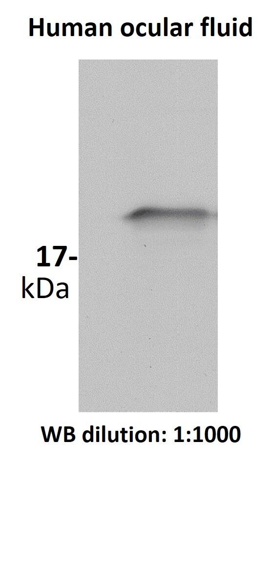
Western Blot: SOD1/Cu-Zn SOD Antibody [NBP2-24915] - Human ocular fluid. Antibody at 1:1000. Western blot image submitted by a verified customer review.
Western Blot: SOD1/Cu-Zn SOD Antibody - BSA Free [NBP2-24915] -
Production of superoxide radicals (O2–) and expression of antioxidant enzymes during MSC expansion, with or without methylene blue (MB) addition. a Quantification of superoxide radicals was carried out by MitoSOX staining (n = 3; values represent the mean +/- SEM). *p < 0.05. b Mitochondrial (MnSOD) and cytosolic superoxide dismutase (Cu/ZnSOD) Western blots were performed on proteins isolated from the same samples, and c results were normalized to actin Image collected and cropped by CiteAb from the following open publication (https://pubmed.ncbi.nlm.nih.gov/28061861), licensed under a CC-BY license. Not internally tested by Novus Biologicals.
Western Blot: SOD1/Cu-Zn SOD Antibody - BSA Free [NBP2-24915] -
SOD1 does not affect laminin alpha 5 expression in endothelial cells. (A) mRNA levels of human SOD1 in 1G11-mock and 1G11-SOD1 cells, as determined by RT-qPCR. Each dot represents the mean of a triplicate from an independent experiment *** p < 0.001; two-tailed t-test. (B) Human SOD1 protein levels in 1G11-mock and 1G11-SOD1 cells as determined by immunoblotting; HEK-293T cells were used as a human SOD1 reference. Immunoblots were rehybridized with an anti-beta -actin antibody as a loading control. A representative experiment is shown. (C) 1G11-mock and 1G11-SOD1 cells were incubated (14 h) with the conditioned medium from N202.1A tumor cell line cultures (MCT) and then stained with DHE (red); nuclei were DAPI-counterstained (blue). Treatment with the SOD mimetic MnTBAP was used as control (right panels). Scale bars 25 um. (D) SOD activity in 1G11-mock and 1G11-SOD1 cell extracts. (E) Relative LAMA5 mRNA levels in 1G11-mock and 1G11-SOD1 cells. Each dot represents the mean of a triplicate from an independent experiment (n = 5). p = 0.32, two-tailed t-test. (F) Representative images of laminin alpha 5 (green; PAC078MV01) staining in 1G11-mock and 1G11-SOD1 cells; nuclei were DAPI counterstained (blue). Scale bars 50 um. (G) Quantification of laminin alpha 5 mean fluorescence intensity from the images as in (F) (n = 5 fields/condition). p = 0.58; two-tailed t-test. (H) Relative LAMA4 mRNA levels in 1G11-mock, 1G11-SOD3 and 1G11-SOD1 cells. Each dot represents the mean of a triplicate from an independent experiment (n = 6). ** p < 0.01, *** p < 0.001, two-tailed t-test. Image collected and cropped by CiteAb from the following open publication (https://pubmed.ncbi.nlm.nih.gov/35267534), licensed under a CC-BY license. Not internally tested by Novus Biologicals.Applications for SOD1/Cu-Zn SOD Antibody - BSA Free
Application
Recommended Usage
Immunohistochemistry
1:200
Immunohistochemistry-Paraffin
1:200
Western Blot
1 - 2 ug/ml
Reviewed Applications
Read 3 reviews rated 5 using NBP2-24915 in the following applications:
Formulation, Preparation, and Storage
Purification
Immunogen affinity purified
Formulation
PBS
Format
BSA Free
Preservative
0.02% Sodium Azide
Concentration
1.0 mg/ml
Shipping
The product is shipped with polar packs. Upon receipt, store it immediately at the temperature recommended below.
Stability & Storage
Store at 4C short term. Aliquot and store at -20C long term. Avoid freeze-thaw cycles.
Background: SOD1/Cu-Zn SOD
Long Name
Superoxide Dismutase-1
Alternate Names
Cu-Zn SOD, CuZn SOD, Ipo1, IPOA, SOD, cytosolic, SOD, Soluble
Gene Symbol
SOD1
UniProt
Additional SOD1/Cu-Zn SOD Products
Product Documents for SOD1/Cu-Zn SOD Antibody - BSA Free
Certificate of Analysis
To download a Certificate of Analysis, please enter a lot or batch number in the search box below.
Product Specific Notices for SOD1/Cu-Zn SOD Antibody - BSA Free
This product is for research use only and is not approved for use in humans or in clinical diagnosis. Primary Antibodies are guaranteed for 1 year from date of receipt.
Citations for SOD1/Cu-Zn SOD Antibody - BSA Free
 Powered by Bioz
Customer Reviews for SOD1/Cu-Zn SOD Antibody - BSA Free (3)
5 out of 5
3 Customer Ratings
Have you used SOD1/Cu-Zn SOD Antibody - BSA Free?
Submit a review and receive an Amazon gift card!
$25/€18/£15/$25CAN/¥2500 Yen for a review with an image
$10/€7/£6/$10CAN/¥1110 Yen for a review without an image
Submit a review
Customer Images
Showing
1
-
3 of
3 reviews
Showing All
Filter By:
-
Application: Western BlotSample Tested: ocular fluidSpecies: BovineVerified Customer | Posted 09/23/2020works really well. Use at a dilution of 1:1000.use at a dilution of 1:1000

-
Application: Western BlotSample Tested: ocular fluidSpecies: HumanVerified Customer | Posted 09/23/2020works really well at 1:1000 for western blottinguse at dilution 1:1000
-
Application: Western BlotSample Tested: LiverSpecies: MouseVerified Customer | Posted 01/12/2017N/A
There are no reviews that match your criteria.
Protocols
View specific protocols for SOD1/Cu-Zn SOD Antibody - BSA Free (NBP2-24915):
Immunohistochemistry-Paraffin Embedded Sections
Antigen Unmasking:
Bring slides to a boil in 10 mM sodium citrate buffer (pH 6.0) then maintain at a sub-boiling temperature for 10 minutes. Cool slides on bench-top for 30 minutes (keep slides in the sodium citrate buffer at all times).
Staining:
1. Wash sections in deionized water three times for 5 minutes each.
2. Wash sections in PBS for 5 minutes.
3. Block each section with 100-400 ul blocking solution (1% BSA in PBS) for 1 hour at room temperature.
4. Remove blocking solution and add 100-400 ul diluted primary antibody. Incubate overnight at 4 C.
5. Remove antibody solution and wash sections in wash buffer three times for 5 minutes each.
6. Add 100-400 ul HRP polymer conjugated secondary antibody. Incubate 30 minutes at room temperature.
7. Wash sections three times in wash buffer for 5 minutes each.
8. Add 100-400 ul DAB substrate to each section and monitor staining closely.
9. As soon as the sections develop, immerse slides in deionized water.
10. Counterstain sections in hematoxylin.
11. Wash sections in deionized water two times for 5 minutes each.
12. Dehydrate sections.
13. Mount coverslips.
Western Blot Protocol
1. Perform SDS-PAGE on samples to be analyzed, loading 10-25 ug of total protein per lane.
2. Transfer proteins to PVDF membrane according to the instructions provided by the manufacturer of the membrane and transfer apparatus.
3. Stain the membrane with Ponceau S (or similar product) to assess transfer success, and mark molecular weight standards where appropriate.
4. Rinse the blot TBS -0.05% Tween 20 (TBST).
5. Block the membrane in 5% Non-fat milk in TBST (blocking buffer) for at least 1 hour.
6. Wash the membrane in TBST three times for 10 minutes each.
7. Dilute primary antibody in blocking buffer and incubate overnight at 4C with gentle rocking.
8. Wash the membrane in TBST three times for 10 minutes each.
9. Incubate the membrane in diluted HRP conjugated secondary antibody in blocking buffer (as per manufacturer's instructions) for 1 hour at room temperature.
10. Wash the blot in TBST three times for 10 minutes each (this step can be repeated as required to reduce background).
11. Apply the detection reagent of choice in accordance with the manufacturer's instructions.
Find general support by application which include: protocols, troubleshooting, illustrated assays, videos and webinars.
- Antigen Retrieval Protocol (PIER)
- Antigen Retrieval for Frozen Sections Protocol
- Appropriate Fixation of IHC/ICC Samples
- Cellular Response to Hypoxia Protocols
- Chromogenic IHC Staining of Formalin-Fixed Paraffin-Embedded (FFPE) Tissue Protocol
- Chromogenic Immunohistochemistry Staining of Frozen Tissue
- ClariTSA™ Fluorophore Kits
- Detection & Visualization of Antibody Binding
- Fluorescent IHC Staining of Frozen Tissue Protocol
- Graphic Protocol for Heat-induced Epitope Retrieval
- Graphic Protocol for the Preparation and Fluorescent IHC Staining of Frozen Tissue Sections
- Graphic Protocol for the Preparation and Fluorescent IHC Staining of Paraffin-embedded Tissue Sections
- Graphic Protocol for the Preparation of Gelatin-coated Slides for Histological Tissue Sections
- IHC Sample Preparation (Frozen sections vs Paraffin)
- Immunofluorescent IHC Staining of Formalin-Fixed Paraffin-Embedded (FFPE) Tissue Protocol
- Immunohistochemistry (IHC) and Immunocytochemistry (ICC) Protocols
- Immunohistochemistry Frozen Troubleshooting
- Immunohistochemistry Paraffin Troubleshooting
- Preparing Samples for IHC/ICC Experiments
- Preventing Non-Specific Staining (Non-Specific Binding)
- Primary Antibody Selection & Optimization
- Protocol for Heat-Induced Epitope Retrieval (HIER)
- Protocol for Making a 4% Formaldehyde Solution in PBS
- Protocol for VisUCyte™ HRP Polymer Detection Reagent
- Protocol for the Preparation & Fixation of Cells on Coverslips
- Protocol for the Preparation and Chromogenic IHC Staining of Frozen Tissue Sections
- Protocol for the Preparation and Chromogenic IHC Staining of Frozen Tissue Sections - Graphic
- Protocol for the Preparation and Chromogenic IHC Staining of Paraffin-embedded Tissue Sections
- Protocol for the Preparation and Chromogenic IHC Staining of Paraffin-embedded Tissue Sections - Graphic
- Protocol for the Preparation and Fluorescent IHC Staining of Frozen Tissue Sections
- Protocol for the Preparation and Fluorescent IHC Staining of Paraffin-embedded Tissue Sections
- Protocol for the Preparation of Gelatin-coated Slides for Histological Tissue Sections
- R&D Systems Quality Control Western Blot Protocol
- TUNEL and Active Caspase-3 Detection by IHC/ICC Protocol
- The Importance of IHC/ICC Controls
- Troubleshooting Guide: Immunohistochemistry
- Troubleshooting Guide: Western Blot Figures
- Western Blot Conditions
- Western Blot Protocol
- Western Blot Protocol for Cell Lysates
- Western Blot Troubleshooting
- Western Blot Troubleshooting Guide
- View all Protocols, Troubleshooting, Illustrated assays and Webinars
Loading...