SPARC Antibody - BSA Free
Novus Biologicals | Catalog # NBP1-80972
![Western Blot: SPARC Antibody [NBP1-80972]](https://resources.rndsystems.com/images/products/SPARC-Antibody-Western-Blot-NBP1-80972-img0010.jpg "Western Blot: SPARC Antibody [NBP1-80972]")
Loading...
Key Product Details
Validated by
Orthogonal Validation, Independent Antibodies
Species Reactivity
Validated:
Human
Cited:
Mouse
Predicted:
Rat (96%). Backed by our 100% Guarantee.
Applications
Validated:
Immunohistochemistry, Immunohistochemistry-Paraffin, Western Blot
Cited:
Western Blot
Label
Unconjugated
Antibody Source
Polyclonal Rabbit IgG
Format
BSA Free
Loading...
Product Specifications
Immunogen
This antibody was developed against Recombinant Protein corresponding to amino acids: VLVTLYERDEDNNLLTEKQKLRVKKIHENEKRLEAGDHPVELLARDFEKNYNMYIFPVHWQFGQLDQHPIDGYLSHTELAPLRAPLIPMEHCTTRFFETCDLDNDKYIALDEWAGCFGIKQKDIDKDL
Clonality
Polyclonal
Host
Rabbit
Isotype
IgG
Theoretical MW
35 kDa.
Disclaimer note: The observed molecular weight of the protein may vary from the listed predicted molecular weight due to post translational modifications, post translation cleavages, relative charges, and other experimental factors.
Disclaimer note: The observed molecular weight of the protein may vary from the listed predicted molecular weight due to post translational modifications, post translation cleavages, relative charges, and other experimental factors.
Scientific Data Images for SPARC Antibody - BSA Free
![Western Blot: SPARC Antibody [NBP1-80972]](https://resources.rndsystems.com/images/products/SPARC-Antibody-Western-Blot-NBP1-80972-img0013.jpg "Western Blot: SPARC Antibody [NBP1-80972]")
![Immunohistochemistry-Paraffin: SPARC Antibody [NBP1-80972]](https://resources.rndsystems.com/images/products/SPARC-Antibody-Immunohistochemistry-Paraffin-NBP1-80972-img0011.jpg "Immunohistochemistry-Paraffin: SPARC Antibody [NBP1-80972]")
Immunohistochemistry-Paraffin: SPARC Antibody [NBP1-80972]
Immunohistochemistry-Paraffin: SPARC Antibody [NBP1-80972] - Staining of human placenta shows strong positivity in endothelial cells and fibroblasts.Applications for SPARC Antibody - BSA Free
Application
Recommended Usage
Immunohistochemistry
1:200 - 1:500
Immunohistochemistry-Paraffin
1:200-1:500
Western Blot
0.04 - 0.4 ug/ml
Application Notes
IHC-Paraffin, HIER pH 6 retrieval is recommended.
Reviewed Applications
Read 2 reviews rated 5 using NBP1-80972 in the following applications:
Formulation, Preparation, and Storage
Purification
Affinity purified
Formulation
PBS (pH 7.2) and 40% Glycerol
Format
BSA Free
Preservative
0.02% Sodium Azide
Concentration
Concentrations vary lot to lot. See vial label for concentration. If unlisted please contact technical services.
Shipping
The product is shipped with polar packs. Upon receipt, store it immediately at the temperature recommended below.
Stability & Storage
Store at 4C short term. Aliquot and store at -20C long term. Avoid freeze-thaw cycles.
Background: SPARC
Long Name
Secreted Protein Acidic and Rich in Cysteine
Alternate Names
BM-40, Osteonectin
Entrez Gene IDs
6678 (Human)
Gene Symbol
SPARC
UniProt
Additional SPARC Products
Product Documents for SPARC Antibody - BSA Free
Certificate of Analysis
To download a Certificate of Analysis, please enter a lot or batch number in the search box below.
Product Specific Notices for SPARC Antibody - BSA Free
This product is for research use only and is not approved for use in humans or in clinical diagnosis. Primary Antibodies are guaranteed for 1 year from date of receipt.
Citations for SPARC Antibody - BSA Free
 Powered by Bioz
Customer Reviews for SPARC Antibody - BSA Free (2)
5 out of 5
2 Customer Ratings
Have you used SPARC Antibody - BSA Free?
Submit a review and receive an Amazon gift card!
$25/€18/£15/$25CAN/¥2500 Yen for a review with an image
$10/€7/£6/$10CAN/¥1110 Yen for a review without an image
Submit a review
Customer Images
Showing
1
-
2 of
2 reviews
Showing All
Filter By:
-
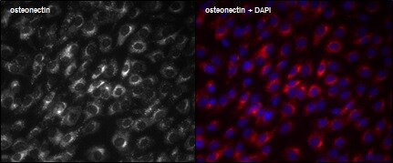
Application: ImmunocytochemistrySample Tested: Human osteosarcoma cells – MG63Species: HumanVerified Customer | Posted 02/02/2015MG63 cells

-
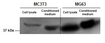
Application: Western BlotSample Tested: MG63 (human) cell lysates and conditioned media, MC3T3 (mouse) cell lysates and conditioned mediaSpecies: HumanVerified Customer | Posted 02/02/2015Western blot analysis of MC3T3 and MG63 cell lysates and conditioned media
There are no reviews that match your criteria.
Protocols
Find general support by application which include: protocols, troubleshooting, illustrated assays, videos and webinars.
- Antigen Retrieval Protocol (PIER)
- Antigen Retrieval for Frozen Sections Protocol
- Appropriate Fixation of IHC/ICC Samples
- Cellular Response to Hypoxia Protocols
- Chromogenic IHC Staining of Formalin-Fixed Paraffin-Embedded (FFPE) Tissue Protocol
- Chromogenic Immunohistochemistry Staining of Frozen Tissue
- ClariTSA™ Fluorophore Kits
- Detection & Visualization of Antibody Binding
- Fluorescent IHC Staining of Frozen Tissue Protocol
- Graphic Protocol for Heat-induced Epitope Retrieval
- Graphic Protocol for the Preparation and Fluorescent IHC Staining of Frozen Tissue Sections
- Graphic Protocol for the Preparation and Fluorescent IHC Staining of Paraffin-embedded Tissue Sections
- Graphic Protocol for the Preparation of Gelatin-coated Slides for Histological Tissue Sections
- IHC Sample Preparation (Frozen sections vs Paraffin)
- Immunofluorescent IHC Staining of Formalin-Fixed Paraffin-Embedded (FFPE) Tissue Protocol
- Immunohistochemistry (IHC) and Immunocytochemistry (ICC) Protocols
- Immunohistochemistry Frozen Troubleshooting
- Immunohistochemistry Paraffin Troubleshooting
- Preparing Samples for IHC/ICC Experiments
- Preventing Non-Specific Staining (Non-Specific Binding)
- Primary Antibody Selection & Optimization
- Protocol for Heat-Induced Epitope Retrieval (HIER)
- Protocol for Making a 4% Formaldehyde Solution in PBS
- Protocol for VisUCyte™ HRP Polymer Detection Reagent
- Protocol for the Preparation & Fixation of Cells on Coverslips
- Protocol for the Preparation and Chromogenic IHC Staining of Frozen Tissue Sections
- Protocol for the Preparation and Chromogenic IHC Staining of Frozen Tissue Sections - Graphic
- Protocol for the Preparation and Chromogenic IHC Staining of Paraffin-embedded Tissue Sections
- Protocol for the Preparation and Chromogenic IHC Staining of Paraffin-embedded Tissue Sections - Graphic
- Protocol for the Preparation and Fluorescent IHC Staining of Frozen Tissue Sections
- Protocol for the Preparation and Fluorescent IHC Staining of Paraffin-embedded Tissue Sections
- Protocol for the Preparation of Gelatin-coated Slides for Histological Tissue Sections
- R&D Systems Quality Control Western Blot Protocol
- TUNEL and Active Caspase-3 Detection by IHC/ICC Protocol
- The Importance of IHC/ICC Controls
- Troubleshooting Guide: Immunohistochemistry
- Troubleshooting Guide: Western Blot Figures
- Western Blot Conditions
- Western Blot Protocol
- Western Blot Protocol for Cell Lysates
- Western Blot Troubleshooting
- Western Blot Troubleshooting Guide
- View all Protocols, Troubleshooting, Illustrated assays and Webinars
Loading...
Associated Pathways
