![Western Blot: Spt6 Antibody [NB100-2582]](https://resources.rndsystems.com/images/products/Spt6-Antibody-Western-Blot-NB100-2582-img0011.jpg "Western Blot: Spt6 Antibody [NB100-2582]")
Loading...
Key Product Details
Validated by
Knockout/Knockdown
Species Reactivity
Validated:
Human, Mouse, Chicken
Cited:
Human, Mouse, Avian - Chicken
Applications
Validated:
Immunohistochemistry, Immunohistochemistry-Paraffin, Western Blot, Immunoprecipitation, Chromatin Immunoprecipitation (ChIP), Knockdown Validated
Cited:
Western Blot, Immunoprecipitation, Chemotaxis, Knockdown Validated
Label
Unconjugated
Antibody Source
Polyclonal Rabbit IgG
Loading...
Product Specifications
Immunogen
The immunogen recognized by this antibody maps to a region between residue 425 and 475 of human Suppressor of Ty 6 Homolog using the numbering given in entry NP_003161.2 (GeneID 6830).
Reactivity Notes
Chicken reactivity reported in scientific literature (PMID: 23008333).
Clonality
Polyclonal
Host
Rabbit
Isotype
IgG
Scientific Data Images for Spt6 Antibody

Spt6 Antibody [NB100-2582] -
Whole cell lysate (50 µg) from HEK293T, K-562,HeLa, TCMK-1, and NIH 3T3 cells prepared using NETNlysis buffer. Antibody: Affinity purified rabbit anti-SUPT6Hantibody used for WB at0.04 µg/ml. Detection: Chemiluminescence with anexposure time of 3 minutes.
Western Blot: Spt6 Antibody [NB100-2582] -
Western Blot: Spt6 Antibody [NB100-2582] - SPT6 Depletion Induces lncRNA Transcription(A) YWHAZ locus showing pre-mRNA (– strand) & PROMPT (+ strand). SPT6 depletion induced lncRNA as shown by ChRNA-seq & mNET-seq analyses. The profile of mNuc-seq/H3K36me3 is shown below.(B) Meta-analysis of strand-specific ChrRNA-seq signals from −3 kb of TSS to +3 kb of transcription end site (TES) for divergent (pre-mRNA-PROMPT) genes.(C) Boxplots of PROMPT mNET-seq/total CTD & T4P signals.(D) Enhancer located 90 kb downstream of NR4A1 gene with neighboring gene ATG101 showing SPT6 depletion-induced eRNA by ChRNA-seq & mNET-seq. mNuc-seq/H3K4me1 & H3K4me3 signals indicate active enhancer & promoter, respectively.(E) Meta-analysis of eRNA from ChrRNA-seq & mNET-seq (Total) −3 kb to +3 kb from TSS (enhancers with highest eRNA levels selected).(F) Boxplots of eRNA mNET-seq/total CTD & T4P signals at TSS (–/+2 kb).(G & H) Western blot (G) & quantitative RT-PCR (H) of chromatin-bound RNA of parental & SETD2 CRISPR KO U2OS cells with indicated siRNA transfection for 48 hr. Data are represented as mean ± SEM.See also Figure S3. Image collected & cropped by CiteAb from the following publication (https://pubmed.ncbi.nlm.nih.gov/30449723), licensed under a CC-BY license. Not internally tested by Novus Biologicals.
Western Blot: Spt6 Antibody [NB100-2582] -
Western Blot: Spt6 Antibody [NB100-2582] - SPT6 Depletion Causes H3K36me3 Redistribution(A) Western blots with indicated antibodies 60 hr post-siSPT6 transfection (versus siLuc nonspecific control). EXOSC3 & H3 profiles shown as loading controls.(B) Meta-analysis of reads density (FPKM) for ratio of mNuc-seq/H3K36me3 with H3 at TSS of divergent PROMPT-pre-mRNA & enhancer RNA (eRNA) following SPT6 depletion. All subsequent transcription images employing siSPT6 versus siLuc are shown in red & blue, respectively.(C) Scatterplots of H3K36me3/H3 on pre-mRNA (0 to +3 kb), PROMPT (−3 kb to 0), & eRNA (−3 kb to +3 kb) regions in siLuc versus siSPT6. The percentage of upregulated regions by SPT6 depletion are indicated.(D) Boxplots of mNuc-seq/H3K36me3 ratio across pre-mRNA gene bodies, PROMPTs (3 kb from TSS), & eRNA (2 kb from center) & across lincRNA gene bodies.(E) Model of redistributed H3K36me3 marks caused by SPT6 depletion.See also Figure S2. Image collected & cropped by CiteAb from the following publication (https://pubmed.ncbi.nlm.nih.gov/30449723), licensed under a CC-BY license. Not internally tested by Novus Biologicals.
Spt6 Antibody [NB100-2582] -
Whole cell lysate (1.0 mgper IP reaction; 20% of IP loaded) from HEK293T cellsprepared using NETN lysis buffer. Antibodies: Affinitypurified rabbit anti-SUPT6H antibody) used for IP at 6 µg per reaction. SUPT6Hwas also immunoprecipitated by a previous lot of thisantibody. For blottingimmunoprecipitated SUPT6H, was used at 0.04µg/ml. Detection: Chemiluminescence with an exposuretime of 75 seconds.
Spt6 Antibody [NB100-2582] -
Section of human breast carcinoma.Antibody: Affinity purified rabbit anti- SUPT6H used at a dilution of 1:200 (1µg/ml).Detection: DAB
Spt6 Antibody [NB100-2582] -
Section of mouse plasmacytoma. Antibody:Affinity purified rabbit anti- SUPT6H used at a dilution of 1:200 (1µg/ml). Detection: DABApplications for Spt6 Antibody
Application
Recommended Usage
Chromatin Immunoprecipitation (ChIP)
Reported in scientific literature (PMID:23503590).
Immunohistochemistry
1:100 to 1:500
Immunohistochemistry-Paraffin
1:100 to 1:500
Immunoprecipitation
2 - 10 µg/mg lysate
Western Blot
1:2000-1:10000
Application Notes
Epitope retrieval with citrate buffer pH 6.0 is recommended for FFPE tissue sections.
Reviewed Applications
Read 2 reviews rated 5 using NB100-2582 in the following applications:
Formulation, Preparation, and Storage
Purification
Immunogen affinity purified
Formulation
TBS, 0.1% BSA
Preservative
0.09% Sodium Azide
Concentration
0.2 mg/ml
Shipping
The product is shipped with polar packs. Upon receipt, store it immediately at the temperature recommended below.
Stability & Storage
Store at 4C. Do not freeze.
Background: Spt6
Alternate Names
emb-5, hSPT6, KIAA0162MGC87943, SPT6HSPT6, suppressor of Ty (S.cerevisiae) 6 homolog, suppressor of Ty 6 homolog (S. cerevisiae), Tat-cotransactivator 2 protein, Tat-CT2 protein, transcription elongation factor SPT6
Entrez Gene IDs
6830 (Human)
Gene Symbol
SUPT6H
UniProt
Additional Spt6 Products
Product Documents for Spt6 Antibody
Certificate of Analysis
To download a Certificate of Analysis, please enter a lot or batch number in the search box below.
Product Specific Notices for Spt6 Antibody
This product is for research use only and is not approved for use in humans or in clinical diagnosis. Primary Antibodies are guaranteed for 1 year from date of receipt.
Citations for Spt6 Antibody
 Powered by Bioz
Customer Reviews for Spt6 Antibody (2)
5 out of 5
2 Customer Ratings
Have you used Spt6 Antibody?
Submit a review and receive an Amazon gift card!
$25/€18/£15/$25CAN/¥2500 Yen for a review with an image
$10/€7/£6/$10CAN/¥1110 Yen for a review without an image
Submit a review
Customer Images
Showing
1
-
2 of
2 reviews
Showing All
Filter By:
-
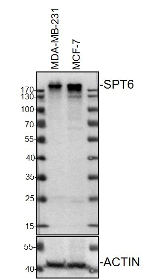
Application: Western BlotSample Tested: breast cancer cell line, Sample Amount: 40Species: HumanVerified Customer | Posted 03/06/2021MDA-MB-231 or MCF-7 cell lysates, 40ug, separated on 10% SDS-PAGE, SPT6 primary antibody (NB100-2582), 1:1000, overnight.

-
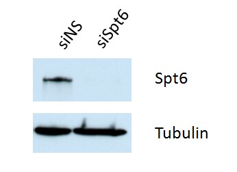
Application: Western BlotSample Tested: Mouse myoblasts (C2C12 cell lysate) and 20 ug whole cell lysateSpecies: MouseVerified Customer | Posted 04/17/2018siRNA knockdown to confirm the specificity of Spt6 antibody (NB100-2582) in C2C12 myoblasts, WB: 1:2000 dilution. 20ug whole cell lysate.
There are no reviews that match your criteria.
Protocols
Find general support by application which include: protocols, troubleshooting, illustrated assays, videos and webinars.
- Antigen Retrieval Protocol (PIER)
- Antigen Retrieval for Frozen Sections Protocol
- Appropriate Fixation of IHC/ICC Samples
- Cellular Response to Hypoxia Protocols
- ChIP Protocol Video
- Chromatin Immunoprecipitation (ChIP) Protocol
- Chromatin Immunoprecipitation Protocol
- Chromogenic IHC Staining of Formalin-Fixed Paraffin-Embedded (FFPE) Tissue Protocol
- Chromogenic Immunohistochemistry Staining of Frozen Tissue
- ClariTSA™ Fluorophore Kits
- Detection & Visualization of Antibody Binding
- Fluorescent IHC Staining of Frozen Tissue Protocol
- Graphic Protocol for Heat-induced Epitope Retrieval
- Graphic Protocol for the Preparation and Fluorescent IHC Staining of Frozen Tissue Sections
- Graphic Protocol for the Preparation and Fluorescent IHC Staining of Paraffin-embedded Tissue Sections
- Graphic Protocol for the Preparation of Gelatin-coated Slides for Histological Tissue Sections
- IHC Sample Preparation (Frozen sections vs Paraffin)
- Immunofluorescent IHC Staining of Formalin-Fixed Paraffin-Embedded (FFPE) Tissue Protocol
- Immunohistochemistry (IHC) and Immunocytochemistry (ICC) Protocols
- Immunohistochemistry Frozen Troubleshooting
- Immunohistochemistry Paraffin Troubleshooting
- Immunoprecipitation Protocol
- Preparing Samples for IHC/ICC Experiments
- Preventing Non-Specific Staining (Non-Specific Binding)
- Primary Antibody Selection & Optimization
- Protocol for Heat-Induced Epitope Retrieval (HIER)
- Protocol for Making a 4% Formaldehyde Solution in PBS
- Protocol for VisUCyte™ HRP Polymer Detection Reagent
- Protocol for the Preparation & Fixation of Cells on Coverslips
- Protocol for the Preparation and Chromogenic IHC Staining of Frozen Tissue Sections
- Protocol for the Preparation and Chromogenic IHC Staining of Frozen Tissue Sections - Graphic
- Protocol for the Preparation and Chromogenic IHC Staining of Paraffin-embedded Tissue Sections
- Protocol for the Preparation and Chromogenic IHC Staining of Paraffin-embedded Tissue Sections - Graphic
- Protocol for the Preparation and Fluorescent IHC Staining of Frozen Tissue Sections
- Protocol for the Preparation and Fluorescent IHC Staining of Paraffin-embedded Tissue Sections
- Protocol for the Preparation of Gelatin-coated Slides for Histological Tissue Sections
- R&D Systems Quality Control Western Blot Protocol
- TUNEL and Active Caspase-3 Detection by IHC/ICC Protocol
- The Importance of IHC/ICC Controls
- Troubleshooting Guide: Immunohistochemistry
- Troubleshooting Guide: Western Blot Figures
- Western Blot Conditions
- Western Blot Protocol
- Western Blot Protocol for Cell Lysates
- Western Blot Troubleshooting
- Western Blot Troubleshooting Guide
- View all Protocols, Troubleshooting, Illustrated assays and Webinars
Loading...