SRp55 Antibody - BSA Free
Novus Biologicals | Catalog # NBP2-04142
![Western Blot: SRp55 Antibody [NBP2-04142]](https://resources.rndsystems.com/images/products/SRp55-Antibody-Western-Blot-NBP2-04142-img0013.jpg "Western Blot: SRp55 Antibody [NBP2-04142]")
Key Product Details
Species Reactivity
Validated:
Human, Mouse
Cited:
Human
Predicted:
Bovine (100%). Backed by our 100% Guarantee.
Applications
Validated:
Immunohistochemistry, Immunohistochemistry-Paraffin, Western Blot, Immunocytochemistry/ Immunofluorescence, Immunoprecipitation
Cited:
Western Blot
Label
Unconjugated
Antibody Source
Polyclonal Rabbit IgG
Format
BSA Free
Loading...
Product Specifications
Immunogen
The immunogen recognized by this antibody maps to this antibody using the numbering given in entry NP_006266.2 (GeneID 6431).
Clonality
Polyclonal
Host
Rabbit
Isotype
IgG
Scientific Data Images for SRp55 Antibody - BSA Free
Western Blot: SRp55 Antibody [NBP2-04142]
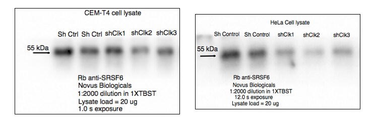
Western Blot: SRp55 Antibody [NBP2-04142] - CEM-T4 and HeLa whole cell lysates. Image from verified customer review.![Immunocytochemistry/ Immunofluorescence: SRp55 Antibody [NBP2-04142]](https://resources.rndsystems.com/images/products/SRp55-Antibody-Immunocytochemistry-Immunofluorescence-NBP2-04142-img0014.jpg "Immunocytochemistry/ Immunofluorescence: SRp55 Antibody [NBP2-04142]")
Immunocytochemistry/ Immunofluorescence: SRp55 Antibody [NBP2-04142]
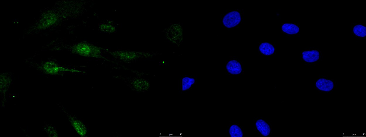
Immunocytochemistry/Immunofluorescence: SRp55 Antibody [NBP2-04142] - Staining in human fibroblasts. 1X PBS/5% normal serum/0.3% Triton™ X-100. Diluted 1:200 in 1X PBS/1% BSA/0.3% Triton™ X-100, 16 hours at 4C incubation. 2 hours (25C) secondary (1.5:1000 dilution). Image submitted by a verified customer review.![Immunohistochemistry-Paraffin: SRp55 Antibody [NBP2-04142]](https://resources.rndsystems.com/images/products/SRp55-Antibody-Immunohistochemistry-NBP2-04142-img0012.jpg "Immunohistochemistry-Paraffin: SRp55 Antibody [NBP2-04142]")
Immunohistochemistry-Paraffin: SRp55 Antibody [NBP2-04142]
Immunohistochemistry-Paraffin: SRp55 Antibody [NBP2-04142] - Sample: FFPE section of mouse teratoma. Antibody: Affinity purified rabbit anti- SRp55 used at a dilution of 1:1,000 (1ug/ml). Detection: DAB. Counterstain: Hematoxylin (blue).![Western Blot: SRp55 Antibody [NBP2-04142]](https://resources.rndsystems.com/images/products/SRp55-Antibody-Western-Blot-NBP2-04142-img0010.jpg "Western Blot: SRp55 Antibody [NBP2-04142]")
Western Blot: SRp55 Antibody [NBP2-04142]
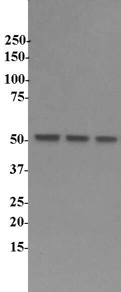
Western Blot: SRp55 Antibody [NBP2-04142] - analysis of SRp55 in human lymphocytes using anti-SRp55 antibody. Each lane loaded with 14ug of whole cell lysate with 3 different human B-cell lines. Image from verified customer review.![Immunohistochemistry-Paraffin: SRp55 Antibody [NBP2-04142]](https://resources.rndsystems.com/images/products/SRp55-Antibody-Immunohistochemistry-NBP2-04142-img0011.jpg "Immunohistochemistry-Paraffin: SRp55 Antibody [NBP2-04142]")
Immunohistochemistry-Paraffin: SRp55 Antibody [NBP2-04142]
Immunohistochemistry-Paraffin: SRp55 Antibody [NBP2-04142] - Sample: FFPE section of human prostate carcinoma. Antibody: Affinity purified rabbit anti- SRp55 used at a dilution of 1:1,000 (1ug/ml). Detection: DAB. Counterstain: Hematoxylin (blue).![Immunoprecipitation: SRp55 Antibody [NBP2-04142]](https://resources.rndsystems.com/images/products/SRp55-Antibody-Immunoprecipitation-NBP2-04142-img0002.jpg "Immunoprecipitation: SRp55 Antibody [NBP2-04142]")
Immunoprecipitation: SRp55 Antibody [NBP2-04142]
Immunoprecipitation: SRp55 Antibody [NBP2-04142] - Samples: Whole cell lysate from HeLa (15 and 50 ug for WB; 1 mg for IP, 20% of IP loaded), 293T (T; 50 ug), Jurkat (J; 50 ug) and mouse NIH3T3 (M; 50 ug) cells.Antibodies: Affinity purified rabbit anti-SRp55 antibody NBP2-04142 used for WB at 0.1 ug/ml (A) and 1 ug/ml (B) and used for IP at 6 ug/mg lysate. SRp55 was also immunoprecipitated by rabbit anti-SRp55 antibody, which recognizes an upstream epitope. Detection: Chemiluminescence with exposure times of 3 minutes (A) and 30 seconds (B).Applications for SRp55 Antibody - BSA Free
Application
Recommended Usage
Immunohistochemistry-Paraffin
1:500 - 1:2000
Immunoprecipitation
2-10 ug/mg lysate
Western Blot
1:2000-1:10000
Application Notes
Epitope retrieval with citrate buffer pH6.0 is recommended for FFPE tissue sections. SRp55 antibody validated for ICC/IF from a verified customer review.
Reviewed Applications
Read 3 reviews rated 4.3 using NBP2-04142 in the following applications:
Formulation, Preparation, and Storage
Purification
Immunogen affinity purified
Formulation
Tris-Citrate/Phosphate (pH 7.0 - 8.0)
Format
BSA Free
Preservative
0.09% Sodium Azide
Concentration
1.0 mg/ml
Shipping
The product is shipped with polar packs. Upon receipt, store it immediately at the temperature recommended below.
Stability & Storage
Store at 4C. Do not freeze.
Background: SRp55
Alternate Names
B52, FLJ08061, pre-mRNA splicing factor SRP55, Pre-mRNA-splicing factor SRP55, serine/arginine-rich splicing factor 6, SFRS6, Splicing factor, arginine/serine-rich 6MGC5045, splicing factor, arginine/serine-rich, 55 kDa, SR splicing factor 6, SRP55arginine/serine-rich splicing factor 6
Gene Symbol
SRSF6
UniProt
Additional SRp55 Products
Product Documents for SRp55 Antibody - BSA Free
Certificate of Analysis
To download a Certificate of Analysis, please enter a lot or batch number in the search box below.
Product Specific Notices for SRp55 Antibody - BSA Free
This product is for research use only and is not approved for use in humans or in clinical diagnosis. Primary Antibodies are guaranteed for 1 year from date of receipt.
Citations for SRp55 Antibody - BSA Free
 Powered by Bioz
Customer Reviews for SRp55 Antibody - BSA Free (3)
4.3 out of 5
3 Customer Ratings
Have you used SRp55 Antibody - BSA Free?
Submit a review and receive an Amazon gift card!
$25/€18/£15/$25CAN/¥2500 Yen for a review with an image
$10/€7/£6/$10CAN/¥1110 Yen for a review without an image
Submit a review
Customer Images
Showing
1
-
3 of
3 reviews
Showing All
Filter By:
-
Application: Western BlotSample Tested: HeLa cell lysateSpecies: HumanVerified Customer | Posted 06/09/2018CEM-T4 and HeLa whole cell lysates probed for SRSF6 (SRp55) antibody

-
Application: ImmunocytochemistrySample Tested: fibroblastsSpecies: HumanVerified Customer | Posted 11/18/20171X PBS/5% normal serum/0.3% Triton™ X-100 Diluted 1:200 in 1X PBS/1% BSA/0.3% Triton™ X-100, 16 hours at 4C incubation. 2 hours (25C) secondary (1.5:1000 dilution) SRp55 in fibroblasts1X PBS/5% normal serum/0.3% Triton™ X-100 Diluted 1:200 in 1X PBS/1% BSA/0.3% Triton™ X-100, 16 hours at 4C incubation. 2 hours (25C) secondary (1.5:1000 dilution)
-
Application: Western BlotSample Tested: Human lymphocytesSpecies: HumanVerified Customer | Posted 01/21/2016SRp55 in Human Lymphocytes
There are no reviews that match your criteria.
Protocols
Find general support by application which include: protocols, troubleshooting, illustrated assays, videos and webinars.
- Antigen Retrieval Protocol (PIER)
- Antigen Retrieval for Frozen Sections Protocol
- Appropriate Fixation of IHC/ICC Samples
- Cellular Response to Hypoxia Protocols
- Chromogenic IHC Staining of Formalin-Fixed Paraffin-Embedded (FFPE) Tissue Protocol
- Chromogenic Immunohistochemistry Staining of Frozen Tissue
- ClariTSA™ Fluorophore Kits
- Detection & Visualization of Antibody Binding
- Fluorescent IHC Staining of Frozen Tissue Protocol
- Graphic Protocol for Heat-induced Epitope Retrieval
- Graphic Protocol for the Preparation and Fluorescent IHC Staining of Frozen Tissue Sections
- Graphic Protocol for the Preparation and Fluorescent IHC Staining of Paraffin-embedded Tissue Sections
- Graphic Protocol for the Preparation of Gelatin-coated Slides for Histological Tissue Sections
- ICC Cell Smear Protocol for Suspension Cells
- ICC Immunocytochemistry Protocol Videos
- ICC for Adherent Cells
- IHC Sample Preparation (Frozen sections vs Paraffin)
- Immunocytochemistry (ICC) Protocol
- Immunocytochemistry Troubleshooting
- Immunofluorescence of Organoids Embedded in Cultrex Basement Membrane Extract
- Immunofluorescent IHC Staining of Formalin-Fixed Paraffin-Embedded (FFPE) Tissue Protocol
- Immunohistochemistry (IHC) and Immunocytochemistry (ICC) Protocols
- Immunohistochemistry Frozen Troubleshooting
- Immunohistochemistry Paraffin Troubleshooting
- Immunoprecipitation Protocol
- Preparing Samples for IHC/ICC Experiments
- Preventing Non-Specific Staining (Non-Specific Binding)
- Primary Antibody Selection & Optimization
- Protocol for Heat-Induced Epitope Retrieval (HIER)
- Protocol for Making a 4% Formaldehyde Solution in PBS
- Protocol for VisUCyte™ HRP Polymer Detection Reagent
- Protocol for the Fluorescent ICC Staining of Cell Smears - Graphic
- Protocol for the Fluorescent ICC Staining of Cultured Cells on Coverslips - Graphic
- Protocol for the Preparation & Fixation of Cells on Coverslips
- Protocol for the Preparation and Chromogenic IHC Staining of Frozen Tissue Sections
- Protocol for the Preparation and Chromogenic IHC Staining of Frozen Tissue Sections - Graphic
- Protocol for the Preparation and Chromogenic IHC Staining of Paraffin-embedded Tissue Sections
- Protocol for the Preparation and Chromogenic IHC Staining of Paraffin-embedded Tissue Sections - Graphic
- Protocol for the Preparation and Fluorescent ICC Staining of Cells on Coverslips
- Protocol for the Preparation and Fluorescent ICC Staining of Non-adherent Cells
- Protocol for the Preparation and Fluorescent ICC Staining of Stem Cells on Coverslips
- Protocol for the Preparation and Fluorescent IHC Staining of Frozen Tissue Sections
- Protocol for the Preparation and Fluorescent IHC Staining of Paraffin-embedded Tissue Sections
- Protocol for the Preparation of Gelatin-coated Slides for Histological Tissue Sections
- Protocol for the Preparation of a Cell Smear for Non-adherent Cell ICC - Graphic
- R&D Systems Quality Control Western Blot Protocol
- TUNEL and Active Caspase-3 Detection by IHC/ICC Protocol
- The Importance of IHC/ICC Controls
- Troubleshooting Guide: Immunohistochemistry
- Troubleshooting Guide: Western Blot Figures
- Western Blot Conditions
- Western Blot Protocol
- Western Blot Protocol for Cell Lysates
- Western Blot Troubleshooting
- Western Blot Troubleshooting Guide
- View all Protocols, Troubleshooting, Illustrated assays and Webinars
Loading...