![Immunocytochemistry/ Immunofluorescence: TAO Kinase 1 Antibody [NBP1-89864]](https://resources.rndsystems.com/images/products/TAO-Kinase-1-Antibody-Immunocytochemistry-Immunofluorescence-NBP1-89864-img0005.jpg "Immunocytochemistry/ Immunofluorescence: TAO Kinase 1 Antibody [NBP1-89864]")
Loading...
Key Product Details
Species Reactivity
Validated:
Human
Predicted:
Mouse (98%), Rat (96%). Backed by our 100% Guarantee.
Applications
Immunohistochemistry, Immunohistochemistry-Paraffin, ELISA, Immunocytochemistry/ Immunofluorescence
Label
Unconjugated
Antibody Source
Polyclonal Rabbit IgG
Format
BSA Free
Loading...
Product Specifications
Immunogen
This antibody was developed against Recombinant Protein corresponding to amino acids: LSPEAFSHSYPGASGWSHNPTGGPGPHWGHPMGGPPQAWGHPMQGGPQPWGHPSGPMQGVPRGSSMGVRNSPQALRRTASGGRTEQGMSRSTSVTSQISNGSH
Clonality
Polyclonal
Host
Rabbit
Isotype
IgG
Scientific Data Images for TAO Kinase 1 Antibody - BSA Free
Immunocytochemistry/ Immunofluorescence: TAO Kinase 1 Antibody [NBP1-89864]
Immunocytochemistry/Immunofluorescence: TAO Kinase 1 Antibody [NBP1-89864] - Staining of human cell line A-431 shows localization to vesicles.![Immunocytochemistry/ Immunofluorescence: TAO Kinase 1 Antibody [NBP1-89864]](https://resources.rndsystems.com/images/products/TAO-Kinase-1-Antibody-Immunocytochemistry-Immunofluorescence-NBP1-89864-img0006.jpg "Immunocytochemistry/ Immunofluorescence: TAO Kinase 1 Antibody [NBP1-89864]")
Immunocytochemistry/ Immunofluorescence: TAO Kinase 1 Antibody [NBP1-89864]
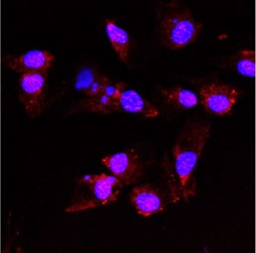
Immunocytochemistry/Immunofluorescence: TAO Kinase 1 Antibody [NBP1-89864] - TAO1 staining in human SH-SY5Y cell line. ICC/IF image submitted by a verified customer review.![TAO Kinase 1 Antibody - BSA Free Immunohistochemistry-Paraffin: TAO Kinase 1 Antibody - BSA Free [NBP1-89864]](https://resources.rndsystems.com/images/products/nbp1-89864_rabbit-polyclonal-tao-kinase-1-antibody-15520258564397.jpg "Immunohistochemistry-Paraffin: TAO Kinase 1 Antibody - BSA Free [NBP1-89864]")
Immunohistochemistry-Paraffin: TAO Kinase 1 Antibody - BSA Free [NBP1-89864]
Staining of human placenta shows moderate cytoplasmic positivity in trophoblastic cells.![TAO Kinase 1 Antibody - BSA Free Immunohistochemistry-Paraffin: TAO Kinase 1 Antibody - BSA Free [NBP1-89864]](https://resources.rndsystems.com/images/products/nbp1-89864_rabbit-polyclonal-tao-kinase-1-antibody-15520258231221.jpg "Immunohistochemistry-Paraffin: TAO Kinase 1 Antibody - BSA Free [NBP1-89864]")
Immunohistochemistry-Paraffin: TAO Kinase 1 Antibody - BSA Free [NBP1-89864]
Staining of human testis shows moderate cytoplasmic positivity in Leydig cells.![TAO Kinase 1 Antibody - BSA Free Immunohistochemistry-Paraffin: TAO Kinase 1 Antibody - BSA Free [NBP1-89864]](https://resources.rndsystems.com/images/products/nbp1-89864_rabbit-polyclonal-tao-kinase-1-antibody-135202517442176.jpg "Immunohistochemistry-Paraffin: TAO Kinase 1 Antibody - BSA Free [NBP1-89864]")
Immunohistochemistry-Paraffin: TAO Kinase 1 Antibody - BSA Free [NBP1-89864]
Staining of human pancreas shows very weak cytoplasmic positivity in exocrine glandular cells.![TAO Kinase 1 Antibody - BSA Free Immunohistochemistry-Paraffin: TAO Kinase 1 Antibody - BSA Free [NBP1-89864]](https://resources.rndsystems.com/images/products/nbp1-89864_rabbit-polyclonal-tao-kinase-1-antibody-1352025167129.jpg "Immunohistochemistry-Paraffin: TAO Kinase 1 Antibody - BSA Free [NBP1-89864]")
Immunohistochemistry-Paraffin: TAO Kinase 1 Antibody - BSA Free [NBP1-89864]
Staining of human cerebral cortex shows strong cytoplasmic positivity in neurons.Applications for TAO Kinase 1 Antibody - BSA Free
Application
Recommended Usage
Immunocytochemistry/ Immunofluorescence
0.25 - 2 ug/mL
Immunohistochemistry
1:20 - 1:50
Immunohistochemistry-Paraffin
1:20 - 1:50
Application Notes
For IHC-Paraffin, HIER pH 6 retrieval is recommended. ICC/IF Fixation/Permeabilization: PFA/Triton X-100. This TAO Kinase 1 Antibody is validated for ELISA from a verified customer review.
Reviewed Applications
Read 2 reviews rated 5 using NBP1-89864 in the following applications:
Formulation, Preparation, and Storage
Purification
Affinity purified
Formulation
PBS (pH 7.2) and 40% Glycerol
Format
BSA Free
Preservative
0.02% Sodium Azide
Concentration
Concentrations vary lot to lot. See vial label for concentration. If unlisted please contact technical services.
Shipping
The product is shipped with polar packs. Upon receipt, store it immediately at the temperature recommended below.
Stability & Storage
Store at 4C short term. Aliquot and store at -20C long term. Avoid freeze-thaw cycles.
Background: TAO Kinase 1
Long Name
Serine/threonine-protein kinase TAO1
Alternate Names
hKFC-B, hTAOK1, KIAA1361, MAP3K16, MARK Kinase, MARKK, PSK-2, PSK2, TAOK1
Gene Symbol
TAOK1
Additional TAO Kinase 1 Products
Product Documents for TAO Kinase 1 Antibody - BSA Free
Certificate of Analysis
To download a Certificate of Analysis, please enter a lot or batch number in the search box below.
Product Specific Notices for TAO Kinase 1 Antibody - BSA Free
This product is for research use only and is not approved for use in humans or in clinical diagnosis. Primary Antibodies are guaranteed for 1 year from date of receipt.
Customer Reviews for TAO Kinase 1 Antibody - BSA Free (2)
5 out of 5
2 Customer Ratings
Have you used TAO Kinase 1 Antibody - BSA Free?
Submit a review and receive an Amazon gift card!
$25/€18/£15/$25CAN/¥2500 Yen for a review with an image
$10/€7/£6/$10CAN/¥1110 Yen for a review without an image
Submit a review
Customer Images
Showing
1
-
2 of
2 reviews
Showing All
Filter By:
-
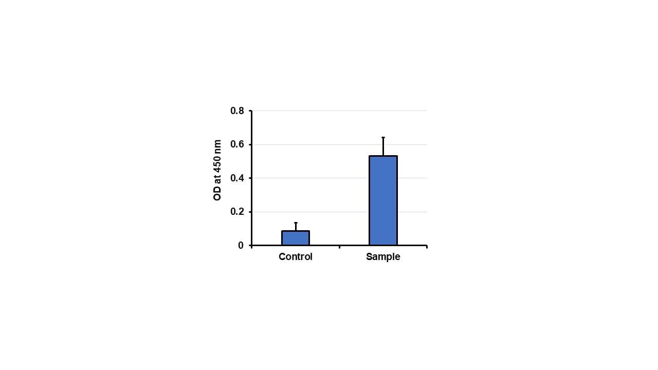
Application: ELISASample Tested: SH-SY5Y Cell lysatesSpecies: HumanVerified Customer | Posted 09/18/2020Determining TAO1 levels in cell lysates.

-
Application: ImmunocytochemistrySample Tested: SH-SY5Y Cell LineSpecies: HumanVerified Customer | Posted 08/20/2020TAO1 staining in SH-SY5Y Cell Line
There are no reviews that match your criteria.
Protocols
Find general support by application which include: protocols, troubleshooting, illustrated assays, videos and webinars.
- Antigen Retrieval Protocol (PIER)
- Antigen Retrieval for Frozen Sections Protocol
- Appropriate Fixation of IHC/ICC Samples
- Cellular Response to Hypoxia Protocols
- Chromogenic IHC Staining of Formalin-Fixed Paraffin-Embedded (FFPE) Tissue Protocol
- Chromogenic Immunohistochemistry Staining of Frozen Tissue
- ClariTSA™ Fluorophore Kits
- Detection & Visualization of Antibody Binding
- ELISA Sample Preparation & Collection Guide
- ELISA Troubleshooting Guide
- Fluorescent IHC Staining of Frozen Tissue Protocol
- Graphic Protocol for Heat-induced Epitope Retrieval
- Graphic Protocol for the Preparation and Fluorescent IHC Staining of Frozen Tissue Sections
- Graphic Protocol for the Preparation and Fluorescent IHC Staining of Paraffin-embedded Tissue Sections
- Graphic Protocol for the Preparation of Gelatin-coated Slides for Histological Tissue Sections
- How to Run an R&D Systems DuoSet ELISA
- How to Run an R&D Systems Quantikine ELISA
- How to Run an R&D Systems Quantikine™ QuicKit™ ELISA
- ICC Cell Smear Protocol for Suspension Cells
- ICC Immunocytochemistry Protocol Videos
- ICC for Adherent Cells
- IHC Sample Preparation (Frozen sections vs Paraffin)
- Immunocytochemistry (ICC) Protocol
- Immunocytochemistry Troubleshooting
- Immunofluorescence of Organoids Embedded in Cultrex Basement Membrane Extract
- Immunofluorescent IHC Staining of Formalin-Fixed Paraffin-Embedded (FFPE) Tissue Protocol
- Immunohistochemistry (IHC) and Immunocytochemistry (ICC) Protocols
- Immunohistochemistry Frozen Troubleshooting
- Immunohistochemistry Paraffin Troubleshooting
- Preparing Samples for IHC/ICC Experiments
- Preventing Non-Specific Staining (Non-Specific Binding)
- Primary Antibody Selection & Optimization
- Protocol for Heat-Induced Epitope Retrieval (HIER)
- Protocol for Making a 4% Formaldehyde Solution in PBS
- Protocol for VisUCyte™ HRP Polymer Detection Reagent
- Protocol for the Fluorescent ICC Staining of Cell Smears - Graphic
- Protocol for the Fluorescent ICC Staining of Cultured Cells on Coverslips - Graphic
- Protocol for the Preparation & Fixation of Cells on Coverslips
- Protocol for the Preparation and Chromogenic IHC Staining of Frozen Tissue Sections
- Protocol for the Preparation and Chromogenic IHC Staining of Frozen Tissue Sections - Graphic
- Protocol for the Preparation and Chromogenic IHC Staining of Paraffin-embedded Tissue Sections
- Protocol for the Preparation and Chromogenic IHC Staining of Paraffin-embedded Tissue Sections - Graphic
- Protocol for the Preparation and Fluorescent ICC Staining of Cells on Coverslips
- Protocol for the Preparation and Fluorescent ICC Staining of Non-adherent Cells
- Protocol for the Preparation and Fluorescent ICC Staining of Stem Cells on Coverslips
- Protocol for the Preparation and Fluorescent IHC Staining of Frozen Tissue Sections
- Protocol for the Preparation and Fluorescent IHC Staining of Paraffin-embedded Tissue Sections
- Protocol for the Preparation of Gelatin-coated Slides for Histological Tissue Sections
- Protocol for the Preparation of a Cell Smear for Non-adherent Cell ICC - Graphic
- Quantikine HS ELISA Kit Assay Principle, Alkaline Phosphatase
- Quantikine HS ELISA Kit Principle, Streptavidin-HRP Polymer
- Sandwich ELISA (Colorimetric) – Biotin/Streptavidin Detection Protocol
- Sandwich ELISA (Colorimetric) – Direct Detection Protocol
- TUNEL and Active Caspase-3 Detection by IHC/ICC Protocol
- The Importance of IHC/ICC Controls
- Troubleshooting Guide: ELISA
- Troubleshooting Guide: Immunohistochemistry
- View all Protocols, Troubleshooting, Illustrated assays and Webinars
Loading...