TFIIIC Antibody - BSA Free
Novus Biologicals | Catalog # NB100-60657
![Western Blot: TFIIIC Antibody [NB100-60657]](https://resources.rndsystems.com/images/products/TFIIIC-Antibody-Western-Blot-NB100-60657-img0001.jpg "Western Blot: TFIIIC Antibody [NB100-60657]")
Key Product Details
Species Reactivity
Validated:
Human, Mouse
Cited:
Human, Mouse
Applications
Validated:
Immunohistochemistry, Immunohistochemistry-Paraffin, Western Blot, Chromatin Immunoprecipitation (ChIP), Immunoprecipitation (Negative)
Cited:
Western Blot, Chemotaxis
Label
Unconjugated
Antibody Source
Polyclonal Rabbit IgG
Format
BSA Free
Loading...
Product Specifications
Immunogen
The immunogen recognized by this antibody maps to a region between residue 2059 and 2109 of human general transcription factor IIIC, polypeptide 1 (general transcription factor IIIC, 220kDa subunit) using the numbering given in entry NP_001511.2 (GeneID 2
Reactivity Notes
Mouse reactivity reported in scientific literature (PMID: 23966877).
Clonality
Polyclonal
Host
Rabbit
Isotype
IgG
Scientific Data Images for TFIIIC Antibody - BSA Free
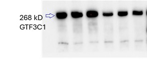
Western Blot: TFIIIC Antibody [NB100-60657]
Western Blot: TFIIIC Antibody [NB100-60657] - Detection of Human GTF3C1/TFIIIC220 on HeLa whole cell lysate using NB100-60657. GTF3C1/TFIIIC220 was efficiently IPed by rabbit anti-GTF3C1/TFIIIC220 antibodies NB100-60655 and NB100-60656.![Immunohistochemistry-Paraffin: TFIIIC Antibody [NB100-60657]](https://resources.rndsystems.com/images/products/TFIIIC-Antibody-Immunohistochemistry-Paraffin-NB100-60657-img0007.jpg "Immunohistochemistry-Paraffin: TFIIIC Antibody [NB100-60657]")
Immunohistochemistry-Paraffin: TFIIIC Antibody [NB100-60657]
Immunohistochemistry-Paraffin: TFIIIC Antibody [NB100-60657] - Human prostate carcinoma. Antibody used at a dilution of 1:1,000.
Western Blot: Rabbit Polyclonal TFIIIC Antibody [NB100-60657] -
Western Blot: Rabbit Polyclonal TFIIIC Antibody [NB100-60657] - Western blot using GTF3C1 Antibody at concentration 1:1000. Image from a verified customer review.Applications for TFIIIC Antibody - BSA Free
Application
Recommended Usage
Immunohistochemistry
1:500 -1:2000
Immunohistochemistry-Paraffin
1:500 - 1:2000
Western Blot
1:2000-1:10000
Application Notes
Use in chromatin immunoprecipitation reported in scientific literature (PMID: 31759822).
Reviewed Applications
Read 1 review rated 4 using NB100-60657 in the following applications:
Formulation, Preparation, and Storage
Purification
Immunogen affinity purified
Formulation
Tris-Citrate/Phosphate (pH 7.0 - 8.0)
Format
BSA Free
Preservative
0.09% Sodium Azide
Concentration
1.0 mg/ml
Shipping
The product is shipped with polar packs. Upon receipt, store it immediately at the temperature recommended below.
Stability & Storage
Store at 4C. Do not freeze.
Background: TFIIIC
Alternate Names
general transcription factor 3C polypeptide 1, general transcription factor IIIC, polypeptide 1 (alpha subunit), general transcription factor IIIC, polypeptide 1 (alpha subunit, 220kD ), general transcription factor IIIC, polypeptide 1, alpha 220kDa, TF3C-alpha, TFIIIC, TFIIIC 220 kDa subunit, TFIIIC box B-binding subunit, TFIIIC220DKFZp686A111, TFIIICalpha, Transcription factor IIIC 220 kDa subunit, Transcription factor IIIC subunit alpha
Entrez Gene IDs
2975 (Human)
Gene Symbol
GTF3C1
UniProt
Additional TFIIIC Products
Product Documents for TFIIIC Antibody - BSA Free
Certificate of Analysis
To download a Certificate of Analysis, please enter a lot or batch number in the search box below.
Product Specific Notices for TFIIIC Antibody - BSA Free
This product is for research use only and is not approved for use in humans or in clinical diagnosis. Primary Antibodies are guaranteed for 1 year from date of receipt.
Citations for TFIIIC Antibody - BSA Free
 Powered by Bioz
Customer Reviews for TFIIIC Antibody - BSA Free (1)
4 out of 5
1 Customer Rating
Have you used TFIIIC Antibody - BSA Free?
Submit a review and receive an Amazon gift card!
$25/€18/£15/$25CAN/¥2500 Yen for a review with an image
$10/€7/£6/$10CAN/¥1110 Yen for a review without an image
Submit a review
Customer Images
Showing
1
-
1 of
1 review
Showing All
Filter By:
-
Application: Western BlotSample Tested: Cell LysatesSpecies: HumanVerified Customer | Posted 01/30/2025Western blot using GTF3C1 Antibody at concentration 1:1000.I have used the antibody for western blot at concentration 1:1000.

There are no reviews that match your criteria.
Protocols
Find general support by application which include: protocols, troubleshooting, illustrated assays, videos and webinars.
- Antigen Retrieval Protocol (PIER)
- Antigen Retrieval for Frozen Sections Protocol
- Appropriate Fixation of IHC/ICC Samples
- Cellular Response to Hypoxia Protocols
- ChIP Protocol Video
- Chromatin Immunoprecipitation (ChIP) Protocol
- Chromatin Immunoprecipitation Protocol
- Chromogenic IHC Staining of Formalin-Fixed Paraffin-Embedded (FFPE) Tissue Protocol
- Chromogenic Immunohistochemistry Staining of Frozen Tissue
- ClariTSA™ Fluorophore Kits
- Detection & Visualization of Antibody Binding
- Fluorescent IHC Staining of Frozen Tissue Protocol
- Graphic Protocol for Heat-induced Epitope Retrieval
- Graphic Protocol for the Preparation and Fluorescent IHC Staining of Frozen Tissue Sections
- Graphic Protocol for the Preparation and Fluorescent IHC Staining of Paraffin-embedded Tissue Sections
- Graphic Protocol for the Preparation of Gelatin-coated Slides for Histological Tissue Sections
- IHC Sample Preparation (Frozen sections vs Paraffin)
- Immunofluorescent IHC Staining of Formalin-Fixed Paraffin-Embedded (FFPE) Tissue Protocol
- Immunohistochemistry (IHC) and Immunocytochemistry (ICC) Protocols
- Immunohistochemistry Frozen Troubleshooting
- Immunohistochemistry Paraffin Troubleshooting
- Preparing Samples for IHC/ICC Experiments
- Preventing Non-Specific Staining (Non-Specific Binding)
- Primary Antibody Selection & Optimization
- Protocol for Heat-Induced Epitope Retrieval (HIER)
- Protocol for Making a 4% Formaldehyde Solution in PBS
- Protocol for VisUCyte™ HRP Polymer Detection Reagent
- Protocol for the Preparation & Fixation of Cells on Coverslips
- Protocol for the Preparation and Chromogenic IHC Staining of Frozen Tissue Sections
- Protocol for the Preparation and Chromogenic IHC Staining of Frozen Tissue Sections - Graphic
- Protocol for the Preparation and Chromogenic IHC Staining of Paraffin-embedded Tissue Sections
- Protocol for the Preparation and Chromogenic IHC Staining of Paraffin-embedded Tissue Sections - Graphic
- Protocol for the Preparation and Fluorescent IHC Staining of Frozen Tissue Sections
- Protocol for the Preparation and Fluorescent IHC Staining of Paraffin-embedded Tissue Sections
- Protocol for the Preparation of Gelatin-coated Slides for Histological Tissue Sections
- R&D Systems Quality Control Western Blot Protocol
- TUNEL and Active Caspase-3 Detection by IHC/ICC Protocol
- The Importance of IHC/ICC Controls
- Troubleshooting Guide: Immunohistochemistry
- Troubleshooting Guide: Western Blot Figures
- Western Blot Conditions
- Western Blot Protocol
- Western Blot Protocol for Cell Lysates
- Western Blot Troubleshooting
- Western Blot Troubleshooting Guide
- View all Protocols, Troubleshooting, Illustrated assays and Webinars
Loading...