TLR8 Antibody (307D3.01) [Alexa Fluor® 488]
Novus Biologicals | Catalog # DDX0481A488-100
Key Product Details
Species Reactivity
Human, Mouse
Applications
Flow Cytometry, Immunocytochemistry/ Immunofluorescence
Label
Alexa Fluor 488 (Excitation = 488 nm, Emission = 515-545 nm)
Antibody Source
Monoclonal Mouse IgG1 Clone # 307D3.01
Loading...
Product Specifications
Immunogen
TLR8/CD288 was used as immunogen.
Reactivity Notes
Human, Mouse
Clonality
Monoclonal
Host
Mouse
Isotype
IgG1
Scientific Data Images for TLR8 Antibody (307D3.01) [Alexa Fluor® 488]
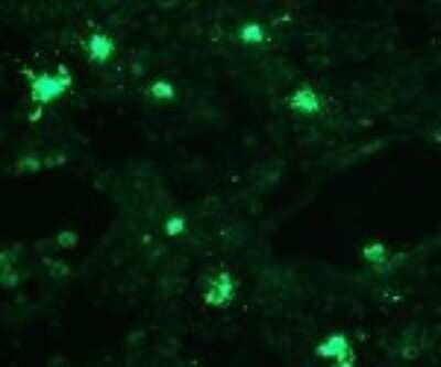
Immunocytochemistry/Immunofluorescence: TLR8 Antibody (307D3.01) [Alexa Fluor (R) 488] [DDX0481A488-100] - Staining of human tonsil frozen section.
Applications for TLR8 Antibody (307D3.01) [Alexa Fluor® 488]
Application
Recommended Usage
Flow Cytometry
1:10-1:1000
Immunocytochemistry/ Immunofluorescence
1:10-1:2000
Spectra Viewer
Plan Your Experiments
Use our spectra viewer to interactively plan your experiments, assessing multiplexing options. View the excitation and emission spectra for our fluorescent dye range and other commonly used dyes.
Spectra Viewer
Flow Cytometry Panel Builder
Bio-Techne Knows Flow Cytometry
Save time and reduce costly mistakes by quickly finding compatible reagents using the Panel Builder Tool.
Advanced Features
- Spectra Viewer - Custom analysis of spectra from multiple fluorochromes
- Spillover Popups - Visualize the spectra of individual fluorochromes
- Antigen Density Selector - Match fluorochrome brightness with antigen density

Formulation, Preparation, and Storage
Purification
Ion exchange chromatography
Formulation
PBS and 50% Glycerol
Preservative
No Preservative
Concentration
Please see the vial label for concentration. If unlisted please contact technical services.
Shipping
The product is shipped with polar packs. Upon receipt, store it immediately at the temperature recommended below.
Stability & Storage
Store at -20C in the dark. Avoid freeze-thaw cycles.
Background: TLR8
TLR8 is highly similar to TLR7 and both pathways are mediated by the adapter protein MyD88 to signal through IFN regulatory factor 7 (IRF7) and nuclear factor (NF)-kappaB (1-3,5). However, TLR7 recognizes guanosine and GU-rich ssRNA, while TLR8 recognizes uridine and AU-rich sequences (2,5). TLR7/TLR8 agonists, including derivatives of the immunostimulatory imiquimod, have been shown to be a promising cancer therapy capable of providing anticancer signals to antigen presenting cells (APCs), with many agonists being tested in both pre-clinical and clinical trials (6). Similarly, studies suggest that agonists for TLR8, in combination with other individual TLR agonists and antagonists, may also be useful for treating inflammatory allergic diseases, such as allergic rhinitis (7).
References
1. Sakaniwa, K., & Shimizu, T. (2020). Targeting the innate immune receptor TLR8 using small-molecule agents. Acta crystallographica. Section D, Structural biology, 76(Pt 7). https://doi.org/10.1107/S2059798320006518
2. Cervantes, J. L., Weinerman, B., Basole, C., & Salazar, J. C. (2012). TLR8: the forgotten relative revindicated. Cellular & molecular immunology. https://doi.org/10.1038/cmi.2012.38
3. Ohto, U., Tanji, H., & Shimizu, T. (2014). Structure and function of toll-like receptor 8. Microbes and infection. https://doi.org/10.1016/j.micinf.2014.01.007
4. Uniprot (Q9NR97)
5. Jannuzzi, G. P., de Almeida, J., Paulo, L., de Almeida, S. R., & Ferreira, K. S. (2020). Intracellular PRRs Activation in Targeting the Immune Response Against Fungal Infections. Frontiers in cellular and infection microbiology. https://doi.org/10.3389/fcimb.2020.591970
6. Frega, G., Wu, Q., Le Naour, J., Vacchelli, E., Galluzzi, L., Kroemer, G., & Kepp, O. (2020). Trial Watch: experimental TLR7/TLR8 agonists for oncological indications. Oncoimmunology. https://doi.org/10.1080/2162402X.2020.1796002
7. Golshiri-Isfahani, A., Amizadeh, M., & Arababadi, M. K. (2018). The roles of toll like receptor 3, 7 and 8 in allergic rhinitis pathogenesis. Allergologia et immunopathologia. https://doi.org/10.1016/j.aller.2017.09.026
Long Name
Toll-like Receptor 8
Alternate Names
CD288
Gene Symbol
TLR8
Additional TLR8 Products
Product Documents for TLR8 Antibody (307D3.01) [Alexa Fluor® 488]
Certificate of Analysis
To download a Certificate of Analysis, please enter a lot or batch number in the search box below.
Product Specific Notices for TLR8 Antibody (307D3.01) [Alexa Fluor® 488]
This product is manufactured by Eurobio Scientific (formerly Dendritics) and distributed by Novus Biologicals.
This product is for research use only and is not approved for use in humans or in clinical diagnosis. Primary Antibodies are guaranteed for 1 year from date of receipt.
Customer Reviews for TLR8 Antibody (307D3.01) [Alexa Fluor® 488]
There are currently no reviews for this product. Be the first to review TLR8 Antibody (307D3.01) [Alexa Fluor® 488] and earn rewards!
Have you used TLR8 Antibody (307D3.01) [Alexa Fluor® 488]?
Submit a review and receive an Amazon gift card!
$25/€18/£15/$25CAN/¥2500 Yen for a review with an image
$10/€7/£6/$10CAN/¥1110 Yen for a review without an image
Submit a review
Protocols
Find general support by application which include: protocols, troubleshooting, illustrated assays, videos and webinars.
- 7-Amino Actinomycin D (7-AAD) Cell Viability Flow Cytometry Protocol
- Appropriate Fixation of IHC/ICC Samples
- Cellular Response to Hypoxia Protocols
- ClariTSA™ Fluorophore Kits
- Detection & Visualization of Antibody Binding
- Extracellular Membrane Flow Cytometry Protocol
- Flow Cytometry Protocol for Cell Surface Markers
- Flow Cytometry Protocol for Staining Membrane Associated Proteins
- Flow Cytometry Staining Protocols
- Flow Cytometry Troubleshooting Guide
- ICC Cell Smear Protocol for Suspension Cells
- ICC Immunocytochemistry Protocol Videos
- ICC for Adherent Cells
- Immunocytochemistry (ICC) Protocol
- Immunocytochemistry Troubleshooting
- Immunofluorescence of Organoids Embedded in Cultrex Basement Membrane Extract
- Immunohistochemistry (IHC) and Immunocytochemistry (ICC) Protocols
- Intracellular Flow Cytometry Protocol Using Alcohol (Methanol)
- Intracellular Flow Cytometry Protocol Using Detergents
- Intracellular Nuclear Staining Flow Cytometry Protocol Using Detergents
- Intracellular Staining Flow Cytometry Protocol Using Alcohol Permeabilization
- Intracellular Staining Flow Cytometry Protocol Using Detergents to Permeabilize Cells
- Preparing Samples for IHC/ICC Experiments
- Preventing Non-Specific Staining (Non-Specific Binding)
- Primary Antibody Selection & Optimization
- Propidium Iodide Cell Viability Flow Cytometry Protocol
- Protocol for Liperfluo
- Protocol for VisUCyte™ HRP Polymer Detection Reagent
- Protocol for the Characterization of Human Th22 Cells
- Protocol for the Characterization of Human Th9 Cells
- Protocol for the Fluorescent ICC Staining of Cell Smears - Graphic
- Protocol for the Fluorescent ICC Staining of Cultured Cells on Coverslips - Graphic
- Protocol for the Preparation and Fluorescent ICC Staining of Cells on Coverslips
- Protocol for the Preparation and Fluorescent ICC Staining of Non-adherent Cells
- Protocol for the Preparation and Fluorescent ICC Staining of Stem Cells on Coverslips
- Protocol for the Preparation of a Cell Smear for Non-adherent Cell ICC - Graphic
- Protocol: Annexin V and PI Staining by Flow Cytometry
- Protocol: Annexin V and PI Staining for Apoptosis by Flow Cytometry
- TUNEL and Active Caspase-3 Detection by IHC/ICC Protocol
- The Importance of IHC/ICC Controls
- Troubleshooting Guide: Fluorokine Flow Cytometry Kits
- View all Protocols, Troubleshooting, Illustrated assays and Webinars


