ATF4 is an approximately 38 kDa member of the bzip family of transcription factors. It plays an important role autophagy, the unfolded protein response, and the response to oxygen and nutrient deprivation. These functions are regulated by the dimerization of ATF4 with the leucine zipper protein FIAT. ATF4 contains a basic DNA-binding domain (aa 280-300) and a leucine zipper domain (aa 306-334).
TMEM119 Antibody - BSA Free
Novus Biologicals | Catalog # NBP2-30551
![Immunohistochemistry-Paraffin: TMEM119 Antibody [NBP2-30551]](https://resources.rndsystems.com/images/products/TMEM119-Antibody-Immunohistochemistry-Paraffin-NBP2-30551-img0010.jpg "Immunohistochemistry-Paraffin: TMEM119 Antibody [NBP2-30551]")
Loading...
Key Product Details
Validated by
Orthogonal Validation, Independent Antibodies
Species Reactivity
Validated:
Human
Cited:
Human, Rat
Applications
Validated:
Immunohistochemistry, Immunohistochemistry-Paraffin
Cited:
Immunohistochemistry, Immunohistochemistry-Paraffin, Immunocytochemistry/ Immunofluorescence, IF/IHC
Label
Unconjugated
Antibody Source
Polyclonal Rabbit IgG
Format
BSA Free
Loading...
Product Specifications
Immunogen
This antibody was developed against a recombinant protein corresponding to amino acids: ITRQKQKASAYYPSSFPKKKYVDQSDRAGGPRAFSEVPDRAPDSRPEEALD
Clonality
Polyclonal
Host
Rabbit
Isotype
IgG
Scientific Data Images for TMEM119 Antibody - BSA Free
![Immunohistochemistry-Paraffin: TMEM119 Antibody [NBP2-30551]](https://resources.rndsystems.com/images/products/TMEM119-Antibody-Immunohistochemistry-Paraffin-NBP2-30551-img0014.jpg "Immunohistochemistry-Paraffin: TMEM119 Antibody [NBP2-30551]")
Immunohistochemistry-Paraffin: TMEM119 Antibody [NBP2-30551]
Immunohistochemistry-Paraffin: TMEM119 Antibody [NBP2-30551] - Staining of human cerebral cortex, liver, lymph node and testis using Anti-TMEM119 antibody NBP2-30551 (A) shows similar protein distribution across tissues to independent antibody NBP2-30792 (B).![Immunohistochemistry-Paraffin: TMEM119 Antibody [NBP2-30551]](https://resources.rndsystems.com/images/products/TMEM119-Antibody-Immunohistochemistry-Paraffin-NBP2-30551-img0006.jpg "Immunohistochemistry-Paraffin: TMEM119 Antibody [NBP2-30551]")
Immunohistochemistry-Paraffin: TMEM119 Antibody [NBP2-30551]
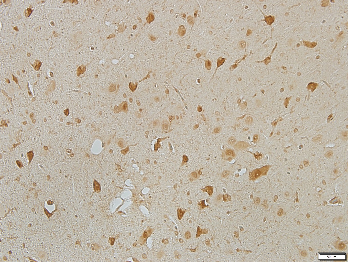
Immunohistochemistry-Paraffin: TMEM119 Antibody [NBP2-30551] - Staining of human cerebral cortex shows weak positivity in microglia.![Immunohistochemistry-Paraffin: TMEM119 Antibody [NBP2-30551]](https://resources.rndsystems.com/images/products/TMEM119-Antibody-Immunohistochemistry-Paraffin-NBP2-30551-img0007.jpg "Immunohistochemistry-Paraffin: TMEM119 Antibody [NBP2-30551]")
Immunohistochemistry-Paraffin: TMEM119 Antibody [NBP2-30551]
Immunohistochemistry-Paraffin: TMEM119 Antibody [NBP2-30551] - Staining of human liver shows no positivity in hepatocytes as expected.![Immunohistochemistry-Paraffin: TMEM119 Antibody [NBP2-30551]](https://resources.rndsystems.com/images/products/TMEM119-Antibody-Immunohistochemistry-Paraffin-NBP2-30551-img0008.jpg "Immunohistochemistry-Paraffin: TMEM119 Antibody [NBP2-30551]")
Immunohistochemistry-Paraffin: TMEM119 Antibody [NBP2-30551]
Immunohistochemistry-Paraffin: TMEM119 Antibody [NBP2-30551] - Staining of human lymph node shows moderate membranous positivity in germinal center cells.![Immunohistochemistry-Paraffin: TMEM119 Antibody [NBP2-30551]](https://resources.rndsystems.com/images/products/TMEM119-Antibody-Immunohistochemistry-Paraffin-NBP2-30551-img0009.jpg "Immunohistochemistry-Paraffin: TMEM119 Antibody [NBP2-30551]")
Immunohistochemistry-Paraffin: TMEM119 Antibody [NBP2-30551]
Immunohistochemistry-Paraffin: TMEM119 Antibody [NBP2-30551] - Staining of human testis shows weak membranous positivity in cells in lamina propria.Applications for TMEM119 Antibody - BSA Free
Application
Recommended Usage
Immunohistochemistry
1:50 - 1:200
Immunohistochemistry-Paraffin
1:50 - 1:200
Application Notes
IHC-Paraffin, HIER pH 6 retrieval is recommended.
Reviewed Applications
Read 5 reviews rated 2.6 using NBP2-30551 in the following applications:
Formulation, Preparation, and Storage
Purification
Affinity purified
Formulation
PBS (pH 7.2) and 40% Glycerol
Format
BSA Free
Preservative
0.02% Sodium Azide
Concentration
Concentrations vary lot to lot. See vial label for concentration. If unlisted please contact technical services.
Shipping
The product is shipped with polar packs. Upon receipt, store it immediately at the temperature recommended below.
Stability & Storage
Store at 4C short term. Aliquot and store at -20C long term. Avoid freeze-thaw cycles.
Background: TMEM119
Additional TMEM119 Products
Product Documents for TMEM119 Antibody - BSA Free
Certificate of Analysis
To download a Certificate of Analysis, please enter a lot or batch number in the search box below.
Product Specific Notices for TMEM119 Antibody - BSA Free
This product is for research use only and is not approved for use in humans or in clinical diagnosis. Primary Antibodies are guaranteed for 1 year from date of receipt.
Related Research Areas
Citations for TMEM119 Antibody - BSA Free
 Powered by Bioz
Customer Reviews for TMEM119 Antibody - BSA Free (5)
2.6 out of 5
5 Customer Ratings
Have you used TMEM119 Antibody - BSA Free?
Submit a review and receive an Amazon gift card!
$25/€18/£15/$25CAN/¥2500 Yen for a review with an image
$10/€7/£6/$10CAN/¥1110 Yen for a review without an image
Submit a review
Customer Images


Showing
1
-
5 of
5 reviews
Showing All
Filter By:
-
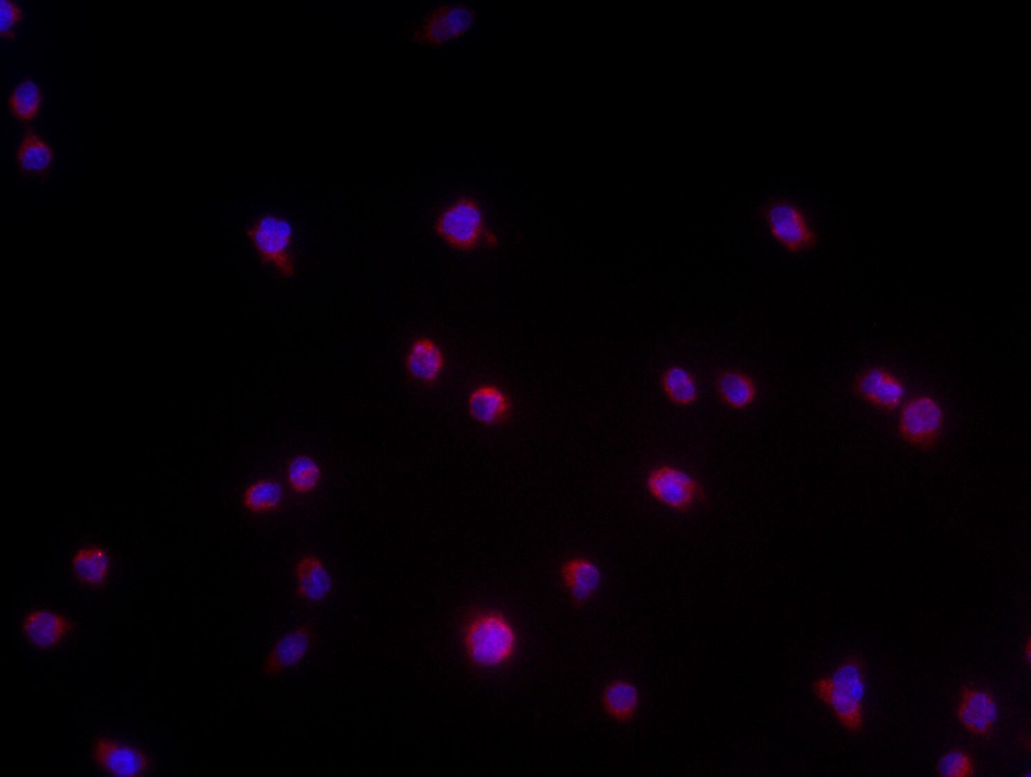
Application: ImmunocytochemistrySample Tested: Human microgliaSpecies: HumanVerified Customer | Posted 05/20/2021Tmem staining with dapi in human microglai cell line

-
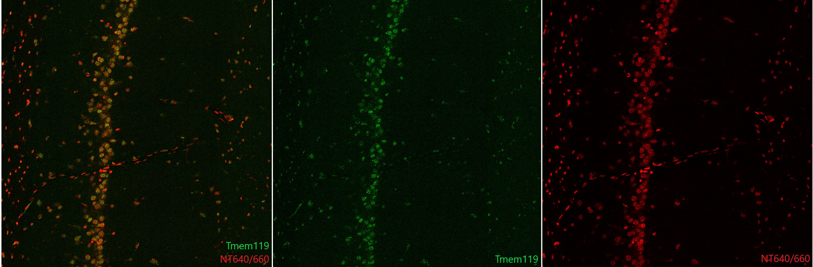
Application: Immunohistochemistry-FrozenSample Tested: Coronal rat brain slices and Adult brainSpecies: RatVerified Customer | Posted 07/28/2020Confocal images from the rat hippocampal CA1 area. Green: Tmem119 1:100 + a-rabbit A488 1:200. Red: NT640/660 1:250. Tmem119 labelled most cell bodies in the hippocampus, esp. neuronal somas. Thus, the antibody is not selective for microglial cells.Rats were perfused with 4% paraformaldehyde in PBS (pH=7.6). The brain was removed and immersed in PFA then in 30% sucrose. The brain was sliced with a freezing microtome. A mixture of 1% Triton-X and 10% donkey serum (in TRIS) was used for antigen retrieval and blocking. Tmem119 antibody was used in 1:100 dilution and was visualized by an Alexa-488 conjugated anti-rabbit secondary antibody (1:200). Cell bodies were stained with NeuroTrace 640/660 (1:250).
Bio-Techne ResponseThank you for reviewing our product. We are sorry to hear that this product did not perform as expected. We have been in touch with the customer to resolve this issue according to our Product Guarantee and to the customer’s satisfaction.
-
Application: Immunohistochemistry-FrozenSample Tested: Brain (hypothalamus) tissueSpecies: SheepVerified Customer | Posted 03/13/2020Photomicrograph at 40x magnification of a sheep hypothalamic section stained using Rabbit polyclonal TMEM119 antibody NBP2-30551. Results showed no positive staining in our tissue using this antibody.To test, we used free-floating sheep hypothalamic brain sections from two animals. We did dilutions of 1:100 to 1:1000 and did not see any staining in our tissue using this antibody NBP2-30551. We also employed an antigen retrieval step in one of our runs, and it did not yield any positive staining.
Bio-Techne ResponseThis review was submitted through the legacy Novus Innovators Program, reflecting a new species or application tested on a primary antibody.
-
Application: Immunohistochemistry-ParaffinSample Tested: Brain (cerebral cortex), Brain (hippocampus) tissue, brain (thalamus) and brain (aqueduct)Species: SwineVerified Customer | Posted 04/23/2018Pig cerebral cortex1:100 dilution. 5 minute DAB stain. Polymer secondary. pH 6 Citrate buffer used for antigen retrieval by steam heat.
-
Application: Immunohistochemistry-FrozenSample Tested: optic nerve and Nerve pia materSpecies: FelineVerified Customer | Posted 01/31/20171:200 concentration. Green-TMEM119, Blue-DAPIPFA fixed and cryoprotected tissue.
There are no reviews that match your criteria.
Protocols
Find general support by application which include: protocols, troubleshooting, illustrated assays, videos and webinars.
- Antigen Retrieval Protocol (PIER)
- Antigen Retrieval for Frozen Sections Protocol
- Appropriate Fixation of IHC/ICC Samples
- Cellular Response to Hypoxia Protocols
- Chromogenic IHC Staining of Formalin-Fixed Paraffin-Embedded (FFPE) Tissue Protocol
- Chromogenic Immunohistochemistry Staining of Frozen Tissue
- ClariTSA™ Fluorophore Kits
- Detection & Visualization of Antibody Binding
- Fluorescent IHC Staining of Frozen Tissue Protocol
- Graphic Protocol for Heat-induced Epitope Retrieval
- Graphic Protocol for the Preparation and Fluorescent IHC Staining of Frozen Tissue Sections
- Graphic Protocol for the Preparation and Fluorescent IHC Staining of Paraffin-embedded Tissue Sections
- Graphic Protocol for the Preparation of Gelatin-coated Slides for Histological Tissue Sections
- IHC Sample Preparation (Frozen sections vs Paraffin)
- Immunofluorescent IHC Staining of Formalin-Fixed Paraffin-Embedded (FFPE) Tissue Protocol
- Immunohistochemistry (IHC) and Immunocytochemistry (ICC) Protocols
- Immunohistochemistry Frozen Troubleshooting
- Immunohistochemistry Paraffin Troubleshooting
- Preparing Samples for IHC/ICC Experiments
- Preventing Non-Specific Staining (Non-Specific Binding)
- Primary Antibody Selection & Optimization
- Protocol for Heat-Induced Epitope Retrieval (HIER)
- Protocol for Making a 4% Formaldehyde Solution in PBS
- Protocol for VisUCyte™ HRP Polymer Detection Reagent
- Protocol for the Preparation & Fixation of Cells on Coverslips
- Protocol for the Preparation and Chromogenic IHC Staining of Frozen Tissue Sections
- Protocol for the Preparation and Chromogenic IHC Staining of Frozen Tissue Sections - Graphic
- Protocol for the Preparation and Chromogenic IHC Staining of Paraffin-embedded Tissue Sections
- Protocol for the Preparation and Chromogenic IHC Staining of Paraffin-embedded Tissue Sections - Graphic
- Protocol for the Preparation and Fluorescent IHC Staining of Frozen Tissue Sections
- Protocol for the Preparation and Fluorescent IHC Staining of Paraffin-embedded Tissue Sections
- Protocol for the Preparation of Gelatin-coated Slides for Histological Tissue Sections
- TUNEL and Active Caspase-3 Detection by IHC/ICC Protocol
- The Importance of IHC/ICC Controls
- Troubleshooting Guide: Immunohistochemistry
- View all Protocols, Troubleshooting, Illustrated assays and Webinars
Loading...