TPX2 Antibody - Azide and BSA Free
Novus Biologicals | Catalog # NB500-179
![Immunocytochemistry/ Immunofluorescence: TPX2 Antibody [NB500-179]](https://resources.rndsystems.com/images/products/TPX2-Antibody-Immunocytochemistry-Immunofluorescence-NB500-179-img0004.jpg "Immunocytochemistry/ Immunofluorescence: TPX2 Antibody [NB500-179]")
Key Product Details
Species Reactivity
Validated:
Cited:
Applications
Validated:
Cited:
Label
Antibody Source
Format
Product Specifications
Immunogen
Localization
Clonality
Host
Isotype
Theoretical MW
Disclaimer note: The observed molecular weight of the protein may vary from the listed predicted molecular weight due to post translational modifications, post translation cleavages, relative charges, and other experimental factors.
Scientific Data Images for TPX2 Antibody - Azide and BSA Free
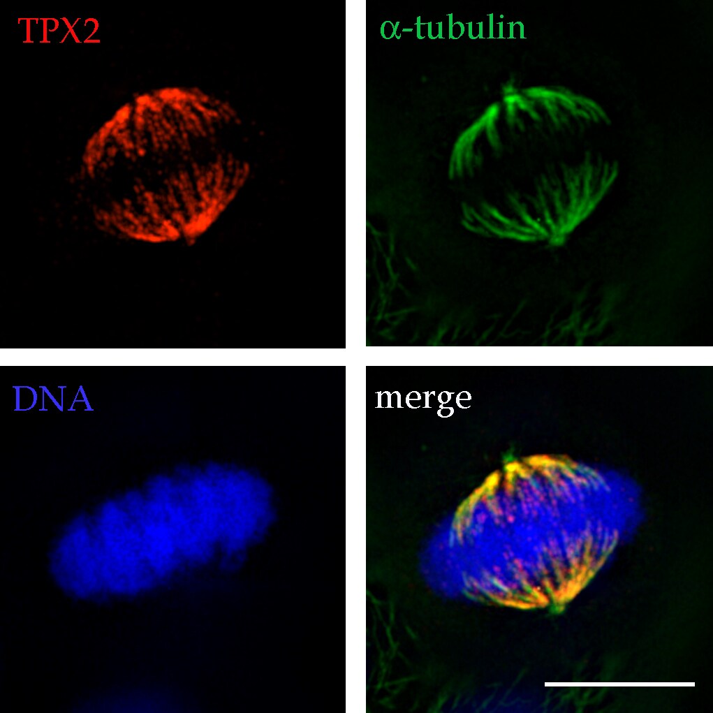
Immunocytochemistry/ Immunofluorescence: TPX2 Antibody [NB500-179]
Immunocytochemistry/Immunofluorescence: TPX2 Antibody [NB500-179] - Analysis of TPX2 at the mitotic spindle microtubules and poles in a HeLa metaphase cell. ICC/IF image submitted by a verified customer review.![Flow Cytometry: TPX2 Antibody [NB500-179]](https://resources.rndsystems.com/images/products/TPX2-Antibody-Flow-Cytometry-NB500-179-img0005.jpg "Flow Cytometry: TPX2 Antibody [NB500-179]")
Flow Cytometry: TPX2 Antibody [NB500-179]
TPX2-Antibody-Flow-Cytometry-NB500-179-img0005.jpg![Western Blot: TPX2 Antibody [NB500-179]](https://resources.rndsystems.com/images/products/TPX2-Antibody-Western-Blot-NB500-179-img0001.jpg "Western Blot: TPX2 Antibody [NB500-179]")
Western Blot: TPX2 Antibody [NB500-179]
Western Blot: TPX2 Antibody [NB500-179] - Analysis of TPX2 expression in 1) HeLa, 2) Ntera2, 3) K-562 and 4) Raji whole cell lysates using NB500-179.![Immunocytochemistry/ Immunofluorescence: TPX2 Antibody [NB500-179]](https://resources.rndsystems.com/images/products/TPX2-Antibody-Immunocytochemistry-Immunofluorescence-NB500-179-img0003.jpg "Immunocytochemistry/ Immunofluorescence: TPX2 Antibody [NB500-179]")
Immunocytochemistry/ Immunofluorescence: TPX2 Antibody [NB500-179]
Immunocytochemistry/Immunofluorescence: TPX2 Antibody [NB500-179] - Staining of HeLa cells fixed in 3.5% paraformaldehyde using NB 500-179 (1:1,000). Nuclear staining during interphase and spindle staining during mitosis.![Simple Western: TPX2 Antibody [NB500-179]](https://resources.rndsystems.com/images/products/TPX2-Antibody-Simple-Western-NB500-179-img0006.jpg "Simple Western: TPX2 Antibody [NB500-179]")
Simple Western: TPX2 Antibody [NB500-179]
Simple Western: TPX2 Antibody [NB500-179] - Lane view shows a specific band for TPX2 using HeLa cell lysate and antibody at 1:250. Electropherogram image of corresponding Simple Western lane view. Image reported by internal validation.
Immunocytochemistry/ Immunofluorescence: TPX2 Antibody [NB500-179] -
Immunocytochemistry/ Immunofluorescence: TPX2 Antibody [NB500-179] - Depletion of TPX2 or Aurora-A reduces cell viability of BRCA2-deficient breast cancer cells. a HCC1806-shBRCA2dox, HCC38-shBRCA2dox, SUM149-shBRCA2dox, & MB231-shBRCA2dox were grown on coverslips & treated with doxycycline (3 days) and/or irradiated (IR, 5 Gy) as indicated. Subsequently, cells were stained for RAD51 & gamma H2AX. Scale bars represent 5 μm. b Quantification of results from a. Percentages of cells with ≥5 RAD51 foci per nucleus are indicated (n ≥ 31). c Percentages of cell survival of doxycycline-treated cells vs untreated cells, transfected with indicated siRNAs. Unpaired two-tailed t tests were used to test for statistical significance (*p ≤ 0.05, **p ≤ 0.01, ***p ≤ 0.001). d BT-549 cells were transfected with siTPX2 or control siRNA (CTR). Cells were grown on coverslips for 3 days after which they were incubated with EdU conjugated to azide-Alexa 488 (10 μM) for 15 min. Subsequently, cells were fixed & stained for 53BP1 & gamma H2AX. Amounts of 53BP1 & gamma H2AX foci per cell of at least 30 EdU-positive cells were counted. Means & standard deviations are depicted. Mann–Whitney U tests were used to analyze statistical significance (*p ≤ 0.05, ** = p ≤ 0.01, ***p ≤ 0.001, ns not significant). e BT-549 cells were transfected as in d, irradiated (IR, 5 Gy), & fixated 0.5 or 6 h after irradiation. Amounts of 53BP1 & gamma H2AX foci per cell were counted. Means & standard deviations are depicted. Mann–Whitney U tests were used to analyze statistical significance (*p ≤ 0.05, **p ≤ 0.01, ***p ≤ 0.001, ns = not significant) Image collected & cropped by CiteAb from the following publication (https://pubmed.ncbi.nlm.nih.gov/30177840), licensed under a CC-BY license. Not internally tested by Novus Biologicals.
Immunocytochemistry/ Immunofluorescence: TPX2 Antibody [NB500-179] -
Immunocytochemistry/ Immunofluorescence: TPX2 Antibody [NB500-179] - TPX2 depletion preferentially affects cell viability in BRCA2-deficient cancer cells. a BT-549-shBRCA2dox cells were left untreated or were treated with doxycycline (2 or 4 days), & subsequently harvested for western blotting for BRCA2 & actin. b BT-549-shBRCA2dox cells were treated as in panel A, & mRNA expression levels of BRCA2 were analyzed relative to GAPDH using qRT-PCR. c BT-549-shBRCA2dox cells were grown on coverslips & treated with doxycycline (3 days) and/or irradiated (IR, 5 Gy) as indicated. At 3 h after irradiation, cells were fixed & analyzed for RAD51 & gamma H2AX foci formation. Scale bars represent 5 μm. d Percentages of cells with ≥5 RAD51 foci per nucleus are indicated. (n ≥ 50 per condition). e BT-549-shBRCA2dox cells were treated with doxycycline (3 days) & were subsequently transfected with indicated siRNAs. A total of 30,000 cells were plated 48 h following transfection. Viable cells were counted 5 days later. Percentages of cell survival of doxycycline-treated vs untreated cells are depicted. Error bars indicate standard deviations of two experimental replicates. Unpaired two-tailed t tests were used to test for statistical significance (*p ≤ 0.05, **p ≤ 0.01, ***p ≤ 0.001) Image collected & cropped by CiteAb from the following publication (https://pubmed.ncbi.nlm.nih.gov/30177840), licensed under a CC-BY license. Not internally tested by Novus Biologicals.
Western Blot: TPX2 Antibody - Azide and BSA Free [NB500-179] -
CtIP depletion causes improper progression of mitosis. (A) The progression of mitosis in HeLa cells was monitored by time-lapse microscopy. HeLa cells were transfected with control or CtIP-1 or siRNA. After 48 h, control and CtIP-depleted HeLa cells were seeded in 12-well plates and transfected with GFP-tagged histone H2B. Fluorescent images were obtained every 5 min starting at the stage of nuclear envelope breakdown. (B) A quantification of the time from nuclear envelope breakdown to anaphase onset in control cells and CtIP-depleted cells. Bars represent the mean +/- SD from three independent experiments. **, p < 0.01, compared to control cells. (C) Delayed mitosis progression in CtIP-depleted cells was confirmed by prolonged phosphorylation of histone H3 (pH3S10). Control and CtIP-depleted HeLa cells were synchronized with a double thymidine block to arrest at the G1/S boundary and released from this block for indicated times. Total proteins collected at the indicated times after release were analyzed by Western blotting using anti-pH3S10 antibody. Histone H3 antibody was used as a loading control. Image collected and cropped by CiteAb from the following open publication (https://www.mdpi.com/2073-4409/11/18/2814), licensed under a CC-BY license. Not internally tested by Novus Biologicals.
Western Blot: TPX2 Antibody - Azide and BSA Free [NB500-179] -
CtIP interacts with TPX2. (A,B) CtIP coimmunoprecipitates with TPX2. Total cell lysates (1 mg) from HEK293T cells transfected with Flag-tagged full length CtIP and HA-tagged full length TPX2 were immunoprecipitated with anti-Flag (A) or anti-HA (B) antibodies. Immunoblotting was then performed with the indicated antibodies. (C) Total cell lysates from HEK293T cells transfected with control siRNA and CtIP siRNA were immunoprecipitated with anti-CtIP or anti-TPX2 antibodies. Immunoblotting was then performed with the indicated antibodies. (D) The cellular localization of CtIP and TPX2 during mitosis was monitored using Immunofluorescence microcopy. Asynchronous HeLa cells were fixed and stained with anti-CtIP and anti-TPX2 antibodies. Representative images show that CtIP colocalizes with TPX2 at the kinetochore from prometaphase through anaphase. Image collected and cropped by CiteAb from the following open publication (https://www.mdpi.com/2073-4409/11/18/2814), licensed under a CC-BY license. Not internally tested by Novus Biologicals.Applications for TPX2 Antibody - Azide and BSA Free
Flow Cytometry
Immunocytochemistry/ Immunofluorescence
Immunoprecipitation
Knockdown Validated
Simple Western
Western Blot
See Simple Western Antibody Database for Simple Western validation: Tested in HeLa lysate 0.2 mg/mL, separated by Size, antibody dilution of 1:250, apparent MW was 89 kDa. Separated by Size-Wes, Sally Sue/Peggy Sue.
Reviewed Applications
Read 1 review rated 5 using NB500-179 in the following applications:
Flow Cytometry Panel Builder
Bio-Techne Knows Flow Cytometry
Save time and reduce costly mistakes by quickly finding compatible reagents using the Panel Builder Tool.
Advanced Features
- Spectra Viewer - Custom analysis of spectra from multiple fluorochromes
- Spillover Popups - Visualize the spectra of individual fluorochromes
- Antigen Density Selector - Match fluorochrome brightness with antigen density

Formulation, Preparation, and Storage
Purification
Formulation
Format
Preservative
Concentration
Shipping
Stability & Storage
Background: TPX2
Alternate Names
Gene Symbol
UniProt
Additional TPX2 Products
Product Documents for TPX2 Antibody - Azide and BSA Free
Certificate of Analysis
To download a Certificate of Analysis, please enter a lot or batch number in the search box below.
Product Specific Notices for TPX2 Antibody - Azide and BSA Free
This product is for research use only and is not approved for use in humans or in clinical diagnosis. Primary Antibodies are guaranteed for 1 year from date of receipt.
Citations for TPX2 Antibody - Azide and BSA Free
 Powered by Bioz
Customer Reviews for TPX2 Antibody - Azide and BSA Free (1)
Have you used TPX2 Antibody - Azide and BSA Free?
Submit a review and receive an Amazon gift card!
$25/€18/£15/$25CAN/¥2500 Yen for a review with an image
$10/€7/£6/$10CAN/¥1110 Yen for a review without an image
Submit a review
Customer Images
-
Application: ImmunofluorescenceSample Tested: See PDFSpecies: HumanVerified Customer | Posted 01/16/2015TPX2 at the mitotic spindle microtubules and poles in a HeLa metaphase cell.

There are no reviews that match your criteria.
Protocols
Find general support by application which include: protocols, troubleshooting, illustrated assays, videos and webinars.
- 7-Amino Actinomycin D (7-AAD) Cell Viability Flow Cytometry Protocol
- Appropriate Fixation of IHC/ICC Samples
- Cellular Response to Hypoxia Protocols
- ClariTSA™ Fluorophore Kits
- Detection & Visualization of Antibody Binding
- Extracellular Membrane Flow Cytometry Protocol
- Flow Cytometry Protocol for Cell Surface Markers
- Flow Cytometry Protocol for Staining Membrane Associated Proteins
- Flow Cytometry Staining Protocols
- Flow Cytometry Troubleshooting Guide
- ICC Cell Smear Protocol for Suspension Cells
- ICC Immunocytochemistry Protocol Videos
- ICC for Adherent Cells
- Immunocytochemistry (ICC) Protocol
- Immunocytochemistry Troubleshooting
- Immunofluorescence of Organoids Embedded in Cultrex Basement Membrane Extract
- Immunohistochemistry (IHC) and Immunocytochemistry (ICC) Protocols
- Immunoprecipitation Protocol
- Intracellular Flow Cytometry Protocol Using Alcohol (Methanol)
- Intracellular Flow Cytometry Protocol Using Detergents
- Intracellular Nuclear Staining Flow Cytometry Protocol Using Detergents
- Intracellular Staining Flow Cytometry Protocol Using Alcohol Permeabilization
- Intracellular Staining Flow Cytometry Protocol Using Detergents to Permeabilize Cells
- Preparing Samples for IHC/ICC Experiments
- Preventing Non-Specific Staining (Non-Specific Binding)
- Primary Antibody Selection & Optimization
- Propidium Iodide Cell Viability Flow Cytometry Protocol
- Protocol for Liperfluo
- Protocol for VisUCyte™ HRP Polymer Detection Reagent
- Protocol for the Characterization of Human Th22 Cells
- Protocol for the Characterization of Human Th9 Cells
- Protocol for the Fluorescent ICC Staining of Cell Smears - Graphic
- Protocol for the Fluorescent ICC Staining of Cultured Cells on Coverslips - Graphic
- Protocol for the Preparation and Fluorescent ICC Staining of Cells on Coverslips
- Protocol for the Preparation and Fluorescent ICC Staining of Non-adherent Cells
- Protocol for the Preparation and Fluorescent ICC Staining of Stem Cells on Coverslips
- Protocol for the Preparation of a Cell Smear for Non-adherent Cell ICC - Graphic
- Protocol: Annexin V and PI Staining by Flow Cytometry
- Protocol: Annexin V and PI Staining for Apoptosis by Flow Cytometry
- R&D Systems Quality Control Western Blot Protocol
- TUNEL and Active Caspase-3 Detection by IHC/ICC Protocol
- The Importance of IHC/ICC Controls
- Troubleshooting Guide: Fluorokine Flow Cytometry Kits
- Troubleshooting Guide: Western Blot Figures
- Western Blot Conditions
- Western Blot Protocol
- Western Blot Protocol for Cell Lysates
- Western Blot Troubleshooting
- Western Blot Troubleshooting Guide
- View all Protocols, Troubleshooting, Illustrated assays and Webinars
FAQs for TPX2 Antibody - Azide and BSA Free
-
Q: In what assays cross-reactivity of NB500-179 with mouse has been confirmed? Do you have any information, or are there by chance any mouse images that just haven't made it onto the datasheet yet?
A: We tested this ab on mouse cells in a Western blot with good results.
-
Q: We know that the immunogen is a recombinant segment of the C-terminal domain of human TPX2. If it is possible, we would be interested to know the exact epitope within this domain.
A: Unfortunately, the exact immunogen sequence for NB500-179 is considered proprietary. It comes from the last 100 amino acids of TPX2.
-
Q: In what assays cross-reactivity of NB500-179 with mouse has been confirmed? Do you have any information, or are there by chance any mouse images that just haven't made it onto the datasheet yet?
A: We tested this ab on mouse cells in a Western blot with good results.
-
Q: We know that the immunogen is a recombinant segment of the C-terminal domain of human TPX2. If it is possible, we would be interested to know the exact epitope within this domain.
A: Unfortunately, the exact immunogen sequence for NB500-179 is considered proprietary. It comes from the last 100 amino acids of TPX2.