Key Product Details
Species Reactivity
Human, Mouse, Rat, Porcine, Bovine, Canine, Guinea Pig, Rabbit, Sheep, Zebrafish
Applications
Western Blot, Immunocytochemistry/ Immunofluorescence
Label
Unconjugated
Antibody Source
Monoclonal Mouse IgG2a Kappa Clone # SPM575
Loading...
Product Specifications
Immunogen
UCH-L1/PGP9.5 protein from brain (Uniprot: P09936)
Localization
Cytoplasmic. Endoplasmic Reticulum membrane
Marker
pan-Neuronal Marker
Clonality
Monoclonal
Host
Mouse
Isotype
IgG2a Kappa
Description
200ug/ml of antibody purified from Bioreactor Concentrate by Protein A or G. Prepared in 10 mM PBS with 0.05% BSA & 0.05% azide. Also available WITHOUT BSA & azide at 1.0 mg/ml. (NBP2-34808)
Antibody with azide - store at 2 to 8C. Antibody without azide - store at -20 to -80C.
Antibody with azide - store at 2 to 8C. Antibody without azide - store at -20 to -80C.
Scientific Data Images for UCH-L1/PGP9.5 Antibody (SPM575)
![Western Blot: UCH-L1/PGP9.5 Antibody (SPM575) [NBP2-32896]](https://resources.rndsystems.com/images/products/UCH-L1-PGP9-5-Antibody-SPM575-Western-Blot-NBP2-32896-img0003.jpg "Western Blot: UCH-L1/PGP9.5 Antibody (SPM575) [NBP2-32896]")
Western Blot: UCH-L1/PGP9.5 Antibody (SPM575) [NBP2-32896]
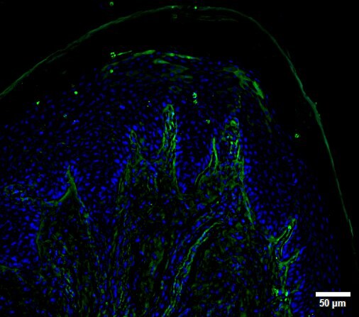
Western Blot: UCH-L1/PGP9.5 Antibody (SPM575) [NBP2-32896] - Western Blot Analysis of human brain tissue lysate using UCH-L1/PGP9.5 Antibody (SPM575).![Immunocytochemistry/ Immunofluorescence: UCH-L1/PGP9.5 Antibody (SPM575) [NBP2-32896]](https://resources.rndsystems.com/images/products/UCH-L1-PGP9-5-Antibody-SPM575-Immunocytochemistry-Immunofluorescence-NBP2-32896-img0002.jpg "Immunocytochemistry/ Immunofluorescence: UCH-L1/PGP9.5 Antibody (SPM575) [NBP2-32896]")
Immunocytochemistry/ Immunofluorescence: UCH-L1/PGP9.5 Antibody (SPM575) [NBP2-32896]
Immunocytochemistry/Immunofluorescence: UCH-L1/PGP9.5 Antibody (SPM575) [NBP2-32896] - Immunofluorescence of skin innervation using anti-PGP9.5. Antibody at dilution 1:500. This image was submitted via customer Review.Applications for UCH-L1/PGP9.5 Antibody (SPM575)
Application
Recommended Usage
Immunocytochemistry/ Immunofluorescence
1-2ug/ml
Western Blot
1-2 ug/ml
Application Notes
Optimal dilution for a specific application should be determined.
Reviewed Applications
Read 1 review rated 4 using NBP2-32896 in the following applications:
Formulation, Preparation, and Storage
Purification
Protein A or G purified
Formulation
10 mM PBS with 0.05% BSA
Preservative
0.05% Sodium Azide
Concentration
0.2 mg/ml
Shipping
The product is shipped with polar packs. Upon receipt, store it immediately at the temperature recommended below.
Stability & Storage
Store at 4C.
Background: UCH-L1/PGP9.5
UCH L1 is down-regulated in brains from Parkinson disease and Alzheimer disease patients, and certian site specific mutations in the UCHL1 gene can either increase or decrese the risk of Parkinson's and/or Alzheimer's neurodegenerative diseases.
Human UCHL 1 and the closely related UCHL3 protein have one of the most complicated knot structures ever discovered, with five knot crossings. This knot structure is expected to help the protein resist degradation in the proteasome.
Long Name
Ubiquitin C-terminal Hydrolase L1
Alternate Names
PARK5, PGP9.5, UCHL1
Gene Symbol
UCHL1
UniProt
Additional UCH-L1/PGP9.5 Products
Product Documents for UCH-L1/PGP9.5 Antibody (SPM575)
Certificate of Analysis
To download a Certificate of Analysis, please enter a lot or batch number in the search box below.
Product Specific Notices for UCH-L1/PGP9.5 Antibody (SPM575)
This product is for research use only and is not approved for use in humans or in clinical diagnosis. Primary Antibodies are guaranteed for 1 year from date of receipt.
Related Research Areas
Customer Reviews for UCH-L1/PGP9.5 Antibody (SPM575) (1)
4 out of 5
1 Customer Rating
Have you used UCH-L1/PGP9.5 Antibody (SPM575)?
Submit a review and receive an Amazon gift card!
$25/€18/£15/$25CAN/¥2500 Yen for a review with an image
$10/€7/£6/$10CAN/¥1110 Yen for a review without an image
Submit a review
Customer Images
Showing
1
-
1 of
1 review
Showing All
Filter By:
-
Application: ImmunocytochemistrySample Tested: mouse skinSpecies: MouseVerified Customer | Posted 03/10/2017Immunofluorescence of skin innervation using anti-PGP9.5 antibody.Blocking and antibody solution: BSA1%/Triton0.2% in PBS Dilution PGP: 1/500 Secondary antibody: Alexa 488

There are no reviews that match your criteria.
Protocols
Find general support by application which include: protocols, troubleshooting, illustrated assays, videos and webinars.
- Appropriate Fixation of IHC/ICC Samples
- Cellular Response to Hypoxia Protocols
- ClariTSA™ Fluorophore Kits
- Detection & Visualization of Antibody Binding
- ICC Cell Smear Protocol for Suspension Cells
- ICC Immunocytochemistry Protocol Videos
- ICC for Adherent Cells
- Immunocytochemistry (ICC) Protocol
- Immunocytochemistry Troubleshooting
- Immunofluorescence of Organoids Embedded in Cultrex Basement Membrane Extract
- Immunohistochemistry (IHC) and Immunocytochemistry (ICC) Protocols
- Preparing Samples for IHC/ICC Experiments
- Preventing Non-Specific Staining (Non-Specific Binding)
- Primary Antibody Selection & Optimization
- Protocol for VisUCyte™ HRP Polymer Detection Reagent
- Protocol for the Fluorescent ICC Staining of Cell Smears - Graphic
- Protocol for the Fluorescent ICC Staining of Cultured Cells on Coverslips - Graphic
- Protocol for the Preparation and Fluorescent ICC Staining of Cells on Coverslips
- Protocol for the Preparation and Fluorescent ICC Staining of Non-adherent Cells
- Protocol for the Preparation and Fluorescent ICC Staining of Stem Cells on Coverslips
- Protocol for the Preparation of a Cell Smear for Non-adherent Cell ICC - Graphic
- R&D Systems Quality Control Western Blot Protocol
- TUNEL and Active Caspase-3 Detection by IHC/ICC Protocol
- The Importance of IHC/ICC Controls
- Troubleshooting Guide: Western Blot Figures
- Western Blot Conditions
- Western Blot Protocol
- Western Blot Protocol for Cell Lysates
- Western Blot Troubleshooting
- Western Blot Troubleshooting Guide
- View all Protocols, Troubleshooting, Illustrated assays and Webinars
Loading...