UCH-L1/PGP9.5 Antibody - BSA Free
Novus Biologicals | Catalog # NB300-675
![Western Blot: UCH-L1/PGP9.5 Antibody [NB300-675]](https://resources.rndsystems.com/images/products/PGP9.5---UCHL-1-Antibody-Western-Blot-NB300-675-img0012.jpg "Western Blot: UCH-L1/PGP9.5 Antibody [NB300-675]")
Key Product Details
Species Reactivity
Validated:
Human, Mouse, Guinea Pig
Cited:
Mouse, Guinea Pig
Predicted:
Bovine (95%), Equine (96%), Porcine (96%), Primate (98%), Rat (95%). Backed by our 100% Guarantee.
Applications
Validated:
Immunohistochemistry, Immunohistochemistry-Paraffin, Western Blot, Immunocytochemistry/ Immunofluorescence
Cited:
IF/IHC
Label
Unconjugated
Antibody Source
Polyclonal Rabbit IgG
Format
BSA Free
Loading...
Product Specifications
Immunogen
A synthetic peptide made to an internal portion of the human UCHL1 protein (between residues 100-200). [UniProt# P09936]
Reactivity Notes
Guinea Pig reactivity reported in scientific literature (PMID: 31739508).
Localization
Cytoplasmic
Marker
pan-Neuronal Marker
Clonality
Polyclonal
Host
Rabbit
Isotype
IgG
Theoretical MW
25 kDa.
Disclaimer note: The observed molecular weight of the protein may vary from the listed predicted molecular weight due to post translational modifications, post translation cleavages, relative charges, and other experimental factors.
Disclaimer note: The observed molecular weight of the protein may vary from the listed predicted molecular weight due to post translational modifications, post translation cleavages, relative charges, and other experimental factors.
Scientific Data Images for UCH-L1/PGP9.5 Antibody - BSA Free
Western Blot: UCH-L1/PGP9.5 Antibody [NB300-675]
Western Blot: UCH-L1/PGP9.5 Antibody [NB300-675] - Analysis of extracts from C6 cells using PGP9.5 / UCHL-1 antibody (NB300-675, 1:200). Image from verified customer review.![Immunocytochemistry/ Immunofluorescence: UCH-L1/PGP9.5 Antibody [NB300-675]](https://resources.rndsystems.com/images/products/PGP9.5---UCHL-1-Antibody-Immunofluorescence-NB300-675-img0014.jpg "Immunocytochemistry/ Immunofluorescence: UCH-L1/PGP9.5 Antibody [NB300-675]")
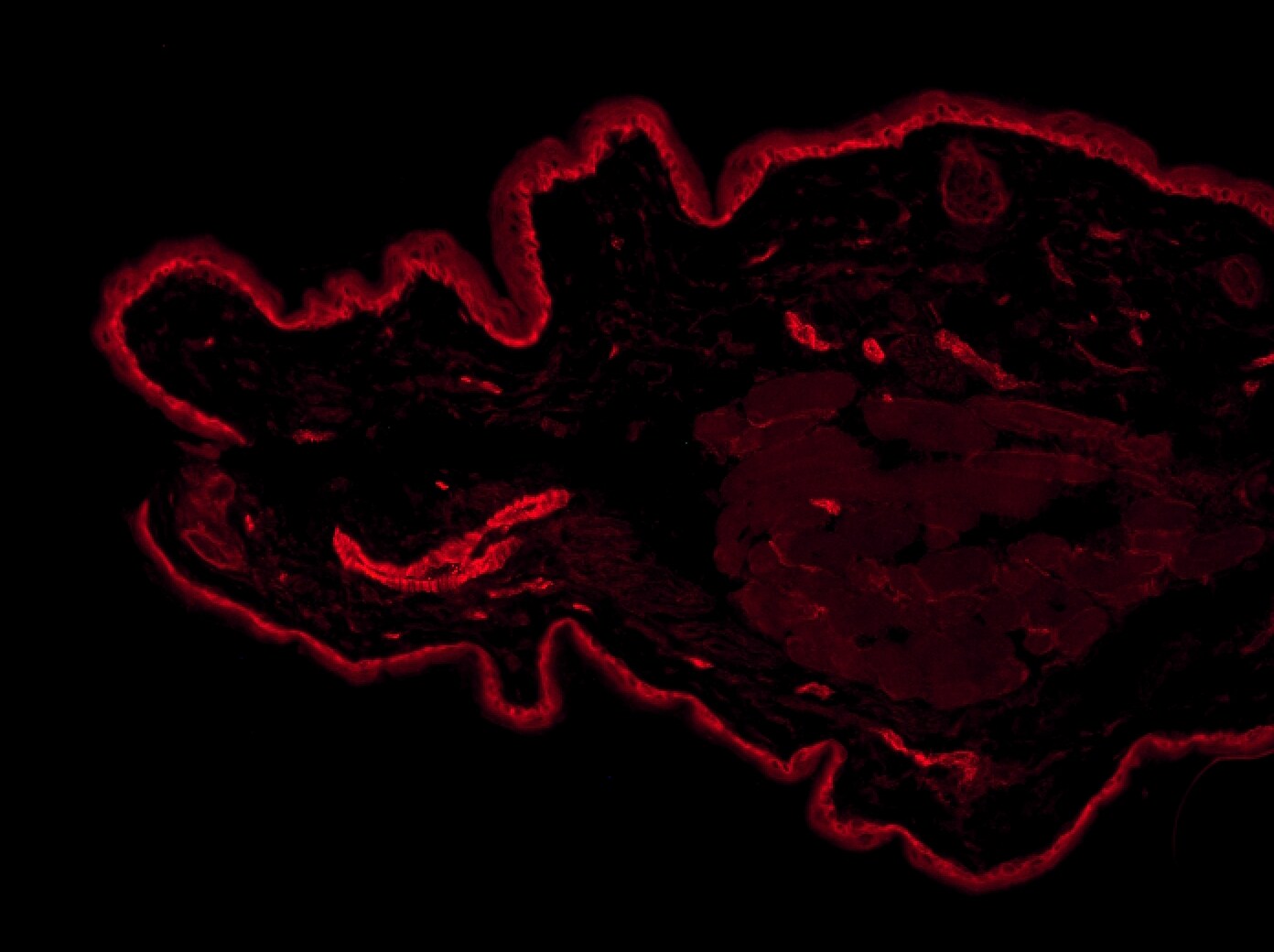
Immunocytochemistry/ Immunofluorescence: UCH-L1/PGP9.5 Antibody [NB300-675]
Immunocytochemistry/Immunofluorescence: UCH-L1/PGP9.5 Antibody [NB300-675] - Analysis of acetone-fixed, frozen mouse ear skin section using anti-PGP9.5 antibody. Image from verified customer review.![Immunohistochemistry: UCH-L1/PGP9.5 Antibody [NB300-675]](https://resources.rndsystems.com/images/products/PGP9.5---UCHL-1-Antibody-Immunohistochemistry-NB300-675-img0010.jpg "Immunohistochemistry: UCH-L1/PGP9.5 Antibody [NB300-675]")
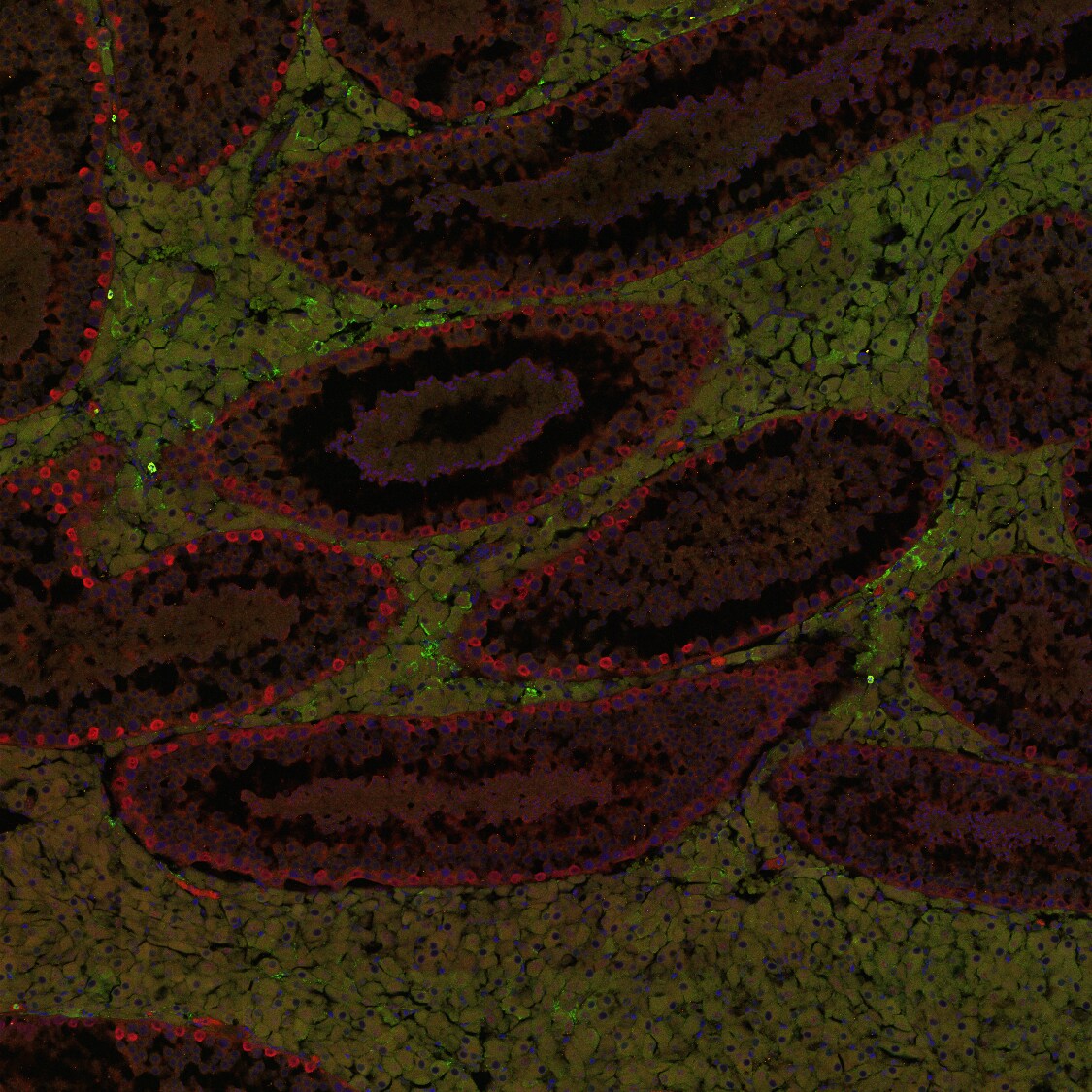
Immunohistochemistry: UCH-L1/PGP9.5 Antibody [NB300-675]
Immunohistochemistry: PGP9.5 / UCHL-1 Antibody [NB300-675] - Human brain, Substantia Nigra, Pigmented Neuron, 40X.![Western Blot: UCH-L1/PGP9.5 Antibody [NB300-675]](https://resources.rndsystems.com/images/products/PGP9.5---UCHL-1-Antibody-Western-Blot-NB300-675-img0011.jpg "Western Blot: UCH-L1/PGP9.5 Antibody [NB300-675]")
Western Blot: UCH-L1/PGP9.5 Antibody [NB300-675]
Western Blot: PGP9.5 / UCHL-1 Antibody [NB300-675] - Detection of UCHL1 in mouse brain lysate.![Immunocytochemistry/ Immunofluorescence: UCH-L1/PGP9.5 Antibody [NB300-675]](https://resources.rndsystems.com/images/products/PGP9.5---UCHL-1-Antibody-Immunocytochemistry-Immunofluorescence-NB300-675-img0009.jpg "Immunocytochemistry/ Immunofluorescence: UCH-L1/PGP9.5 Antibody [NB300-675]")
Immunocytochemistry/ Immunofluorescence: UCH-L1/PGP9.5 Antibody [NB300-675]
Immunocytochemistry/Immunofluorescence: UCH-L1/PGP9.5 Antibody [NB300-675] - In Neuro2a cells with FITC (green). Nuclei were counterstained with DAPI (blue).![Immunocytochemistry/ Immunofluorescence: UCH-L1/PGP9.5 Antibody [NB300-675]](https://resources.rndsystems.com/images/products/PGP9.5---UCHL-1-Antibody-Immunocytochemistry-Immunofluorescence-NB300-675-img0013.jpg "Immunocytochemistry/ Immunofluorescence: UCH-L1/PGP9.5 Antibody [NB300-675]")
Immunocytochemistry/ Immunofluorescence: UCH-L1/PGP9.5 Antibody [NB300-675]
Immunocytochemistry/Immunofluorescence: UCH-L1/PGP9.5 Antibody [NB300-675] - Confocal analysis of C6 cells using PGP9.5 / UCHL-1 antibody (NB300-675, 1:5). An Alexa Fluor 488-conjugated Goat to rabbit IgG was used as secondary antibody (green). Actin filaments were labeled with Alexa Fluor 568 phalloidin (red). DAPI was used to stain the cell nuclei (blue). Image from verified customer review.Applications for UCH-L1/PGP9.5 Antibody - BSA Free
Application
Recommended Usage
Immunocytochemistry/ Immunofluorescence
1:100-1:200
Immunohistochemistry
2.5-5.0 ug/ml
Immunohistochemistry-Paraffin
2.5-5.0 ug/ml
Western Blot
0.5-2.0 ug/ml
Application Notes
This UCHL1 antibody is useful for Immunocytochemistry/Immunofluorescence, Western blot and Immunohistochemistry-Paraffin. By Western blot a band at ~25 kDa is seen.
Reviewed Applications
Read 5 reviews rated 5 using NB300-675 in the following applications:
Formulation, Preparation, and Storage
Purification
Immunogen affinity purified
Formulation
PBS
Format
BSA Free
Preservative
0.02% Sodium Azide
Concentration
1 mg/ml
Shipping
The product is shipped with polar packs. Upon receipt, store it immediately at the temperature recommended below.
Stability & Storage
Store at 4C short term. Aliquot and store at -20C long term. Avoid freeze-thaw cycles.
Background: UCH-L1/PGP9.5
Long Name
Ubiquitin C-terminal Hydrolase L1
Alternate Names
PARK5, PGP9.5, UCHL1
Gene Symbol
UCHL1
Additional UCH-L1/PGP9.5 Products
Product Documents for UCH-L1/PGP9.5 Antibody - BSA Free
Certificate of Analysis
To download a Certificate of Analysis, please enter a lot or batch number in the search box below.
Product Specific Notices for UCH-L1/PGP9.5 Antibody - BSA Free
This product is for research use only and is not approved for use in humans or in clinical diagnosis. Primary Antibodies are guaranteed for 1 year from date of receipt.
Related Research Areas
Citations for UCH-L1/PGP9.5 Antibody - BSA Free
 Powered by Bioz
Customer Reviews for UCH-L1/PGP9.5 Antibody - BSA Free (5)
5 out of 5
5 Customer Ratings
Have you used UCH-L1/PGP9.5 Antibody - BSA Free?
Submit a review and receive an Amazon gift card!
$25/€18/£15/$25CAN/¥2500 Yen for a review with an image
$10/€7/£6/$10CAN/¥1110 Yen for a review without an image
Submit a review
Customer Images
-(01-ml)_NB300-675_8661.jpg)
-(01-ml)_NB300-675_8656.bmp)
Showing
1
-
5 of
5 reviews
Showing All
Filter By:
-
Application: Immunohistochemistry-FrozenSample Tested: TestisSpecies: OpossumVerified Customer | Posted 01/04/2017immunofluorescence staining of gray short-tailed opossum (Monodelphis domestica) in testis from 8-month animal using Uch-L1 antibody.Tested in gray short-tailed opossum (Monodelphis domestica) in testis from 8-month animal. Tissue was fixed in 4% Paraformaldehyde overnight, incubated in 30% sucrose/PBS overnight and embedded in OCT. Sections were cut on a cryostat at 10um. For immunofluorescence staining: Slides were dried 15 minutes, washed 3x3 min TBST (0.1% Triton X-100), permeabilized for 10 minutes in 2% Triton X-100 in TBS, rinsed 3x3 min TBST, blocked in 10% heat inactivated goat serum in TBST for 30 min, and primary added at 1:100 in 10% blocking solution. Slides were coverslipped with parafilm overnight at 4C. The next day slides were washed with TBST 4x5 min, secondaries (AlexaFluor 568 goat anti rabbit and AlexaFluor 488 goat anti mouse 1:500) for 1 hour, 3x3 washes TBST, and mounted with VectaShield HardSet (+ DAPI). Intense staining at periphery of seminiferous tubules of Uchl-1 (red in image) with these conditions. 1:200 also worked though staining was less intense (not shown). The same conditions with 1% Triton X-100 for permeabiliziation resulted in weak staining at 1:100 and 1:200. However, ID-4 did not work well under any conditions. Shown in green is some faint staining in interstitial cells but this was not consistent between samples and did not appear to be specific. ID-4 was tested in 4 and 8 month opossum testes, and at dilutions ranging from 1:25 to 1:500. Single-antibody staining was also performed for each antibody and successful in Uchl-1, not ID-4.

-
Application: Immunohistochemistry-FrozenSample Tested: Rat brainSpecies: RatVerified Customer | Posted 09/15/2015
-
Application: ImmunofluorescenceSample Tested: Mouse Ear Skin (frozen section)Species: MouseVerified Customer | Posted 08/29/2015Mouse Ear Skin
-
Application: Western BlotSample Tested:Species: HumanVerified Customer | Posted 07/01/2014Western blot analysis of extracts from C6 cells using PGP9.5 / UCHL-1 antibody (NB300-675, 1:200).
-
Application: ImmunocytochemistrySample Tested:Species: HumanVerified Customer | Posted 07/01/2014IF Confocal analysis of C6 cells using PGP9.5 / UCHL-1 antibody (NB300-675, 1:5).
There are no reviews that match your criteria.
Protocols
View specific protocols for UCH-L1/PGP9.5 Antibody - BSA Free (NB300-675):
UCH-L1/PGP9.5 Antibody:
Western Blot Protocol
1. Perform SDS-PAGE (4-12%) on samples to be analyzed, loading 40 ug of total protein per lane.
2. Transfer proteins to Nitrocellulose according to the instructions provided by the manufacturer of the transfer apparatus.
3. Rinse membrane with dH2O and then stain the blot using ponceau S for 1-2 minutes to access the transfer of proteins onto the nitrocellulose membrane. Rinse the blot in water to remove excess stain and mark the lane locations and locations of molecular weight markers using a pencil.
4. Rinse the blot in TBS for approximately 5 minutes.
5. Block the membrane using 5% non-fat dry milk + 1% BSA in TBS for 2 hours at room temperature.
6. Rinse the membrane in dH2O and then wash the membrane in wash buffer [TBS + 0.1% Tween] 3 times for 10 minutes each.
7. Dilute the rabbit anti-UCHL1 primary antibody (NB 300-675) in blocking buffer and incubate 1 hour at room temperature.
8. Rinse the membrane in dH2O and then wash the membrane in wash buffer [TBS + 0.1% Tween] 3 times for 10 minutes each.
9. Apply the diluted rabbit-IgG HRP-conjugated secondary antibody in blocking buffer (as per manufacturer's instructions) and incubate 1 hour at room temperature.
10. Wash the blot in wash buffer [TBS + 0.1% Tween] 3 times for 10 minutes each (this step can be repeated as required to reduce background).
11. Apply the detection reagent of choice in accordance with the manufacturer's instructions (Pierce's ECL). Note: Tween-20 can be added to the blocking or antibody dilution buffer at a final concentration of 0.05-0.2%, provided it does not interfere with antibody-antigen binding.
IHC-FFPE sections
I. Deparaffinization:
A. Treat slides with Xylene: 3 changes for 5 minutes each. Drain slides for 10 seconds between changes.
B. Treat slides with 100% Reagent Alcohol: 3 changes for 5 minutes each. Drain slides for 10 seconds between changes.
II. Quench Endogenous Peroxidase:
A. Place slides in peroxidase quenching solution: 15-30 minutes. To Prepare 200 ml of Quenching Solution: Add 3 ml of 30% Hydrogen Peroxide to 200 ml of Methanol.
Use within 4 hours of preparation
B. Place slides in distilled water: 2 changes for 2 minutes each.
III. Retrieve Epitopes:
A. Preheat Citrate Buffer. Place 200 ml of Citrate Buffer Working Solution into container, cover and place into steamer. Heat to 90-96 degrees Celsius.
B. Place rack of slides into hot Citrate Buffer for 20 minutes. Cover.
C. Carefully remove container with slides from steamer and cool on bench, uncovered, for 20 minutes.
D. Slowly add distilled water to further cool for 5 minutes.
E. Rinse slides with distilled water. 2 changes for 2 minutes each.
IV. Immunostaining Procedure:
A. Remove each slide from rack and circle tissue section with a hydrophobic barrier pen (e.g. Liquid Blocker-Super Pap-Pen).
B. Flood slide with Wash Solution. Do not allow tissue sections to dry for the rest of the procedure.
C. Drain wash solution and apply 4 drops of Blocking Reagent to each slide and incubate for 15 minutes.
D. Drain Blocking Reagent (do not wash off the Blocking Reagent), apply 200 ul of Primary Antibody solution to each slide, and incubate for 1 hour.
E. Wash slides with Wash Solution: 3 changes for 5 minutes each.
F. Drain wash solution, apply 4 drops of Secondary antibody to each slide and incubate for 1 hour.
G. Wash slides with Wash Solution: 3 changes for 5 minutes each.
H. Drain wash solution, apply 4 drops of DAB Substrate to each slide and develop for 5-10 minutes. Check development with microscope.
I. Wash slides with Wash Solution: 3 changes for 5 minutes each. Wash slides with Wash Solution: 3 changes for 5 minutes each
J. Drain wash solution, apply 4 drops of Hematoxylin to each slide and stain for 1-3 minutes. Increase time if darker counterstaining is desired.
K. Wash slides with Wash Solution: 2-3 changes for 2 minutes each.
L. Drain wash solution and apply 4 drops of Bluing Solution to each slide for 1-2 minutes.
M. Rinse slides in distilled water.
N. Soak slides in 70% reagent alcohol: 3 minutes with intermittent agitation.
O. Soak slides in 95% reagent alcohol: 2 changes for 3 minutes each with intermittent agitation.
P. Soak slides in 100% reagent alcohol: 3 changes for 3 minutes each with intermittent agitation. Drain slides for 10 seconds between each change.
Q. Soak slides in Xylene: 3 changes for 3 minutes each with intermittent agitation. Drain slides for 10 seconds between each change.
R. Apply 2-3 drops of non-aqueous mounting media to each slide and mount coverslip.
S. Lay slides on a flat surface to dry prior to viewing under microscope.
NOTES:
-Use treated slides (e.g. HistoBond) to assure adherence of FFPE sections to slide.
-Prior to deparaffinization, heat slides overnight in a 60 degrees Celsius oven.
-All steps in which Xylene is used should be performed in a fume hood.
-For Epitope Retrieval, a microwave or pressure cooker may be substituted for the steamer method. Adjust times as necessary depending on conditions.
-For the initial IHC run with a new primary antibody, test tissues with and without Epitope Retrieval. In some instances, Epitope Retrieval may not be necessary.
-200 ul is the recommended maximum volume to apply to a slide for full coverage. Using more than 200 ul may allow solutions to wick off the slide and create drying artifacts. For small tissue sections less than 200 ul may be used.
-5 minutes of development with DAB Substrate should be sufficient. Do not develop for more than 10 minutes. If 5 minutes of development causes background staining, further dilution of the primary antibody may be necessary.
-Hematoxylin should produce a light nuclear counterstain so as not to obscure the DAB staining. Counterstain for 1-1.5 minutes for nuclear antigens. Counterstain for 2-3 minutes for cytoplasmic and membranous antigens. If darker counterstaining is desired increase time (up to 10 minutes).
Find general support by application which include: protocols, troubleshooting, illustrated assays, videos and webinars.
- Antigen Retrieval Protocol (PIER)
- Antigen Retrieval for Frozen Sections Protocol
- Appropriate Fixation of IHC/ICC Samples
- Cellular Response to Hypoxia Protocols
- Chromogenic IHC Staining of Formalin-Fixed Paraffin-Embedded (FFPE) Tissue Protocol
- Chromogenic Immunohistochemistry Staining of Frozen Tissue
- ClariTSA™ Fluorophore Kits
- Detection & Visualization of Antibody Binding
- Fluorescent IHC Staining of Frozen Tissue Protocol
- Graphic Protocol for Heat-induced Epitope Retrieval
- Graphic Protocol for the Preparation and Fluorescent IHC Staining of Frozen Tissue Sections
- Graphic Protocol for the Preparation and Fluorescent IHC Staining of Paraffin-embedded Tissue Sections
- Graphic Protocol for the Preparation of Gelatin-coated Slides for Histological Tissue Sections
- ICC Cell Smear Protocol for Suspension Cells
- ICC Immunocytochemistry Protocol Videos
- ICC for Adherent Cells
- IHC Sample Preparation (Frozen sections vs Paraffin)
- Immunocytochemistry (ICC) Protocol
- Immunocytochemistry Troubleshooting
- Immunofluorescence of Organoids Embedded in Cultrex Basement Membrane Extract
- Immunofluorescent IHC Staining of Formalin-Fixed Paraffin-Embedded (FFPE) Tissue Protocol
- Immunohistochemistry (IHC) and Immunocytochemistry (ICC) Protocols
- Immunohistochemistry Frozen Troubleshooting
- Immunohistochemistry Paraffin Troubleshooting
- Preparing Samples for IHC/ICC Experiments
- Preventing Non-Specific Staining (Non-Specific Binding)
- Primary Antibody Selection & Optimization
- Protocol for Heat-Induced Epitope Retrieval (HIER)
- Protocol for Making a 4% Formaldehyde Solution in PBS
- Protocol for VisUCyte™ HRP Polymer Detection Reagent
- Protocol for the Fluorescent ICC Staining of Cell Smears - Graphic
- Protocol for the Fluorescent ICC Staining of Cultured Cells on Coverslips - Graphic
- Protocol for the Preparation & Fixation of Cells on Coverslips
- Protocol for the Preparation and Chromogenic IHC Staining of Frozen Tissue Sections
- Protocol for the Preparation and Chromogenic IHC Staining of Frozen Tissue Sections - Graphic
- Protocol for the Preparation and Chromogenic IHC Staining of Paraffin-embedded Tissue Sections
- Protocol for the Preparation and Chromogenic IHC Staining of Paraffin-embedded Tissue Sections - Graphic
- Protocol for the Preparation and Fluorescent ICC Staining of Cells on Coverslips
- Protocol for the Preparation and Fluorescent ICC Staining of Non-adherent Cells
- Protocol for the Preparation and Fluorescent ICC Staining of Stem Cells on Coverslips
- Protocol for the Preparation and Fluorescent IHC Staining of Frozen Tissue Sections
- Protocol for the Preparation and Fluorescent IHC Staining of Paraffin-embedded Tissue Sections
- Protocol for the Preparation of Gelatin-coated Slides for Histological Tissue Sections
- Protocol for the Preparation of a Cell Smear for Non-adherent Cell ICC - Graphic
- R&D Systems Quality Control Western Blot Protocol
- TUNEL and Active Caspase-3 Detection by IHC/ICC Protocol
- The Importance of IHC/ICC Controls
- Troubleshooting Guide: Immunohistochemistry
- Troubleshooting Guide: Western Blot Figures
- Western Blot Conditions
- Western Blot Protocol
- Western Blot Protocol for Cell Lysates
- Western Blot Troubleshooting
- Western Blot Troubleshooting Guide
- View all Protocols, Troubleshooting, Illustrated assays and Webinars
Loading...