Uromodulin Antibody (10.32) - BSA Free
Novus Biologicals | Catalog # NBP1-50321
![Immunohistochemistry-Frozen: Uromodulin Antibody (10.32) - BSA Free [NBP1-50321]](https://resources.rndsystems.com/images/products/Uromodulin-Antibody-10-32-Immunohistochemistry-Frozen-NBP1-50321-img0003.jpg "Immunohistochemistry-Frozen: Uromodulin Antibody (10.32) - BSA Free [NBP1-50321]")
Loading...
Key Product Details
Species Reactivity
Validated:
Human, Canine
Cited:
Human
Applications
Validated:
Immunohistochemistry, Immunohistochemistry-Paraffin, Immunohistochemistry-Frozen, Immunofluorescence
Cited:
Hydrolysis Assay
Label
Unconjugated
Antibody Source
Monoclonal Mouse IgG2B Clone # 10.32
Format
BSA Free
Loading...
Product Specifications
Immunogen
Human Uromodulin protein.
Specificity
Anti-human Uromodulin is a monoclonal antibody which reacts with an epitope of the urinary mucoprotein. Uromodulin is a glycoprotein of approximately 80 kDa containing up to 25% carbohydrate by weight.
Clonality
Monoclonal
Host
Mouse
Isotype
IgG2B
Scientific Data Images for Uromodulin Antibody (10.32) - BSA Free
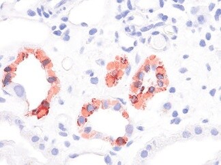
Immunohistochemistry-Frozen: Uromodulin Antibody (10.32) - BSA Free [NBP1-50321]
Immunohistochemistry-Frozen: Uromodulin Antibody (10.32) [NBP1-50321] - Human kidney tissue section stained with Uromodulin antibody. IHC-Fr image submitted by a verified customer review.![Immunohistochemistry-Paraffin: Uromodulin Antibody (10.32) - BSA Free [NBP1-50321]](https://resources.rndsystems.com/images/products/Uromodulin-Antibody-10-32-Immunohistochemistry-Paraffin-NBP1-50321-img0001.jpg "Immunohistochemistry-Paraffin: Uromodulin Antibody (10.32) - BSA Free [NBP1-50321]")
Immunohistochemistry-Paraffin: Uromodulin Antibody (10.32) - BSA Free [NBP1-50321]
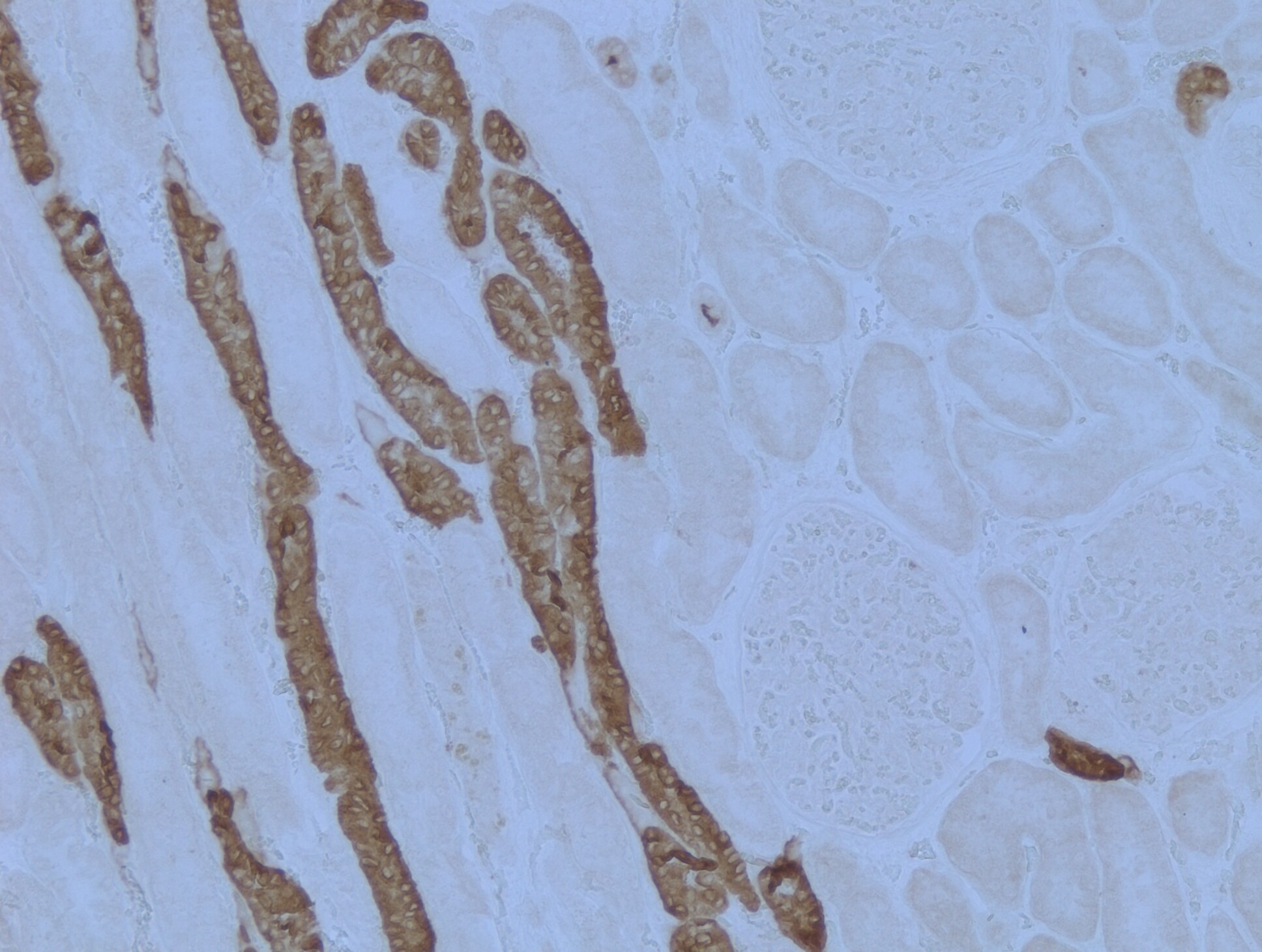
Immunohistochemistry-Paraffin: Uromodulin Antibody (10.32) [NBP1-50321] - Analysis of anti-UMOD / Uromodulin antibody with human kidney at concentration 20 ug/mL.![Immunohistochemistry-Paraffin: Uromodulin Antibody (10.32) - BSA Free [NBP1-50321]](https://resources.rndsystems.com/images/products/Uromodulin-Antibody-10-32-Immunohistochemistry-Paraffin-NBP1-50321-img0002.jpg "Immunohistochemistry-Paraffin: Uromodulin Antibody (10.32) - BSA Free [NBP1-50321]")
Immunohistochemistry-Paraffin: Uromodulin Antibody (10.32) - BSA Free [NBP1-50321]
Immunohistochemistry-Paraffin: Uromodulin Antibody (10.32) [NBP1-50321] - Human kidney tissue, staining in kidney tubules. Heat mediated antigen retrieval was performed by heating in citrate buffer (pH 6) at 95C for 20 minutes. IHC-P image submitted by a verified customer review.Applications for Uromodulin Antibody (10.32) - BSA Free
Application
Recommended Usage
Immunofluorescence
1:10 - 1:500
Immunohistochemistry
5 - 10 ug/mL
Immunohistochemistry-Frozen
1:10 - 1:500
Immunohistochemistry-Paraffin
5 - 10 ug/mL
Application Notes
This Uromodulin antibody is validated for IHC-P from a verified customer review..
Reviewed Applications
Read 2 reviews rated 5 using NBP1-50321 in the following applications:
Formulation, Preparation, and Storage
Purification
Protein G purified
Formulation
PBS
Format
BSA Free
Preservative
0.02% Sodium Azide
Concentration
1 mg/ml
Shipping
The product is shipped with polar packs. Upon receipt, store it immediately at the temperature recommended below.
Stability & Storage
Store at 4C short term. Aliquot and store at -20C long term. Avoid freeze-thaw cycles.
Background: Uromodulin
Alternate Names
ADMCKD2, FJHN, HNFJ, MCKD2, THGP, THP, UMOD, Uromucoid
Entrez Gene IDs
7369 (Human)
Gene Symbol
UMOD
UniProt
Additional Uromodulin Products
Product Documents for Uromodulin Antibody (10.32) - BSA Free
Certificate of Analysis
To download a Certificate of Analysis, please enter a lot or batch number in the search box below.
Product Specific Notices for Uromodulin Antibody (10.32) - BSA Free
This product is for research use only and is not approved for use in humans or in clinical diagnosis. Primary Antibodies are guaranteed for 1 year from date of receipt.
Citations for Uromodulin Antibody (10.32) - BSA Free
 Powered by Bioz
Customer Reviews for Uromodulin Antibody (10.32) - BSA Free (2)
5 out of 5
2 Customer Ratings
Have you used Uromodulin Antibody (10.32) - BSA Free?
Submit a review and receive an Amazon gift card!
$25/€18/£15/$25CAN/¥2500 Yen for a review with an image
$10/€7/£6/$10CAN/¥1110 Yen for a review without an image
Submit a review
Customer Images
Showing
1
-
2 of
2 reviews
Showing All
Filter By:
-
Application: Immunohistochemistry-FrozenSample Tested: Human kidneySpecies: HumanVerified Customer | Posted 08/12/2021Kidney Uromodulin staining

-
Application: Immunohistochemistry-ParaffinSample Tested: Human kidney tissueSpecies: HumanVerified Customer | Posted 05/22/2018Staining observed in kidney tubules.Heat mediated antigen retrieval was performed by heating in citrate buffer (pH6) at 95C for 20 minutes
There are no reviews that match your criteria.
Protocols
Find general support by application which include: protocols, troubleshooting, illustrated assays, videos and webinars.
- Antigen Retrieval Protocol (PIER)
- Antigen Retrieval for Frozen Sections Protocol
- Appropriate Fixation of IHC/ICC Samples
- Cellular Response to Hypoxia Protocols
- Chromogenic IHC Staining of Formalin-Fixed Paraffin-Embedded (FFPE) Tissue Protocol
- Chromogenic Immunohistochemistry Staining of Frozen Tissue
- ClariTSA™ Fluorophore Kits
- Detection & Visualization of Antibody Binding
- Fluorescent IHC Staining of Frozen Tissue Protocol
- Graphic Protocol for Heat-induced Epitope Retrieval
- Graphic Protocol for the Preparation and Fluorescent IHC Staining of Frozen Tissue Sections
- Graphic Protocol for the Preparation and Fluorescent IHC Staining of Paraffin-embedded Tissue Sections
- Graphic Protocol for the Preparation of Gelatin-coated Slides for Histological Tissue Sections
- ICC Cell Smear Protocol for Suspension Cells
- ICC Immunocytochemistry Protocol Videos
- ICC for Adherent Cells
- IHC Sample Preparation (Frozen sections vs Paraffin)
- Immunocytochemistry (ICC) Protocol
- Immunocytochemistry Troubleshooting
- Immunofluorescence of Organoids Embedded in Cultrex Basement Membrane Extract
- Immunofluorescent IHC Staining of Formalin-Fixed Paraffin-Embedded (FFPE) Tissue Protocol
- Immunohistochemistry (IHC) and Immunocytochemistry (ICC) Protocols
- Immunohistochemistry Frozen Troubleshooting
- Immunohistochemistry Paraffin Troubleshooting
- Preparing Samples for IHC/ICC Experiments
- Preventing Non-Specific Staining (Non-Specific Binding)
- Primary Antibody Selection & Optimization
- Protocol for Heat-Induced Epitope Retrieval (HIER)
- Protocol for Making a 4% Formaldehyde Solution in PBS
- Protocol for VisUCyte™ HRP Polymer Detection Reagent
- Protocol for the Fluorescent ICC Staining of Cell Smears - Graphic
- Protocol for the Fluorescent ICC Staining of Cultured Cells on Coverslips - Graphic
- Protocol for the Preparation & Fixation of Cells on Coverslips
- Protocol for the Preparation and Chromogenic IHC Staining of Frozen Tissue Sections
- Protocol for the Preparation and Chromogenic IHC Staining of Frozen Tissue Sections - Graphic
- Protocol for the Preparation and Chromogenic IHC Staining of Paraffin-embedded Tissue Sections
- Protocol for the Preparation and Chromogenic IHC Staining of Paraffin-embedded Tissue Sections - Graphic
- Protocol for the Preparation and Fluorescent ICC Staining of Cells on Coverslips
- Protocol for the Preparation and Fluorescent ICC Staining of Non-adherent Cells
- Protocol for the Preparation and Fluorescent ICC Staining of Stem Cells on Coverslips
- Protocol for the Preparation and Fluorescent IHC Staining of Frozen Tissue Sections
- Protocol for the Preparation and Fluorescent IHC Staining of Paraffin-embedded Tissue Sections
- Protocol for the Preparation of Gelatin-coated Slides for Histological Tissue Sections
- Protocol for the Preparation of a Cell Smear for Non-adherent Cell ICC - Graphic
- TUNEL and Active Caspase-3 Detection by IHC/ICC Protocol
- The Importance of IHC/ICC Controls
- Troubleshooting Guide: Immunohistochemistry
- View all Protocols, Troubleshooting, Illustrated assays and Webinars
Loading...