![Western Blot: ZEB1 Antibody [NBP1-68930]](https://resources.rndsystems.com/images/products/ZEB1-Antibody-Western-Blot-NBP1-68930-img0005.jpg "Western Blot: ZEB1 Antibody [NBP1-68930]")
Key Product Details
Species Reactivity
Applications
Label
Antibody Source
Format
Product Specifications
Immunogen
Marker
Clonality
Host
Isotype
Theoretical MW
Disclaimer note: The observed molecular weight of the protein may vary from the listed predicted molecular weight due to post translational modifications, post translation cleavages, relative charges, and other experimental factors.
Description
Scientific Data Images for ZEB1 Antibody - BSA Free
Western Blot: ZEB1 Antibody [NBP1-68930]
Western Blot: ZEB1 Antibody [NBP1-68930] - Lanes: 1. 45ug capan1 cell lysate 2. 45 ug HPAF cell lysate Primary, Antibody Dilution: 1 : 1000 Secondary Antibody: Anti-Rabbit HRP Secondary, Antibody Dilution: 1 : 5000 Gene name: ZEB1.![Immunohistochemistry: ZEB1 Antibody [NBP1-68930]](https://resources.rndsystems.com/images/products/ZEB1-Antibody-Immunohistochemistry-NBP1-68930-img0004.jpg "Immunohistochemistry: ZEB1 Antibody [NBP1-68930]")
Immunohistochemistry: ZEB1 Antibody [NBP1-68930]
Immunohistochemistry: ZEB1 Antibody [NBP1-68930] - Human Capan1 cells (Pancreatic cancer cell line) Primary Antibody Dilution: 1 : 300 Secondary Antibody: Anti-rabbit-AlexaFluor-488 Secondary Antibody Dilution: 1 : 200Color/Signal.![Western Blot: ZEB1 Antibody [NBP1-68930]](https://resources.rndsystems.com/images/products/ZEB1-Antibody-Western-Blot-NBP1-68930-img0003.jpg "Western Blot: ZEB1 Antibody [NBP1-68930]")
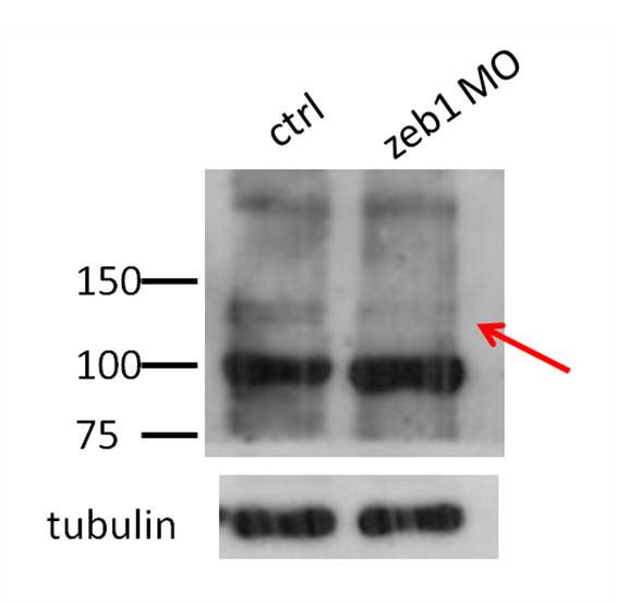
Western Blot: ZEB1 Antibody [NBP1-68930]
Western Blot: ZEB1 Antibody [NBP1-68930] - Lane 1: Control, Lane 2: ZEB1 morphants. Upper panel: ZEB1 blot; Bottom panel: tubulin blot![Western Blot: ZEB1 Antibody [NBP1-68930]](https://resources.rndsystems.com/images/products/ZEB1-Antibody-Western-Blot-NBP1-68930-img0002.jpg "Western Blot: ZEB1 Antibody [NBP1-68930]")
Western Blot: ZEB1 Antibody [NBP1-68930]
Western Blot: ZEB1 Antibody [NBP1-68930] - 293T cells lysate, concentration 0.2-1 ug/ml.Applications for ZEB1 Antibody - BSA Free
Western Blot
Reviewed Applications
Read 1 review rated 3 using NBP1-68930 in the following applications:
Formulation, Preparation, and Storage
Purification
Formulation
Format
Preservative
Concentration
Shipping
Stability & Storage
Background: ZEB1
Long Name
Alternate Names
Gene Symbol
UniProt
Additional ZEB1 Products
Product Documents for ZEB1 Antibody - BSA Free
Certificate of Analysis
To download a Certificate of Analysis, please enter a lot or batch number in the search box below.
Product Specific Notices for ZEB1 Antibody - BSA Free
This product is for research use only and is not approved for use in humans or in clinical diagnosis. Primary Antibodies are guaranteed for 1 year from date of receipt.
Customer Reviews for ZEB1 Antibody - BSA Free (1)
Have you used ZEB1 Antibody - BSA Free?
Submit a review and receive an Amazon gift card!
$25/€18/£15/$25CAN/¥2500 Yen for a review with an image
$10/€7/£6/$10CAN/¥1110 Yen for a review without an image
Submit a review
Customer Images
-
Application: Western BlotSample Tested: zebrafish embryonic trunk at 3 days post fertilizationSpecies: OtherVerified Customer | Posted 11/05/2013lane 1: control, lane 2: zeb1 morphants. Upper panel: zeb1 blot; bottom panel: tubulin blot

There are no reviews that match your criteria.
Protocols
Find general support by application which include: protocols, troubleshooting, illustrated assays, videos and webinars.
- Antigen Retrieval Protocol (PIER)
- Antigen Retrieval for Frozen Sections Protocol
- Appropriate Fixation of IHC/ICC Samples
- Cellular Response to Hypoxia Protocols
- Chromogenic IHC Staining of Formalin-Fixed Paraffin-Embedded (FFPE) Tissue Protocol
- Chromogenic Immunohistochemistry Staining of Frozen Tissue
- ClariTSA™ Fluorophore Kits
- Detection & Visualization of Antibody Binding
- Fluorescent IHC Staining of Frozen Tissue Protocol
- Graphic Protocol for Heat-induced Epitope Retrieval
- Graphic Protocol for the Preparation and Fluorescent IHC Staining of Frozen Tissue Sections
- Graphic Protocol for the Preparation and Fluorescent IHC Staining of Paraffin-embedded Tissue Sections
- Graphic Protocol for the Preparation of Gelatin-coated Slides for Histological Tissue Sections
- IHC Sample Preparation (Frozen sections vs Paraffin)
- Immunofluorescent IHC Staining of Formalin-Fixed Paraffin-Embedded (FFPE) Tissue Protocol
- Immunohistochemistry (IHC) and Immunocytochemistry (ICC) Protocols
- Immunohistochemistry Frozen Troubleshooting
- Immunohistochemistry Paraffin Troubleshooting
- Preparing Samples for IHC/ICC Experiments
- Preventing Non-Specific Staining (Non-Specific Binding)
- Primary Antibody Selection & Optimization
- Protocol for Heat-Induced Epitope Retrieval (HIER)
- Protocol for Making a 4% Formaldehyde Solution in PBS
- Protocol for VisUCyte™ HRP Polymer Detection Reagent
- Protocol for the Preparation & Fixation of Cells on Coverslips
- Protocol for the Preparation and Chromogenic IHC Staining of Frozen Tissue Sections
- Protocol for the Preparation and Chromogenic IHC Staining of Frozen Tissue Sections - Graphic
- Protocol for the Preparation and Chromogenic IHC Staining of Paraffin-embedded Tissue Sections
- Protocol for the Preparation and Chromogenic IHC Staining of Paraffin-embedded Tissue Sections - Graphic
- Protocol for the Preparation and Fluorescent IHC Staining of Frozen Tissue Sections
- Protocol for the Preparation and Fluorescent IHC Staining of Paraffin-embedded Tissue Sections
- Protocol for the Preparation of Gelatin-coated Slides for Histological Tissue Sections
- R&D Systems Quality Control Western Blot Protocol
- TUNEL and Active Caspase-3 Detection by IHC/ICC Protocol
- The Importance of IHC/ICC Controls
- Troubleshooting Guide: Immunohistochemistry
- Troubleshooting Guide: Western Blot Figures
- Western Blot Conditions
- Western Blot Protocol
- Western Blot Protocol for Cell Lysates
- Western Blot Troubleshooting
- Western Blot Troubleshooting Guide
- View all Protocols, Troubleshooting, Illustrated assays and Webinars
FAQs for ZEB1 Antibody - BSA Free
-
Q: I'm looking for an antibody that recognize zeb1 (zinc finger E-box binding homeobox 1) in zebrafish. In particular, I am interested in the detection by WB of zeb1a (in zebrafish there are two different isoforms). Do you have an antibody that is at least predicted to be reactive in zebrafish?
A:
Our ZEB1 antibody #NBP1-68930 would be the best choice for you as its immunogen sequence is 100% identical to Zebrafish Zeb1. Besides WB, our customers have reported the workability of this antibody in ICC-IF application also. If you would be interested in testing this novel species or this novel application, please take a look at our Innovators Reward Program.
-
Q: In looking at the Zeb1 antibodies on your website, the molecular weights of Zeb1 detected by several antibodies are different. Why is this?
A: ZEB1 is a rather large protein and undergoes a lot of post translational modifications such as phosphorylation and glycosylation (please see: UniProt P37275). The reason band patterns are different in the images may be due to sample type and how highly modified the ZEB1 protein is that the antibody is detecting. The more post translational modifications the higher you will detect the protein.
-
Q: I'm looking for an antibody that recognize zeb1 (zinc finger E-box binding homeobox 1) in zebrafish. In particular, I am interested in the detection by WB of zeb1a (in zebrafish there are two different isoforms). Do you have an antibody that is at least predicted to be reactive in zebrafish?
A:
Our ZEB1 antibody #NBP1-68930 would be the best choice for you as its immunogen sequence is 100% identical to Zebrafish Zeb1. Besides WB, our customers have reported the workability of this antibody in ICC-IF application also. If you would be interested in testing this novel species or this novel application, please take a look at our Innovators Reward Program.
-
Q: In looking at the Zeb1 antibodies on your website, the molecular weights of Zeb1 detected by several antibodies are different. Why is this?
A: ZEB1 is a rather large protein and undergoes a lot of post translational modifications such as phosphorylation and glycosylation (please see: UniProt P37275). The reason band patterns are different in the images may be due to sample type and how highly modified the ZEB1 protein is that the antibody is detecting. The more post translational modifications the higher you will detect the protein.