Counterstaining and Mounting in ICC/IF
Immunofluorescence Staining and Counterstaining
In addition to immunofluorescence, cells are typically counterstained with DNA or cytoskeleton-binding dyes to help identify cellular organelles and structures. Fluorescent microscopy is used for individual cell identification and analysis. When designing an ICC experiment, it is critical to ensure that the counterstain and the selected fluorochrome display limited spectral overlap so they can be sufficiently distinguished from one another. For example, DAPI should not be used in combination with other blue emitting fluorochromes like Alexa Fluor® 405 or DyLight™ 405.
DAPI can penetrate the nucleus without permeabilization and intercalates into the DNA helix. However, high concentrations of DAPI are required for viable cells compared to dying cells, where DNA binding is much more efficient, making DAPI a suitable viability dye in IF staining. Other examples include staining the cytoskeletal marker actin with fluorescently-labeled phalloidin, or the plasma membrane using fluorescently-tagged wheat germ agglutinin (WGA). In the example below using DAPI as a nuclear counterstain, DDX21 is shown to be localized to the nucleus.
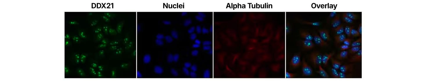
DDX21 was detected in fixed HeLa cells using anti-DDX21 [NB100-1718] and anti-rabbit Dylight™ 488 (green). Alpha tubulin (DM1A) [NB100-690] was used as a co-stain and detected with an anti-mouse DylightTM 550 (red). Nuclei were counterstained with DAPI (blue) [NBP2-31156].
| Dye | Excitation/Emission Max (nm) | Usefulness |
|---|---|---|
| DRAQ5™ | 646/697 | Deep Red Anthraquinone 5 (DRAQ5™) is a nuclear DNA dye that can be used on live or fixed cells. It can be used as an indicator of cell cycle phase. |
| DRAQ7™ | 646/697 | Deep Red Anthraquinone 7 (DRAQ7™) is a new far-red fluorescent DNA dye that will stain the nuclei of permeabilized, dead cells to assess viability of fixed cells. DRAQ7™ will not stain intact, live cells. |
| DRAQ9™ | 655/697 | Deep Red Anthraquinone 9 (DRAQ9™) can be used on live or fixed cells. DRAQ9™ stains the cytoplasm but not the nucleus. DRAQ9™ is for ICC/IF applications. |
| CyTRAK Orange™ | 510/610 | CyTRAK Orange™ preferentially stains the nucleus, but also defines the cytoplasmic area in both live and fixed cells. CyTRAK Orange™ stains arrested/senescent cells compared to healthy cells, thus enabling easy identification and separation of these two distinct cellular populations. This dye can be used to distinguish cell location, cell perimeter, cell shape and cell spread parameters. |
| DAPI | 360/460 | DAPI can penetrate cell membranes, allowing use in live or fixed cells. However, as DAPI intercalates into double stranded nucleic acids, the emission of blue fluorescence is strongest in dying or dead cells and can thus help assess cell viability and requires high concentrations to penetrate live cells. DAPI's blue emission is useful for multiplexing as there is very little overlap between DAPI and green-fluorescent molecules like FITC and GFP. |
| VECTASHIELD® HardSet™ Antifade Mounting Medium with DAPI | 360/460 | VECTASHIELD Mounting Media is compatible with a wide array of fluorochromes, enzymatic substrates, and fluorescent proteins. DAPI produces a blue fluorescence when bound to DNA of dead cells and can help differentiate cells to determine cell viability. |
| VECTASHIELD® Antifade Mounting Medium with DAPI | 360/460 | VECTASHIELD Antifade Mounting Medium with DAPI preserves fluorescence by preventing the rapid photobleaching of fluorescent proteins and fluorescent dyes. |
DRAQ5, DRAQ7, DRAQ9 and CyTRAK Orange are registered trademarks of BioStatus Limited.
Immunofluorescence Mounting Medium
After staining, cells on coverslips must be mounted onto microscope slides prior to imaging. The use of mounting media protects stained cells from dehydration and makes the sample suitable for microscopy. It is important to match the refractive index of your mounting media with microscope objectives to generate high-quality images. Most mounting media contains anti-fading agents to protect samples from photobleaching. Some mounting media also includes DNA stains, such as DAPI, eliminating the need for a separate counterstaining step. While mounted slides can be stored for months in the dark at -20°C, the fluorescence intensity will slowly decay over time.
Related Links
Fluorochromes Selection for Multicolor ICC/IF
Sample Preparation for ICC/IF Experiments
Controls for ICC/IF Experiments