alpha Tubulin Antibody (DM1A) - BSA Free
Novus Biologicals | Catalog # NB100-690
Key Product Details
Validated by
Biological Validation
Species Reactivity
Validated:
Human, Mouse, Rat, Porcine, Avian, Bovine, Canine, Chicken, Chinese Hamster, Drosophila, Fungi, Goat, Guinea Pig, Hamster, Monkey, Parasite, Primate, Rabbit, Xenopus, Yeast
Cited:
Human, Mouse, Rat, Avian - Chicken, Canine, Frog - Xenopus (African Clawed Frog), Fungi, Goat, Hamster, Insect - Drosophila, Primate, Primate - Macaca mulatta (Rhesus Macaque), Rabbit, Yeast
Applications
Validated:
Immunohistochemistry, Immunohistochemistry-Paraffin, Immunohistochemistry-Frozen, Immunomicroscopy, Western Blot, Flow Cytometry, Flow (Intracellular), Immunocytochemistry/ Immunofluorescence, Simple Western, Immunoprecipitation, CyTOF-ready
Cited:
Immunohistochemistry-Paraffin, Immunohistochemistry-Frozen, Western Blot, Flow Cytometry, Immunocytochemistry, Immunocytochemistry/ Immunofluorescence, Simple Western, Immunoprecipitation, IF/IHC
Label
Unconjugated
Antibody Source
Monoclonal Mouse IgG1 kappa Clone # DM1A
Format
BSA Free
Loading...
Product Specifications
Immunogen
This alpha Tubulin Antibody (DM1A) was developed against native chicken brain microtubules.
Reactivity Notes
Use in Mouse reported in scientific literature (PMID:34871568) Use in Mouse reported in scientific literature (PMID:34533563). Yeast reactivity reported in scientific literature (PMID: 25126732). Goat reactivity reported in scientific literature (PMID:31805146). Will likely react with all mammals.
Marker
Microtubule Marker
Specificity
This alpha Tubulin Antibody (DM1A) does not cross-react with beta Tubulin.
Clonality
Monoclonal
Host
Mouse
Isotype
IgG1 kappa
Theoretical MW
50 kDa.
Disclaimer note: The observed molecular weight of the protein may vary from the listed predicted molecular weight due to post translational modifications, post translation cleavages, relative charges, and other experimental factors.
Disclaimer note: The observed molecular weight of the protein may vary from the listed predicted molecular weight due to post translational modifications, post translation cleavages, relative charges, and other experimental factors.
Description
As the TUBA1A gene is conserved evolutionarily and is ubiquitously expressed in most eukaryotic cell lines,
the Alpha tubulin antibody has been shown to be an attractive and effective choice for
a loading control, detecting at approximately 50-55 kDa. Quantitative western blotting requires a loading
control in order to account and adjust for the differences in the loading of samples across wells.
Scientific Data Images for alpha Tubulin Antibody (DM1A) - BSA Free
![Simple Western: alpha Tubulin Antibody (DM1A)BSA Free [NB100-690]](https://resources.rndsystems.com/images/products/alpha-Tubulin-Antibody-DM1A-Simple-Western-NB100-690-img0023.jpg "Simple Western: alpha Tubulin Antibody (DM1A)BSA Free [NB100-690]")
Simple Western: alpha Tubulin Antibody (DM1A)BSA Free [NB100-690]
Simple Western: alpha Tubulin Antibody (DM1A) [NB100-690] - Simple Western lane view shows a specific band for alpha Tubulin in 1.0 mg/ml of HeLa lysate. This experiment was performed under reducing conditions using the 12-230 kDa separation system. Alpha tubulin molecular weight: 50 kDa.![Immunohistochemistry: alpha Tubulin Antibody (DM1A) - BSA Free [NB100-690]](https://resources.rndsystems.com/images/products/alpha-Tubulin-Antibody-DM1A-Immunohistochemistry-NB100-690-img0028.jpg "Immunohistochemistry: alpha Tubulin Antibody (DM1A) - BSA Free [NB100-690]")
Immunohistochemistry: alpha Tubulin Antibody (DM1A) - BSA Free [NB100-690]
Immunohistochemistry: alpha Tubulin Antibody (DM1A) [NB100-690] - Analysis of formalin fixed colon sections. Heat mediated antigen retrieval was performed using sodium citrate buffer for 20 min before incubating with primary antibody at a 0.5ug/ml dilution for 15 min at RT.![Immunohistochemistry: alpha Tubulin Antibody (DM1A) - BSA Free [NB100-690]](https://resources.rndsystems.com/images/products/alpha-Tubulin-Antibody-DM1A-Immunohistochemistry-NB100-690-img0029.jpg "Immunohistochemistry: alpha Tubulin Antibody (DM1A) - BSA Free [NB100-690]")
Immunohistochemistry: alpha Tubulin Antibody (DM1A) - BSA Free [NB100-690]
Immunohistochemistry: alpha Tubulin Antibody (DM1A) [NB100-690] - Analysis of colon tissue. Sections were formalin fixed and embedded with paraffin. Sodium citrate heat mediated antigen retrieval for 20 min. Incubated with primary antibody for 15 min at a 5 ug/ml concentration. Corner image is staining with secondary only.![Immunohistochemistry: alpha Tubulin Antibody (DM1A) - BSA Free [NB100-690]](https://resources.rndsystems.com/images/products/alpha-Tubulin-Antibody-DM1A-Immunohistochemistry-NB100-690-img0031.jpg "Immunohistochemistry: alpha Tubulin Antibody (DM1A) - BSA Free [NB100-690]")
Immunohistochemistry: alpha Tubulin Antibody (DM1A) - BSA Free [NB100-690]
Immunohistochemistry: alpha Tubulin Antibody (DM1A) [NB100-690] - Analysis of formalin fixed paraffin embedded heart sections. Used at a dilution of 1:500.![Western Blot: alpha Tubulin Antibody (DM1A)BSA Free [NB100-690]](https://resources.rndsystems.com/images/products/alpha-Tubulin-Antibody-DM1A-Western-Blot-NB100-690-img0032.jpg "Western Blot: alpha Tubulin Antibody (DM1A)BSA Free [NB100-690]")
Western Blot: alpha Tubulin Antibody (DM1A)BSA Free [NB100-690]
Western Blot: alpha Tubulin Antibody (DM1A) [NB100-690] - Analsis of alpha tubulin in 9 cell lysates. Lane 1. HeLa; Lane 2. JURKAT; Lane 3. COS7; Lane 4. NIH-3T3; Lane 5. PC-12; Lane 6. RAT2; Lane 7. CHO; Lane 8. MDBK; Lane 9. MDCK![Flow Cytometry: alpha Tubulin Antibody (DM1A) - BSA Free [NB100-690]](https://resources.rndsystems.com/images/products/alpha-Tubulin-Antibody-DM1A-Flow-Cytometry-NB100-690-img0016.jpg "Flow Cytometry: alpha Tubulin Antibody (DM1A) - BSA Free [NB100-690]")
Flow Cytometry: alpha Tubulin Antibody (DM1A) - BSA Free [NB100-690]
Flow Cytometry: alpha Tubulin Antibody (DM1A) [NB100-690] - Intracellular flow cytometric staining of 1 x 10^6 CHO (A) and HEK-293 (B) cells using alpha Tubulin antibody (dark blue). Isotype control shown in orange. An antibody concentration of 1 ug/1x10^6 cells was used.![Immunomicroscopy: alpha Tubulin Antibody (DM1A) - BSA Free [NB100-690]](https://resources.rndsystems.com/images/products/alpha-Tubulin-Antibody-DM1A-Immunomicroscopy-NB100-690-img0037.jpg "Immunomicroscopy: alpha Tubulin Antibody (DM1A) - BSA Free [NB100-690]")
Immunomicroscopy: alpha Tubulin Antibody (DM1A) - BSA Free [NB100-690]
Immunomicroscopy: alpha Tubulin Antibody (DM1A) [NB100-690] - Analysis of HeLa cells, green staining is alpha tubulin whereas red is DNA stained with propidium iodide.![Western Blot: alpha Tubulin Antibody (DM1A)BSA Free [NB100-690]](https://resources.rndsystems.com/images/products/alpha-Tubulin-Antibody-DM1A-Western-Blot-NB100-690-img0054.jpg "Western Blot: alpha Tubulin Antibody (DM1A)BSA Free [NB100-690]")
![Immunocytochemistry/ Immunofluorescence: alpha Tubulin Antibody (DM1A) - BSA Free [NB100-690]](https://resources.rndsystems.com/images/products/alpha-Tubulin-Antibody-DM1A-BSA-Free-Immunocytochemistry-Immunofluorescence-NB100-690-img0061.jpg "Immunocytochemistry/ Immunofluorescence: alpha Tubulin Antibody (DM1A) - BSA Free [NB100-690]")
Immunocytochemistry/ Immunofluorescence: alpha Tubulin Antibody (DM1A) - BSA Free [NB100-690]
Immunocytochemistry/Immunofluorescence: alpha Tubulin Antibody (DM1A) - BSA Free [NB100-690] - Mouse MS1 cells were fixed in 4% paraformaldehyde for 10 minutes and permeabilized in 0.05% Triton X-100 in PBS for 5 minutes. The cells were incubated with alpha Tubulin Antibody [DM1A] conjugated to Alexa Fluor 647 (NB100-690AF647) at 2 ug/ml for 1 hour at room temperature. Nuclei were counterstained with DAPI (Blue). Cells were imaged using a 100X objective and digitally deconvolved.![Immunohistochemistry-Paraffin: alpha Tubulin Antibody (DM1A) - BSA Free [NB100-690]](https://resources.rndsystems.com/images/products/alpha-Tubulin-Antibody-DM1A-Immunohistochemistry-Paraffin-NB100-690-img0044.jpg "Immunohistochemistry-Paraffin: alpha Tubulin Antibody (DM1A) - BSA Free [NB100-690]")
Immunohistochemistry-Paraffin: alpha Tubulin Antibody (DM1A) - BSA Free [NB100-690]
Immunohistochemistry-Paraffin: alpha Tubulin Antibody (DM1A) [NB100-690] - IHC analysis of a formalin fixed and paraffin embedded tissue section of mouse prostate using alpha Tubulin Antibody (DM1A) at 1:200 dilution. The signal was developed using HRP labelled secondary and DAB reagent which followed counterstaining with hematoxylin. The antibody generated a specific cytoplasmic/cytoskeletal staining in the prostate epithelial cells.![Flow Cytometry: alpha Tubulin Antibody (DM1A) - BSA Free [NB100-690]](https://resources.rndsystems.com/images/products/alpha-Tubulin-Antibody-DM1A-Flow-Cytometry-NB100-690-img0060.jpg "Flow Cytometry: alpha Tubulin Antibody (DM1A) - BSA Free [NB100-690]")
Flow Cytometry: alpha Tubulin Antibody (DM1A) - BSA Free [NB100-690]
Flow Cytometry: alpha Tubulin Antibody (DM1A) [NB100-690] - An intracellular stain was performed on HeLa cells with alpha Tubulin [DM1A] Antibody NB100-690AF700 (blue) and a matched isotype control (orange). Cells were fixed with 4% PFA and then permeabilized with 0.1% saponin. Cells were incubated in an antibody dilution of 5 ug/mL for 30 minutes at room temperature. Both antibodies were conjugated to Alexa Fluor 700.![Western Blot: alpha Tubulin Antibody (DM1A)BSA Free [NB100-690]](https://resources.rndsystems.com/images/products/alpha-Tubulin-Antibody-DM1A-Western-Blot-NB100-690-img0019.jpg "Western Blot: alpha Tubulin Antibody (DM1A)BSA Free [NB100-690]")
Western Blot: alpha Tubulin Antibody (DM1A)BSA Free [NB100-690]
Western Blot: alpha Tubulin Antibody (DM1A) [NB100-690] - Western blot analysis of extracts from HeLa, COS and C6 cells using alpha Tubulin antibody (NB100-690, 1:1000, Alpha tubulin molecular weight: 50 kDa)![Western Blot: alpha Tubulin Antibody (DM1A)BSA Free [NB100-690]](https://resources.rndsystems.com/images/products/alpha-Tubulin-Antibody-DM1A-Western-Blot-NB100-690-img0025.jpg "Western Blot: alpha Tubulin Antibody (DM1A)BSA Free [NB100-690]")
Western Blot: alpha Tubulin Antibody (DM1A)BSA Free [NB100-690]
Western Blot: alpha Tubulin Antibody (DM1A) [NB100-690] - Analysis of alpha tubulin (molecular weight of 50 kDa) in 9 cell lysates. Lane 1. HeLa; Lane 2. JURKAT; Lane 3. COS7; Lane 4. NIH-3T3; Lane 5. PC-12; Lane 6. RAT2; Lane 7. CHO; Lane 8. MDBK; Lane 9. MDCK![Western Blot: alpha Tubulin Antibody (DM1A)BSA Free [NB100-690]](https://resources.rndsystems.com/images/products/alpha-Tubulin-Antibody-DM1A-Western-Blot-NB100-690-img0035.jpg "Western Blot: alpha Tubulin Antibody (DM1A)BSA Free [NB100-690]")
Western Blot: alpha Tubulin Antibody (DM1A)BSA Free [NB100-690]
Western Blot: alpha Tubulin Antibody (DM1A) [NB100-690] - Analysis of HeLa and COS-7 lysates. Alpha tubulin molecular weight: 50 kDa.![Immunocytochemistry/ Immunofluorescence: alpha Tubulin Antibody (DM1A) - BSA Free [NB100-690]](https://resources.rndsystems.com/images/products/alpha-Tubulin-Antibody-DM1A-Immunocytochemistry-Immunofluorescence-NB100-690-img0018.jpg "Immunocytochemistry/ Immunofluorescence: alpha Tubulin Antibody (DM1A) - BSA Free [NB100-690]")
Immunocytochemistry/ Immunofluorescence: alpha Tubulin Antibody (DM1A) - BSA Free [NB100-690]
Immunocytochemistry/Immunofluorescence: alpha Tubulin Antibody (DM1A) [NB100-690] - IF Confocal analysis of C6 cells using alpha Tubulin antibody (NB100-690, 1:50). An Alexa Fluor 488-conjugated Goat to mouse IgG was used as secondary antibody (green, A). Actin filaments were labeled with Alexa Fluor 568 phalloidin (red, B). DAPI was used to stain the cell nuclei (blue, C).![Immunocytochemistry/ Immunofluorescence: alpha Tubulin Antibody (DM1A) - BSA Free [NB100-690]](https://resources.rndsystems.com/images/products/alpha-Tubulin-Antibody-DM1A-Immunocytochemistry-Immunofluorescence-NB100-690-img0042.jpg "Immunocytochemistry/ Immunofluorescence: alpha Tubulin Antibody (DM1A) - BSA Free [NB100-690]")
Immunocytochemistry/ Immunofluorescence: alpha Tubulin Antibody (DM1A) - BSA Free [NB100-690]
Immunocytochemistry/Immunofluorescence: alpha Tubulin Antibody (DM1A) [NB100-690] - HeLa cells were fixed for 10 minutes using 10% formalin and then permeabilized for 5 minutes using 1X TBS + 0.5% Triton-X100. The cells were incubated with anti-alpha Tubulin (DM1A) (NB100-690) at a 1:200 dilution overnight at 4C and detected with an anti-mouse Dylight 488 (Green) at a 1:500 dilution. Nuclei were counterstained with DAPI (Blue). Cells were imaged using a 40X objective.![Immunocytochemistry/ Immunofluorescence: alpha Tubulin Antibody (DM1A) - BSA Free [NB100-690]](https://resources.rndsystems.com/images/products/alpha-Tubulin-Antibody-DM1A-Immunocytochemistry-Immunofluorescence-NB100-690-img0030.jpg "Immunocytochemistry/ Immunofluorescence: alpha Tubulin Antibody (DM1A) - BSA Free [NB100-690]")
Immunocytochemistry/ Immunofluorescence: alpha Tubulin Antibody (DM1A) - BSA Free [NB100-690]
Immunocytochemistry/Immunofluorescence: alpha Tubulin Antibody (DM1A) [NB100-690] - Staining of skin fibroblasts.![Immunocytochemistry/ Immunofluorescence: alpha Tubulin Antibody (DM1A) - BSA Free [NB100-690]](https://resources.rndsystems.com/images/products/alpha-Tubulin-Antibody-DM1A-Immunocytochemistry-Immunofluorescence-NB100-690-img0033.jpg "Immunocytochemistry/ Immunofluorescence: alpha Tubulin Antibody (DM1A) - BSA Free [NB100-690]")
Immunocytochemistry/ Immunofluorescence: alpha Tubulin Antibody (DM1A) - BSA Free [NB100-690]
Immunocytochemistry/Immunofluorescence: alpha Tubulin Antibody (DM1A) [NB100-690] - Analysis of embryonic fibroblasts in the anaphase portion of mitosis.![Immunocytochemistry/ Immunofluorescence: alpha Tubulin Antibody (DM1A) - BSA Free [NB100-690]](https://resources.rndsystems.com/images/products/alpha-Tubulin-Antibody-DM1A-Immunocytochemistry-Immunofluorescence-NB100-690-img0055.jpg "Immunocytochemistry/ Immunofluorescence: alpha Tubulin Antibody (DM1A) - BSA Free [NB100-690]")
Immunocytochemistry/ Immunofluorescence: alpha Tubulin Antibody (DM1A) - BSA Free [NB100-690]
Immunocytochemistry/Immunofluorescence: alpha Tubulin Antibody (DM1A) [NB100-690] - U-251 MG cells were fixed in 4% paraformaldehyde for 10 minutes and permeabilized in 0.05% Triton X-100 in PBS for 5 minutes. The cells were incubated with anti-alpha Tubulin Antibody [DM1A] conjugated to Alexa Fluor 488 (NB100-690AF488) at 5 ug/ml for 1 hour at room temperature. Nuclei were counterstained with DAPI (Blue). Cells were imaged using a 100X objective and digitally deconvolved.![Immunocytochemistry/ Immunofluorescence: alpha Tubulin Antibody (DM1A) - BSA Free [NB100-690]](https://resources.rndsystems.com/images/products/alpha-Tubulin-Antibody-DM1A-Immunocytochemistry-Immunofluorescence-NB100-690-img0056.jpg "Immunocytochemistry/ Immunofluorescence: alpha Tubulin Antibody (DM1A) - BSA Free [NB100-690]")
Immunocytochemistry/ Immunofluorescence: alpha Tubulin Antibody (DM1A) - BSA Free [NB100-690]
Immunocytochemistry/Immunofluorescence: alpha Tubulin Antibody (DM1A) [NB100-690] - A431 cells were fixed in 4% paraformaldehyde for 10 minutes and permeabilized in 0.05% Triton X-100 in PBS for 5 minutes. The cells were incubated with anti-alpha Tubulin Antibody [DM1A] conjugated to Alexa Fluor 488 (NB100-690AF488) at 5 ug/ml for 1 hour at room temperature. Nuclei were counterstained with DAPI (Blue). Cells were imaged using a 100X objective and digitally deconvolved.![Immunocytochemistry/ Immunofluorescence: alpha Tubulin Antibody (DM1A) - BSA Free [NB100-690]](https://resources.rndsystems.com/images/products/alpha-Tubulin-Antibody-DM1A-Immunocytochemistry-Immunofluorescence-NB100-690-img0057.jpg "Immunocytochemistry/ Immunofluorescence: alpha Tubulin Antibody (DM1A) - BSA Free [NB100-690]")
Immunocytochemistry/ Immunofluorescence: alpha Tubulin Antibody (DM1A) - BSA Free [NB100-690]
Immunocytochemistry/Immunofluorescence: alpha Tubulin Antibody (DM1A) [NB100-690] - NIH3T3 cells were fixed in 4% paraformaldehyde for 10 minutes and permeabilized in 0.05% Triton X-100 in PBS for 5 minutes. The cells were incubated with anti-alpha Tubulin Antibody [DM1A] conjugated to Alexa Fluor 488 (NB100-690AF488) at 5 ug/ml for 1 hour at room temperature. Nuclei were counterstained with DAPI (Blue). Cells were imaged using a 100X objective and digitally deconvolved.![Immunocytochemistry/ Immunofluorescence: alpha Tubulin Antibody (DM1A) - BSA Free [NB100-690]](https://resources.rndsystems.com/images/products/alpha-Tubulin-Antibody-DM1A-Immunocytochemistry-Immunofluorescence-NB100-690-img0058.jpg "Immunocytochemistry/ Immunofluorescence: alpha Tubulin Antibody (DM1A) - BSA Free [NB100-690]")
Immunocytochemistry/ Immunofluorescence: alpha Tubulin Antibody (DM1A) - BSA Free [NB100-690]
Immunocytochemistry/Immunofluorescence: alpha Tubulin Antibody (DM1A) [NB100-690] - HeLa cells were fixed in 4% paraformaldehyde for 10 minutes and permeabilized in 0.05% Triton X-100 in PBS for 5 minutes. The cells were incubated with alpha Tubulin Antibody [DM1A] conjugated to Janelia Fluor 549 (NB100-690JF549) at 5 ug/ml for 1 hour at room temperature. Nuclei were counterstained with DAPI (Blue). Cells were imaged using a 100X objective and digitally deconvolved.![Immunocytochemistry/ Immunofluorescence: alpha Tubulin Antibody (DM1A) - BSA Free [NB100-690]](https://resources.rndsystems.com/images/products/alpha-Tubulin-Antibody-DM1A-Immunocytochemistry-Immunofluorescence-NB100-690-img0059.jpg "Immunocytochemistry/ Immunofluorescence: alpha Tubulin Antibody (DM1A) - BSA Free [NB100-690]")
Immunocytochemistry/ Immunofluorescence: alpha Tubulin Antibody (DM1A) - BSA Free [NB100-690]
Immunocytochemistry/Immunofluorescence: alpha Tubulin Antibody (DM1A) [NB100-690] - HeLa cells were fixed in 4% paraformaldehyde for 10 minutes and permeabilized in 0.05% Triton X-100 in PBS for 5 minutes. The cells were incubated with alpha Tubulin Antibody [DM1A] conjugated to Janelia Fluor 549 (NB100-690JF549) at 5 ug/ml for 1 hour at room temperature. Nuclei were counterstained with DAPI (Blue). Cells were imaged using a 100X objective and digitally deconvolved.![Immunohistochemistry: alpha Tubulin Antibody (DM1A) - BSA Free [NB100-690]](https://resources.rndsystems.com/images/products/alpha-Tubulin-Antibody-DM1A-Immunohistochemistry-NB100-690-img0036.jpg "Immunohistochemistry: alpha Tubulin Antibody (DM1A) - BSA Free [NB100-690]")
Immunohistochemistry: alpha Tubulin Antibody (DM1A) - BSA Free [NB100-690]
Immunohistochemistry: alpha Tubulin Antibody (DM1A) [NB100-690] - Analysis of paraffin embedded colon sections.![Immunohistochemistry: alpha Tubulin Antibody (DM1A) - BSA Free [NB100-690]](https://resources.rndsystems.com/images/products/alpha-Tubulin-Antibody-DM1A-Immunohistochemistry-NB100-690-img0038.jpg "Immunohistochemistry: alpha Tubulin Antibody (DM1A) - BSA Free [NB100-690]")
Immunohistochemistry: alpha Tubulin Antibody (DM1A) - BSA Free [NB100-690]
Immunohistochemistry: alpha Tubulin Antibody (DM1A) [NB100-690] - Analysis of small intestine tissue fixed with formalin and paraffin embedded showing cytoplasmic and cytoskeletal staining of glandular cells.![Immunohistochemistry-Paraffin: alpha Tubulin Antibody (DM1A) - BSA Free [NB100-690]](https://resources.rndsystems.com/images/products/alpha-Tubulin-Antibody-DM1A-Immunohistochemistry-Paraffin-NB100-690-img0039.jpg "Immunohistochemistry-Paraffin: alpha Tubulin Antibody (DM1A) - BSA Free [NB100-690]")
Immunohistochemistry-Paraffin: alpha Tubulin Antibody (DM1A) - BSA Free [NB100-690]
Immunohistochemistry-Paraffin: alpha Tubulin Antibody (DM1A) [NB100-690] - IHC analysis of a formalin fixed paraffin embedded tissue section of mouse skeletal muscle using alpha Tubulin Antibody (DM1A) at 1:100 dilution with HRP-DAB detection and hematoxylin counterstaining. The antibody generated a strong cytoplasmic signal in the muscle cells with cytoplasmic-nuclear signal in the endothelial cells.![Immunohistochemistry-Paraffin: alpha Tubulin Antibody (DM1A) - BSA Free [NB100-690]](https://resources.rndsystems.com/images/products/alpha-Tubulin-Antibody-DM1A-Immunohistochemistry-Paraffin-NB100-690-img0040.jpg "Immunohistochemistry-Paraffin: alpha Tubulin Antibody (DM1A) - BSA Free [NB100-690]")
Immunohistochemistry-Paraffin: alpha Tubulin Antibody (DM1A) - BSA Free [NB100-690]
Immunohistochemistry-Paraffin: alpha Tubulin Antibody (DM1A) [NB100-690] - IHC analysis of a formalin fixed paraffin embedded tissue section of mouse lung using alpha Tubulin Antibody (DM1A) at 1:100 dilution with HRP-DAB detection and hematoxylin counterstaining. The antibody generated chunks of cytoplasmic signal in the alveolar and bronchiolar epithelial cells.![Immunohistochemistry-Paraffin: alpha Tubulin Antibody (DM1A) - BSA Free [NB100-690]](https://resources.rndsystems.com/images/products/alpha-Tubulin-Antibody-DM1A-Immunohistochemistry-Paraffin-NB100-690-img0041.jpg "Immunohistochemistry-Paraffin: alpha Tubulin Antibody (DM1A) - BSA Free [NB100-690]")
Immunohistochemistry-Paraffin: alpha Tubulin Antibody (DM1A) - BSA Free [NB100-690]
Immunohistochemistry-Paraffin: alpha Tubulin Antibody (DM1A) [NB100-690] - IHC analysis of a formalin fixed paraffin embedded tissue section of mouse heart using alpha Tubulin Antibody (DM1A) at 1:100 dilution with HRP-DAB detection and hematoxylin counterstaining. The antibody generated a strong and specific cytoplasmic signal in the muscle cells.![Flow Cytometry: alpha Tubulin Antibody (DM1A) - BSA Free [NB100-690]](https://resources.rndsystems.com/images/products/alpha-Tubulin-Antibody-DM1A-Flow-Cytometry-NB100-690-img0045.jpg "Flow Cytometry: alpha Tubulin Antibody (DM1A) - BSA Free [NB100-690]")
Flow Cytometry: alpha Tubulin Antibody (DM1A) - BSA Free [NB100-690]
Flow Cytometry: alpha Tubulin Antibody (DM1A) [NB100-690] - Analysis of PE conjugate of NB100-690. An intracellular stain was performed on RAW 246.7 cells with Alpha Tubulin antibody (DM1A) NB100-690PE (blue) and a matched isotype control NBP2-27287PE (orange). Cells were fixed with 4% PFA and then permeablized wi![Flow Cytometry: alpha Tubulin Antibody (DM1A) - BSA Free [NB100-690]](https://resources.rndsystems.com/images/products/alpha-Tubulin-Antibody-DM1A-Flow-Cytometry-NB100-690-img0046.jpg "Flow Cytometry: alpha Tubulin Antibody (DM1A) - BSA Free [NB100-690]")
Flow Cytometry: alpha Tubulin Antibody (DM1A) - BSA Free [NB100-690]
Flow Cytometry: alpha Tubulin Antibody (DM1A) [NB100-690] - Analysis of PE conjugate of NB100-690. An intracellular stain was performed on SH-SY5Y cells with Alpha Tubulin antibody (DM1A) NB100-690PE (blue) and a matched isotype control NBP2-27287PE (orange). Cells were fixed with 4% PFA and then permeablized with![Flow (Intracellular): alpha Tubulin Antibody (DM1A) - BSA Free [NB100-690]](https://resources.rndsystems.com/images/products/alpha-Tubulin-Antibody-DM1A-Flow-Intracellular-NB100-690-img0049.jpg "Flow (Intracellular): alpha Tubulin Antibody (DM1A) - BSA Free [NB100-690]")
Flow (Intracellular): alpha Tubulin Antibody (DM1A) - BSA Free [NB100-690]
Flow (Intracellular): alpha Tubulin Antibody (DM1A) [NB100-690] - An intracellular stain was performed on HeLa cells with alpha Tubulin Antibody (DM1A) NB100-690AF488 (blue) and a matched isotype control (orange). Cells were fixed with 4% PFA and then permeablized with 0.1% saponin. Cells were incubated in an antibody dilution of 5 ug/mL for 30 minutes at room temperature. Both antibodies were conjugated to Alexa Fluor 488. Image from the Alexa Fluor 488 version of this antibody.![Flow Cytometry: alpha Tubulin Antibody (DM1A) - BSA Free [NB100-690]](https://resources.rndsystems.com/images/products/alpha-Tubulin-Antibody-DM1A-Flow-Cytometry-NB100-690-img0051.jpg "Flow Cytometry: alpha Tubulin Antibody (DM1A) - BSA Free [NB100-690]")
Flow Cytometry: alpha Tubulin Antibody (DM1A) - BSA Free [NB100-690]
Flow Cytometry: alpha Tubulin Antibody (DM1A) [NB100-690] - An intracellular stain was performed on HeLa cells with alpha Tubulin (DM1A) Antibody NB100-690G (blue) and a matched isotype control (orange). Cells were fixed with 4% PFA and then permeabilized with 0.1% saponin. Cells were incubated in an antibody dilution of 5 ug/mL for 30 minutes at room temperature. Both antibodies were conjugated to DyLight 488.![Flow Cytometry: alpha Tubulin Antibody (DM1A) - BSA Free [NB100-690]](https://resources.rndsystems.com/images/products/alpha-Tubulin-Antibody-DM1A-Flow-Cytometry-NB100-690-img0052.jpg "Flow Cytometry: alpha Tubulin Antibody (DM1A) - BSA Free [NB100-690]")
Flow Cytometry: alpha Tubulin Antibody (DM1A) - BSA Free [NB100-690]
Flow Cytometry: alpha Tubulin Antibody (DM1A) [NB100-690] - An intracellular stain was performed on HeLa cells with alpha Tubulin [DM1A] Antibody NB100-690AF647 (blue) and a matched isotype control (orange). Cells were fixed with 4% PFA and then permeabilized with 0.1% saponin. Cells were incubated in an antibody dilution of 2.5 ug/mL for 30 minutes at room temperature. Both antibodies were conjugated to Alexa Fluor 647.![Flow Cytometry: alpha Tubulin Antibody (DM1A) - BSA Free [NB100-690]](https://resources.rndsystems.com/images/products/alpha-Tubulin-Antibody-DM1A-Flow-Cytometry-NB100-690-img0053.jpg "Flow Cytometry: alpha Tubulin Antibody (DM1A) - BSA Free [NB100-690]")
Flow Cytometry: alpha Tubulin Antibody (DM1A) - BSA Free [NB100-690]
Flow Cytometry: alpha Tubulin Antibody (DM1A) [NB100-690] - An intracellular stain was performed on HeLa cells with alpha Tubulin (DM1A) Antibody NB100-690JF646 (blue) and a matched isotype control (orange). Cells were fixed with 4% PFA and then permeabilized with 0.1% saponin. Cells were incubated in an antibody dilution of 2.5 ug/mL for 30 minutes at room temperature. Both antibodies were conjugated to Janelia Fluor 646.![Immunomicroscopy: alpha Tubulin Antibody (DM1A) - BSA Free [NB100-690]](https://resources.rndsystems.com/images/products/alpha-Tubulin-Antibody-DM1A-Immunomicroscopy-NB100-690-img0034.jpg "Immunomicroscopy: alpha Tubulin Antibody (DM1A) - BSA Free [NB100-690]")
Immunomicroscopy: alpha Tubulin Antibody (DM1A) - BSA Free [NB100-690]
Immunomicroscopy: alpha Tubulin Antibody (DM1A) [NB100-690] - Staining of the marine parasite Cryptocaryon irritans mouth. Large bundles of microtubules form a cytophyrigeal basket. - BSA Free [NB100-690] -")
Western Blot: alpha Tubulin Antibody (DM1A) - BSA Free [NB100-690] -
GS treatment increases markers of beiging in 3T3-L1 adipocytes. GS treatment upregulates markers of beiging, including UCP1 (A), TBX1 (B), and beta -3AR (C) proteins. Data presented as mean ± SEM from n = 4 replicates per group. * p < 0.05, *** p < 0.001 vs. control. Abbreviations: isoproterenol (ISO), uncoupling protein 1 (UCP1), glyceraldehyde 3-phosphate dehydrogenase (GAPDH), T-box protein 1 (TBX1), beta -3 adrenergic receptor ( beta -3AR). in U-251 MG Human Cell Line -")
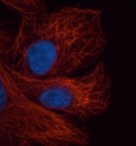
Alpha Tubulin (DM1A) in U-251 MG Human Cell Line -
Alpha Tubulin (DM1A) was detected in immersion fixed U-251 MG human glioblastoma cell line using Mouse anti-alpha Tubulin (DM1A) Protein-G purified Monoclonal Antibody conjugated to Alexa Fluor® 647 (Catalog # NB100-690AF647) (light blue) at 2 µg/mL overnight at 4C. Cells were counterstained with DAPI (dark blue). Cells were imaged using 100X objective and digitally deconvolved. [IMG-80196] [NB100-690]")
Immunohistochemistry-Paraffin: Mouse Monoclonal alpha Tubulin Antibody (DM1A) [IMG-80196] [NB100-690]
Immunohistochemistry-Paraffin: Mouse Monoclonal alpha Tubulin Antibody (DM1A) [IMG-80196] [NB100-690] - Immunofluorescence staining of human tonsil FFPE tissue in a dilution of 1:50 (Catalog # NB100-690AF488) in 3% BSA with overnight incubation at 4°C. Heat mediated antigen retrieval at pH 9. Image from a verified customer review. in U-251 MG Human Cell Line -")
Alpha Tubulin (DM1A) in U-251 MG Human Cell Line -
Alpha Tubulin (DM1A) was detected in immersion fixed U-251 MG human glioblastoma cell line using Mouse anti-alpha Tubulin (DM1A) Protein-G purified Monoclonal Antibody conjugated to Alexa Fluor® 647 (Catalog # NB100-690AF647) (light blue) at 2 µg/mL overnight at 4C. Cells were counterstained with DAPI (dark blue). Cells were imaged using 100X objective and digitally deconvolved. in FR Rat Cell Line.")
alpha Tubulin (DM1A) in FR Rat Cell Line.
alpha Tubulin (DM1A) was detected in immersion FR rat skin fibroblast cell line using Mouse anti-alpha Tubulin (DM1A) Protein G Purified Monoclonal Antibody conjugated to Alexa Fluor® 647 (Catalog # NB100-690AF647) (light blue) at 2 µg/mL overnight at 4C. Cells were counterstained with DAPI (dark blue). Cells were imaged using a 100X objective and digitally deconvolved. in U-251 MG Human Cell Line by Flow Cytometry.")
Detection of alpha Tubulin (DM1A) in U-251 MG Human Cell Line by Flow Cytometry.
U-251 MG human glioblastoma cell line was stained with Mouse anti-alpha Tubulin (DM1A) Protein-G purified Monoclonal Antibody conjugated to Alexa Fluor® 647 (Catalog # NB100-690AF647, blue histogram) or matched control antibody (orange histogram). - BSA Free [NB100-690] -")
Western Blot: alpha Tubulin Antibody (DM1A) - BSA Free [NB100-690] -
Western Blot: alpha Tubulin Antibody (DM1A) - BSA Free [NB100-690] - MiR-375-3p negatively regulates Derlin-1 & blocks EMT in BFTC909 cells. (A) Western blot revealed the restoration of Derlin-1, MMP-2, Snail, & ZEB1 after co-transfection of miR-375-3p mimics & CMV-Derlin-1 compared with cells transfected with miR-375-3p alone in BFTC909 cells with alpha -tubulin as a reference (B) Quantification of the protein levels of Derlin-1, occludin, MMP-2, Snail, & ZEB1 from (A) (N = 3). (C) miR-375-3p suppressed BFTC909 cell migration ability but restored by Derlin-1 overexpression (N = 3). (D) miR-375-3p repressed invasion of BFTC909 cells but restored by Derlin-1 overexpression (N = 3). Data were represented as mean ± SD; * p < 0.05, ** p < 0.01. Image collected & cropped by CiteAb from the following publication (https://pubmed.ncbi.nlm.nih.gov/35205628), licensed under a CC-BY license. Not internally tested by Novus Biologicals. - BSA Free [NB100-690] -")
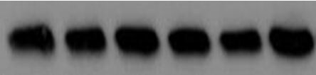
Western Blot: alpha Tubulin Antibody (DM1A) - BSA Free [NB100-690] -
Western Blot: alpha Tubulin Antibody (DM1A) - BSA Free [NB100-690] - Nifedipine stimulated tremendous production of reactive oxygen species (ROS), & KIM-1 in 24 & 48 h. (a) Nifedipine 30 µM-treated group had induced a higher ROS (3.3-fold vs. control, p < 0.01) compared to H2O2 500 μM. (2.7-fold vs. control, p < 0.01). (b,c) Nifedipine 7.5, 15, & 30 μM-treated groups for 24 h (tubulin as internal control) had upregulated KIM-1 in dose dependent fashion (101%, 102%, p < 0.05, & 122%, p < 0.01 respectively) & reduced to 86%, 91%, & 80% in 48 h (actin as internal control), respectively. p-values ≤ 0.05 (marked as *) were considered statistically significant. In addition, p-values ≤ 0.01 are marked as **. Image collected & cropped by CiteAb from the following publication (https://pubmed.ncbi.nlm.nih.gov/30934807), licensed under a CC-BY license. Not internally tested by Novus Biologicals. - BSA Free [NB100-690] -")
Western Blot: alpha Tubulin Antibody (DM1A) - BSA Free [NB100-690] -
Western Blot: alpha Tubulin Antibody (DM1A) - BSA Free [NB100-690] - Pre-treatment with 0.5 mM sodium arsenite (SA) enhances permissivity in a cell-type-specific manner across reovirus strains. (A) CV-1, HeLa, L929, or HPDE cells were left untreated (no SA) or were treated with 0.5 mM SA for 30 min prior to infection (Pre-SA). Following this, cells were infected with T3D such that ~20% to 50% of cells were infected & at 18 h p.i. cells were fixed & immunostained for μNS & DAPI to visualize viral factories (VFs). The percent of cells containing VFs was quantified ((# of cells containing VFs/total # of cells) × 100) from three independent experiments. The expression level of μNS (B) & μ1 (C) was determined in CV-1, L929, or HeLa cells either left untreated (no SA) or treated with 0.5 mM SA for 30 min (Pre-SA) before infection with T3D at MOI = 1. At 18 h p.i., cells were harvested & the expression level of the indicated proteins was determined by immunoblot. M = mock. Densitometry analysis of the band intensity for μNS & μ1 was adjusted to the matched alpha -tubulin loading control for two independent experiments. Columns represent mean ± SEM. (D) CV-1; (E) L929; or (F) HeLa cells were left untreated (no SA) or were treated with 0.5 mM SA prior to infection (Pre-SA). Cells were then infected with the reovirus strains, T3D, T1L, or T3A, as described in (A). At 18 h p.i., cells were fixed & immunostained for μNS & DAPI to detect VFs. The percent of cells containing VFs was quantified ((# of cells containing VFs/total # of cells) × 100) from at least two independent experiments. * p < 0.05; ** p < 0.01; two-tailed unpaired t test. The error bars indicate S.D. Image collected & cropped by CiteAb from the following publication (https://pubmed.ncbi.nlm.nih.gov/31216693), licensed under a CC-BY license. Not internally tested by Novus Biologicals. - BSA Free [NB100-690] -")
Western Blot: alpha Tubulin Antibody (DM1A) - BSA Free [NB100-690] -
Western Blot: alpha Tubulin Antibody (DM1A) - BSA Free [NB100-690] - Simvastatin increases cytotoxicity in lung cancer cells. (A) Relative survival (%) in lung cancer cells treated with simvastatin for 48 h using 3-[4,5-dimethylthiazol-2-yl]-2,5 diphenyl tetrazolium bromide (MTT) assays is shown. (B) The half-maximal inhibitory concentration (IC50) of simvastatin is summarized, & western blots of p53 in low-invasive CL1-0 & high-invasive Bm7 cells are shown with elongation factor 1 alpha (EF1 alpha ) used as a loading control. (C) Z score of statins as well as simvastatin in lung cancer cell lines from NCI-DTP database, z score > 0 for sensitive & <0 resistant. (D) Apoptotic H1299 (null p53), A549 (wild type p53), Bm7-shGFP (mutant p53), & Bm7-shTP53 (knock-down p53) cells treated with simvastatin were detected using flow cytometry, *P < 0.05 & **P < 0.01. (E) Apoptotic HCC827-shGFP (mutant p53) & HCC827-shTP53 (knock-down p53) cells treated with simvastatin & cisplatin were detected using flow cytometry, *P < 0.05 & **P < 0.01. (F) Western blots of indicated proteins involved in apoptosis & autophagy in both Bm7 & HCC827 cells with control (shGFP) & p53 knockdown (shTP53) treated with simvastatin is shown. MDM2, murine double minute 2; AKT, serine–threonine kinase; PARP, poly (ADP-ribose) polymerase; mTOR, mammalian target of rapamycin; WT, wild type. Full-length blots/gels are presented in Supplementary Fig. 1. Image collected & cropped by CiteAb from the following publication (https://pubmed.ncbi.nlm.nih.gov/31892709), licensed under a CC-BY license. Not internally tested by Novus Biologicals. - BSA Free [NB100-690] -")
Western Blot: alpha Tubulin Antibody (DM1A) - BSA Free [NB100-690] -
Western Blot: alpha Tubulin Antibody (DM1A) - BSA Free [NB100-690] - Pre-treatment with 0.5 mM sodium arsenite (SA) enhances permissivity in a cell-type-specific manner across reovirus strains. (A) CV-1, HeLa, L929, or HPDE cells were left untreated (no SA) or were treated with 0.5 mM SA for 30 min prior to infection (Pre-SA). Following this, cells were infected with T3D such that ~20% to 50% of cells were infected & at 18 h p.i. cells were fixed & immunostained for μNS & DAPI to visualize viral factories (VFs). The percent of cells containing VFs was quantified ((# of cells containing VFs/total # of cells) × 100) from three independent experiments. The expression level of μNS (B) & μ1 (C) was determined in CV-1, L929, or HeLa cells either left untreated (no SA) or treated with 0.5 mM SA for 30 min (Pre-SA) before infection with T3D at MOI = 1. At 18 h p.i., cells were harvested & the expression level of the indicated proteins was determined by immunoblot. M = mock. Densitometry analysis of the band intensity for μNS & μ1 was adjusted to the matched alpha -tubulin loading control for two independent experiments. Columns represent mean ± SEM. (D) CV-1; (E) L929; or (F) HeLa cells were left untreated (no SA) or were treated with 0.5 mM SA prior to infection (Pre-SA). Cells were then infected with the reovirus strains, T3D, T1L, or T3A, as described in (A). At 18 h p.i., cells were fixed & immunostained for μNS & DAPI to detect VFs. The percent of cells containing VFs was quantified ((# of cells containing VFs/total # of cells) × 100) from at least two independent experiments. * p < 0.05; ** p < 0.01; two-tailed unpaired t test. The error bars indicate S.D. Image collected & cropped by CiteAb from the following publication (https://pubmed.ncbi.nlm.nih.gov/31216693), licensed under a CC-BY license. Not internally tested by Novus Biologicals. in U-2 OS Human Cell Line.")
Alpha Tubulin (DM1A) in U-2 OS Human Cell Line.
Alpha Tubulin (DM1A) was detected in immersion fixed U-2 OS human osteosarcoma cell line using Mouse anti-Alpha Tubulin (DM1A) Protein G Purified Monoclonal Antibody conjugated to Alexa Fluor ® 488 (Catalog # NB100-690AF488) (green) at 2 µg/mL overnight at 4C. Cells were counterstained with DAPI (dark blue). Cells were imaged using a 100X objective and digitally deconvolved. in U-2 OS Human Cell Line by Flow Cytometry.")
Detection of alpha Tubulin (DM1A) in U-2 OS Human Cell Line by Flow Cytometry.
An intracellular stain was performed on U-2 OS human osteosarcoma cell line with Mouse anti-alpha Tubulin (DM1A) Protein-G purified Monoclonal Antibody conjugated to Alexa Fluor ® 488 (Catalog # NB100-690AF488, blue histogram) or matched control antibody (orange histogram) at 5 µg/mL for 30 minutes at RT. in A431 Human Cell Line by Flow Cytometry.")
Detection of alpha Tubulin (DM1A) in A431 Human Cell Line by Flow Cytometry.
An intracellular stain was performed on A431 human skin carcinoma cell line using Mouse anti-alpha Tubulin (DM1A) Protein-G purified Monoclonal Antibody conjugated to Alexa Fluor ® 647 (Catalog # NB100-690AF647, blue histogram) or matched control antibody (orange histogram) at 2.5 µg/mL for 30 minutes at RT. in NIH-3T3 Mouse Cell Line.")
Alpha Tubulin (DM1A) in NIH-3T3 Mouse Cell Line.
Alpha Tubulin (DM1A) was detected in immersion fixed NIH-3T3 Mouse fibroblast cell line using Mouse anti-Alpha Tubulin (DM1A) Protein G Purified Monoclonal Antibody conjugated to Alexa Fluor® 488 (Catalog # NB100-690AF488) (green) at 2 µg/mL overnight at 4C. Cells were counterstained with DAPI (dark blue). Cells were imaged using a 100X objective and digitally deconvolved. in U-2 OS Human Cell Line.")
Alpha Tubulin (DM1A) in U-2 OS Human Cell Line.
Alpha Tubulin (DM1A) was detected in immersion fixed U-2 OS human osteosarcoma cell line using Mouse anti-Alpha Tubulin (DM1A) Protein G Purified Monoclonal Antibody conjugated to Biotin (Catalog # NB100-690B) at 2 µg/mL overnight at 4C. Cells were stained using Streptavidin conjugated to DyLight 550 (red) and counterstained with DAPI (blue). Cells were imaged using a 100X objective and digitally deconvolved.Applications for alpha Tubulin Antibody (DM1A) - BSA Free
Application
Recommended Usage
Flow Cytometry
1 ug per million cells
Immunocytochemistry/ Immunofluorescence
1:50000-1:100000
Immunohistochemistry
1:100-1:500
Immunohistochemistry-Frozen
1:100-1:500
Immunohistochemistry-Paraffin
1:100-1:500
Immunoprecipitation
1:50-1:100
Simple Western
1:50
Western Blot
1:5000
Application Notes
This alpha Tubulin Antibody (DM1A) is useful as a loading control for Western blot as well as Immunoprecipitation, Immunohistochemistry on paraffin-embedded and frozen sections, Immunocytochemistry/Immunofluorescence and Flow Cytometry.
The DM1A alpha tubulin antibody is ideal for use as a Western blot loading control, where a band can be seen around 50-55 kDa and as a cytoskeletal marker in ICC. For IHC-Paraffin, antigen retrieval is not essential, but may optimize staining.
See Simple Western Antibody Database for Simple Western validation: tested in HeLa lysate (1.0 mg/ml), titrated to saturation using various models; separated by Size-Jess/Wes, Sally Sue/Peggy Sue; separated by size; antibody dilution at 1:50, 6 ug/ml; detects a band at 55 kDa; matrix was 12-230 kDa. Only 10 - 15 ul of the recommended dilution is used per data point.
This antibody is CyTOF ready.
Reviewed Applications
Read 14 reviews rated 4.6 using NB100-690 in the following applications:
Flow Cytometry Panel Builder
Bio-Techne Knows Flow Cytometry
Save time and reduce costly mistakes by quickly finding compatible reagents using the Panel Builder Tool.
Advanced Features
- Spectra Viewer - Custom analysis of spectra from multiple fluorochromes
- Spillover Popups - Visualize the spectra of individual fluorochromes
- Antigen Density Selector - Match fluorochrome brightness with antigen density

Formulation, Preparation, and Storage
Purification
Protein G purified
Formulation
PBS
Format
BSA Free
Preservative
0.05% Sodium Azide
Concentration
1.0 mg/ml
Shipping
The product is shipped with polar packs. Upon receipt, store it immediately at the temperature recommended below.
Stability & Storage
Store at 4C short term. Aliquot and store at -20C long term. Avoid freeze-thaw cycles.
Background: alpha Tubulin
Tyrosine ligase adds a C-terminal tyrosine to monomeric alpha tubulin. Assembled microtubules can again be detyrosinated by a cytoskeleton associated carboxypeptidase. Detyrosinated alpha tubulin is referred to as Glu-tubulin. Another post-translational modification of detyrosinated alpha tubulin is C-terminal polyglutamylation which is characteristic for microtubules in neuronal cells and the mitotic spindle.
Like GAPDH and beta-actin, alpha/beta tubulin is often used as a loading control in immunoblot applications (1). Alpha/beta tubulin is also good for counterstaining microtubules in immunofluorescence (2).
References
1. Hannen, R., Selmansberger, M., Hauswald, M., Pagenstecher, A., Nist, A., Stiewe, T.,... Bartsch, J. W. (2019). Comparative Transcriptomic Analysis of Temozolomide Resistant Primary GBM Stem-Like Cells and Recurrent GBM Identifies Up-Regulation of the Carbonic Anhydrase CA2 Gene as Resistance Factor. Cancers (Basel), 11(7). doi:10.3390/cancers11070921
2. Nel, M., Joubert, A. M., Dohle, W., Potter, B. V., & Theron, A. E. (2018). Modes of cell death induced by tetrahydroisoquinoline-based analogs in MDA-MB-231 breast and A549 lung cancer cell lines. Drug Des Devel Ther, 12, 1881-1904. doi:10.2147/dddt.S152718
Long Name
Tubulin Alpha 1a
Alternate Names
Alpha-Tubulin 3, B-ALPHA-1, LIS3, TUBA1A, TUBA3, Tubulin B-Alpha-1, alpha tubulin loading control, alpha tubulin monoclonal, DM1A, DM1A anti-tubulin, dm1a monoclonal, DM1a tubulin, TUBA1 monoclonal, tubulin monoclonal
Entrez Gene IDs
7846 (Human)
Gene Symbol
TUBA1A
Additional alpha Tubulin Products
Product Documents for alpha Tubulin Antibody (DM1A) - BSA Free
Certificate of Analysis
To download a Certificate of Analysis, please enter a lot or batch number in the search box below.
Product Specific Notices for alpha Tubulin Antibody (DM1A) - BSA Free
This product is for research use only and is not approved for use in humans or in clinical diagnosis. Primary Antibodies are guaranteed for 1 year from date of receipt.
Related Research Areas
Citations for alpha Tubulin Antibody (DM1A) - BSA Free
 Powered by Bioz
Customer Reviews for alpha Tubulin Antibody (DM1A) - BSA Free (14)
4.6 out of 5
14 Customer Ratings
Have you used alpha Tubulin Antibody (DM1A) - BSA Free?
Submit a review and receive an Amazon gift card!
$25/€18/£15/$25CAN/¥2500 Yen for a review with an image
$10/€7/£6/$10CAN/¥1110 Yen for a review without an image
Submit a review
Customer Images
-(01-ml)_NB100-690_8001.jpg)
-(01-ml)_NB100-690_7996.bmp)
Showing
1
-
5 of
14 reviews
Showing All
Filter By:
-
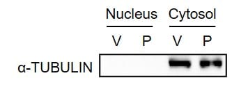
Application: Western BlotSample Tested: Breast cancer cellsSpecies: HumanVerified Customer | Posted 05/18/2020MDA-MB-231 cells were treated with vehicle (V) or paclitaxel (P). Cytosolic and nuclear lysates were prepared, and immunoblot assay was performed.

-
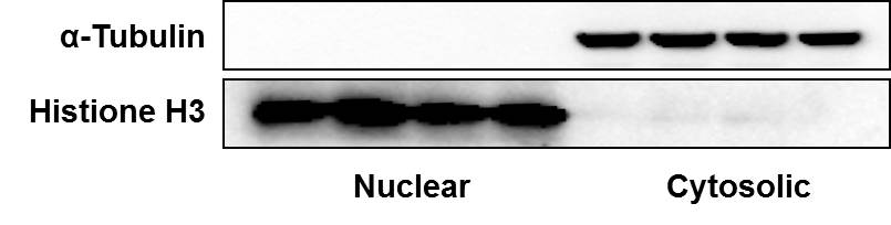
Application: Western BlotSample Tested: Human breast cancer cell linesSpecies: HumanVerified Customer | Posted 10/22/2018Lysates from human breast cancer cells were separated into nuclear and cytosolic proteins and antibodies against alpha-tubulin and Histone H3 were used as markers.
-
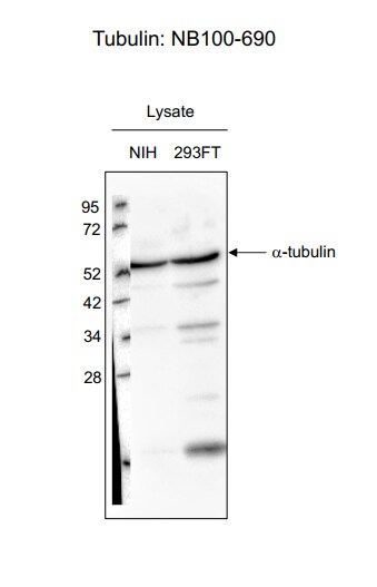
Application: Western BlotSample Tested: 293T cell lysate and nih3t3 cell lysateSpecies: Human and MouseVerified Customer | Posted 06/29/2017
-
Application: Western BlotSample Tested: African Green Monkey Kidney (CV-1) whole cell lysateSpecies: OtherVerified Customer | Posted 07/15/2016a-Tubulin expression in CV-1 cells REPROBE
-
Application: Western BlotSample Tested:Species: HumanVerified Customer | Posted 06/03/2014Western blot analysis of extracts from HeLa, COS and C6 cells using alpha Tubulin antibody (NB100-690, 1:1000).
-
Application: ImmunocytochemistrySample Tested:Species: HumanVerified Customer | Posted 06/03/2014IF Confocal analysis of C6 cells using alpha Tubulin antibody (NB100-690, 1:50).
-
Application: ImmunocytochemistrySample Tested: MDA-MB-231 cellsSpecies: HumanVerified Customer | Posted 11/06/2013MDA-MB-231 cells stained with anti-alpha tubulin
-
Application: Western BlotSample Tested: mouse lung tissueSpecies: MouseVerified Customer | Posted 01/06/2012
-
Application: Western BlotSample Tested: Cell lysateSpecies: MouseVerified Customer | Posted 12/05/2011
-
Application: Western BlotSample Tested: HumanSpecies: HumanVerified Customer | Posted 11/23/2011
-
Application: Western BlotSample Tested: Human cellSpecies: HumanVerified Customer | Posted 11/23/2011
-
Application: Western BlotSample Tested: HumanSpecies: HumanVerified Customer | Posted 11/21/2011
-
Application: Western BlotSample Tested: Human cellSpecies: HumanVerified Customer | Posted 11/18/2011
-
Application: Western BlotSample Tested: HumanSpecies: HumanVerified Customer | Posted 10/31/2011
There are no reviews that match your criteria.
Protocols
Find general support by application which include: protocols, troubleshooting, illustrated assays, videos and webinars.
- 7-Amino Actinomycin D (7-AAD) Cell Viability Flow Cytometry Protocol
- Antigen Retrieval Protocol (PIER)
- Antigen Retrieval for Frozen Sections Protocol
- Appropriate Fixation of IHC/ICC Samples
- Cellular Response to Hypoxia Protocols
- Chromogenic IHC Staining of Formalin-Fixed Paraffin-Embedded (FFPE) Tissue Protocol
- Chromogenic Immunohistochemistry Staining of Frozen Tissue
- ClariTSA™ Fluorophore Kits
- Detection & Visualization of Antibody Binding
- Extracellular Membrane Flow Cytometry Protocol
- Flow Cytometry Protocol for Cell Surface Markers
- Flow Cytometry Protocol for Staining Membrane Associated Proteins
- Flow Cytometry Staining Protocols
- Flow Cytometry Troubleshooting Guide
- Fluorescent IHC Staining of Frozen Tissue Protocol
- Graphic Protocol for Heat-induced Epitope Retrieval
- Graphic Protocol for the Preparation and Fluorescent IHC Staining of Frozen Tissue Sections
- Graphic Protocol for the Preparation and Fluorescent IHC Staining of Paraffin-embedded Tissue Sections
- Graphic Protocol for the Preparation of Gelatin-coated Slides for Histological Tissue Sections
- ICC Cell Smear Protocol for Suspension Cells
- ICC Immunocytochemistry Protocol Videos
- ICC for Adherent Cells
- IHC Sample Preparation (Frozen sections vs Paraffin)
- Immunocytochemistry (ICC) Protocol
- Immunocytochemistry Troubleshooting
- Immunofluorescence of Organoids Embedded in Cultrex Basement Membrane Extract
- Immunofluorescent IHC Staining of Formalin-Fixed Paraffin-Embedded (FFPE) Tissue Protocol
- Immunohistochemistry (IHC) and Immunocytochemistry (ICC) Protocols
- Immunohistochemistry Frozen Troubleshooting
- Immunohistochemistry Paraffin Troubleshooting
- Immunoprecipitation Protocol
- Intracellular Flow Cytometry Protocol Using Alcohol (Methanol)
- Intracellular Flow Cytometry Protocol Using Detergents
- Intracellular Nuclear Staining Flow Cytometry Protocol Using Detergents
- Intracellular Staining Flow Cytometry Protocol Using Alcohol Permeabilization
- Intracellular Staining Flow Cytometry Protocol Using Detergents to Permeabilize Cells
- Preparing Samples for IHC/ICC Experiments
- Preventing Non-Specific Staining (Non-Specific Binding)
- Primary Antibody Selection & Optimization
- Propidium Iodide Cell Viability Flow Cytometry Protocol
- Protocol for Heat-Induced Epitope Retrieval (HIER)
- Protocol for Liperfluo
- Protocol for Making a 4% Formaldehyde Solution in PBS
- Protocol for VisUCyte™ HRP Polymer Detection Reagent
- Protocol for the Characterization of Human Th22 Cells
- Protocol for the Characterization of Human Th9 Cells
- Protocol for the Fluorescent ICC Staining of Cell Smears - Graphic
- Protocol for the Fluorescent ICC Staining of Cultured Cells on Coverslips - Graphic
- Protocol for the Preparation & Fixation of Cells on Coverslips
- Protocol for the Preparation and Chromogenic IHC Staining of Frozen Tissue Sections
- Protocol for the Preparation and Chromogenic IHC Staining of Frozen Tissue Sections - Graphic
- Protocol for the Preparation and Chromogenic IHC Staining of Paraffin-embedded Tissue Sections
- Protocol for the Preparation and Chromogenic IHC Staining of Paraffin-embedded Tissue Sections - Graphic
- Protocol for the Preparation and Fluorescent ICC Staining of Cells on Coverslips
- Protocol for the Preparation and Fluorescent ICC Staining of Non-adherent Cells
- Protocol for the Preparation and Fluorescent ICC Staining of Stem Cells on Coverslips
- Protocol for the Preparation and Fluorescent IHC Staining of Frozen Tissue Sections
- Protocol for the Preparation and Fluorescent IHC Staining of Paraffin-embedded Tissue Sections
- Protocol for the Preparation of Gelatin-coated Slides for Histological Tissue Sections
- Protocol for the Preparation of a Cell Smear for Non-adherent Cell ICC - Graphic
- Protocol: Annexin V and PI Staining by Flow Cytometry
- Protocol: Annexin V and PI Staining for Apoptosis by Flow Cytometry
- R&D Systems Quality Control Western Blot Protocol
- TUNEL and Active Caspase-3 Detection by IHC/ICC Protocol
- The Importance of IHC/ICC Controls
- Troubleshooting Guide: Fluorokine Flow Cytometry Kits
- Troubleshooting Guide: Immunohistochemistry
- Troubleshooting Guide: Western Blot Figures
- Western Blot Conditions
- Western Blot Protocol
- Western Blot Protocol for Cell Lysates
- Western Blot Troubleshooting
- Western Blot Troubleshooting Guide
- View all Protocols, Troubleshooting, Illustrated assays and Webinars
FAQs for alpha Tubulin Antibody (DM1A) - BSA Free
Showing
1
-
3 of
3 FAQs
Showing All
-
Q: I am interested in buying an actin or tubulin antibody for Western blot loading control. We would like to use it on rat heart samples. Please recommend us one of your actin and/or tubulin products, and add it to the NOX2 quotation.
A: NB100-690 is for Alpha Tubulin and has been validated for use in WB and Rat. This will be added to the quote along with our Actin Antibody.
-
Q: I am working on a dissection fixation protocol for a non model organism, and I am using DM1A alpha tubulin as a control. I do not see any sort of Astral projections; does this convince you that it is a working stain ?
A: The staining looks a little off to my eye. What were your fixation conditions? Tubulin immunostaining can be finicky and requires particular fixation conditions for optimal labeling. Depending on your other antigenic sites, it's ideal to use either mixed aldehydes (PFA and Glutaraldehyde) with simultaneous extraction or methanol. PFA alone gives an undesirable hazy staining pattern. Some laboratories also recommend adding a calcium chelator into the fixation buffer as calcium induces depolymerization of microtubules. Lastly, be sure to run negative controls with your staining.
-
Q: We would like to stain cilia with an acetylated alpha tubulin antibody in our cells, but I am unsure if this antibody will be able to conclusively differentiate cilia from other structures such as spindle pole bodies. Does anyone know what acetylated alpha tubulin antibodies might bind to apart from cilia?
A: Acetylated alpha tubulin is found in relatively stable microtubules. It is best practice to use this marker together with a centrosome/centriole marker, which will stain the basal bodies at the base of the cilium. After that, it is relatively straightforward to identify the acetylated alpha tubulin signal that corresponds to the cilium.
-
Q: I am interested in buying an actin or tubulin antibody for Western blot loading control. We would like to use it on rat heart samples. Please recommend us one of your actin and/or tubulin products, and add it to the NOX2 quotation.
A: NB100-690 is for Alpha Tubulin and has been validated for use in WB and Rat. This will be added to the quote along with our Actin Antibody.
-
Q: I am working on a dissection fixation protocol for a non model organism, and I am using DM1A alpha tubulin as a control. I do not see any sort of Astral projections; does this convince you that it is a working stain ?
A: The staining looks a little off to my eye. What were your fixation conditions? Tubulin immunostaining can be finicky and requires particular fixation conditions for optimal labeling. Depending on your other antigenic sites, it's ideal to use either mixed aldehydes (PFA and Glutaraldehyde) with simultaneous extraction or methanol. PFA alone gives an undesirable hazy staining pattern. Some laboratories also recommend adding a calcium chelator into the fixation buffer as calcium induces depolymerization of microtubules. Lastly, be sure to run negative controls with your staining.
-
Q: We would like to stain cilia with an acetylated alpha tubulin antibody in our cells, but I am unsure if this antibody will be able to conclusively differentiate cilia from other structures such as spindle pole bodies. Does anyone know what acetylated alpha tubulin antibodies might bind to apart from cilia?
A: Acetylated alpha tubulin is found in relatively stable microtubules. It is best practice to use this marker together with a centrosome/centriole marker, which will stain the basal bodies at the base of the cilium. After that, it is relatively straightforward to identify the acetylated alpha tubulin signal that corresponds to the cilium.
Loading...