Ki67/MKI67 Antibody (1297A) - BSA Free
Novus Biologicals | Catalog # NBP2-54791
Recombinant Monoclonal Antibody
Key Product Details
Validated by
Knockout/Knockdown
Species Reactivity
Validated:
Human, Mouse
Cited:
Human, Mouse
Applications
Validated:
Knockout Validated, Immunohistochemistry, Immunohistochemistry-Paraffin, Flow Cytometry, Flow (Intracellular), Immunocytochemistry/ Immunofluorescence
Cited:
Immunohistochemistry, Western Blot, Flow Cytometry, IF/IHC
Label
Unconjugated
Antibody Source
Recombinant Monoclonal Rabbit IgG Clone # 1297A expressed in HEK293
Format
BSA Free
Loading...
Product Specifications
Immunogen
The immunogen for this KI67/MKI67 Antibody (1297A) was made using a synthetic peptide from the internal portion of Mouse KI67/MKI67, between amino acids 1850-1950 [UniProt# E9PVX6].
Reactivity Notes
Use in Mouse reported in scientific literature (PMID:33536573).
Clonality
Monoclonal
Host
Rabbit
Isotype
IgG
Theoretical MW
351 kDa.
Disclaimer note: The observed molecular weight of the protein may vary from the listed predicted molecular weight due to post translational modifications, post translation cleavages, relative charges, and other experimental factors.
Disclaimer note: The observed molecular weight of the protein may vary from the listed predicted molecular weight due to post translational modifications, post translation cleavages, relative charges, and other experimental factors.
Scientific Data Images for Ki67/MKI67 Antibody (1297A) - BSA Free
![Immunocytochemistry/ Immunofluorescence: Ki67/MKI67 Antibody (1297A) - BSA Free [NBP2-54791]](https://resources.rndsystems.com/images/products/Ki67-MKI67-Antibody-1297A-Immunocytochemistry-Immunofluorescence-NBP2-54791-img0007.jpg "Immunocytochemistry/ Immunofluorescence: Ki67/MKI67 Antibody (1297A) - BSA Free [NBP2-54791]")
Immunocytochemistry/ Immunofluorescence: Ki67/MKI67 Antibody (1297A) - BSA Free [NBP2-54791]
Immunocytochemistry/Immunofluorescence: Ki67/MKI67 Antibody (1297A) [NBP2-54791] - A431 cells were fixed in 4% paraformaldehyde for 10 minutes and permeabilized in 0.5% Triton X-100 in PBS for 5 minutes. The cells were incubated with anti-Ki67/MKI67 Antibody (1297A) NBP2-54791 at 2 ug/ml overnight at 4C and detected with an anti-rabbit Dylight 488 (Green) at a 1:1000 dilution for 60 minutes. Alpha tubulin (DM1A) NB100-690 was used as a co-stain at a 1:1000 dilution and detected with an anti-mouse Dylight 550 (Red) at a 1:1000 dilution. Nuclei were counterstained with DAPI (Blue). Cells were imaged using a 100X objective and digitally deconvolved.![Knockout Validated: Ki67/MKI67 Antibody (1297A) - BSA Free [NBP2-54791]](https://resources.rndsystems.com/images/products/Ki67-MKI67-Antibody-1297A-Knockout-Validated-NBP2-54791-img0006.jpg "Knockout Validated: Ki67/MKI67 Antibody (1297A) - BSA Free [NBP2-54791]")
Knockout Validated: Ki67/MKI67 Antibody (1297A) - BSA Free [NBP2-54791]
Knockout Validated: Ki67/MKI67 Antibody (1297A) [NBP2-54791] - Ki67/MKI67 was detected in immersion fixed HeLa human cervical epithelial carcinoma cell line but is not detected in Ki67/MKI67 knockout (KO) HeLa cell line using Rabbit Anti-Human Ki67/MKI67 Monoclonal Antibody (Catalog # NBP2-54791) at 1 ug/mL for 3 hours at room temperature. Cells were stained using the NorthernLights(TM) 557-conjugated Anti-Rabbit IgG Secondary Antibody (red; Catalog # NL004) and counterstained with DAPI (blue). Specific staining was localized to nuclei.![Immunocytochemistry/ Immunofluorescence: Ki67/MKI67 Antibody (1297A) - BSA Free [NBP2-54791]](https://resources.rndsystems.com/images/products/Ki67-MKI67-Antibody-1297A-Immunocytochemistry-Immunofluorescence-NBP2-54791-img0009.jpg "Immunocytochemistry/ Immunofluorescence: Ki67/MKI67 Antibody (1297A) - BSA Free [NBP2-54791]")
Immunocytochemistry/ Immunofluorescence: Ki67/MKI67 Antibody (1297A) - BSA Free [NBP2-54791]
Immunocytochemistry/Immunofluorescence: Ki67/MKI67 Antibody (1297A) [NBP2-54791] - NIH3T3 cells were fixed in 4% paraformaldehyde for 10 minutes and permeabilized in 0.5% Triton X-100 in PBS for 5 minutes. The cells were incubated with anti- NBP2-54791 at 2 ug/ml overnight at 4C and detected with an anti-rabbit Dylight 488 (Green) at a 1:1000 dilution for 60 minutes. Alpha tubulin (DM1A) NB100-690 was used as a co-stain at a 1:1000 dilution and detected with an anti-mouse Dylight 550 (Red) at a 1:1000 dilution. Nuclei were counterstained with DAPI (Blue). Cells were imaged using a 100X objective and digitally deconvolved.![Immunocytochemistry/ Immunofluorescence: Ki67/MKI67 Antibody (1297A) - BSA Free [NBP2-54791]](https://resources.rndsystems.com/images/products/Ki67-MKI67-Antibody-1297A-Immunocytochemistry-Immunofluorescence-NBP2-54791-img0010.jpg "Immunocytochemistry/ Immunofluorescence: Ki67/MKI67 Antibody (1297A) - BSA Free [NBP2-54791]")
Immunocytochemistry/ Immunofluorescence: Ki67/MKI67 Antibody (1297A) - BSA Free [NBP2-54791]
Immunocytochemistry/Immunofluorescence: Ki67/MKI67 Antibody (1297A) [NBP2-54791] - HeLa cells were fixed in 4% paraformaldehyde for 10 minutes and permeabilized in 0.5% Triton X-100 in PBS for 5 minutes. The cells were incubated with Ki67/MKI67 Antibody [1297A] conjugated to Alexa Fluor 647 (NBP2-54791AF647) at 2 ug/ml for 1 hour at room temperature. Nuclei were counterstained with DAPI (Blue). Cells were imaged using a 40X objective.![Immunohistochemistry-Paraffin: Ki67/MKI67 Antibody (1297A) - BSA Free [NBP2-54791]](https://resources.rndsystems.com/images/products/Ki67-MKI67-Antibody-1297A-Immunohistochemistry-Paraffin-NBP2-54791-img0002.jpg "Immunohistochemistry-Paraffin: Ki67/MKI67 Antibody (1297A) - BSA Free [NBP2-54791]")
Immunohistochemistry-Paraffin: Ki67/MKI67 Antibody (1297A) - BSA Free [NBP2-54791]
Immunohistochemistry-Paraffin: Ki67/MKI67 Antibody (1297A) [NBP2-54791] - Ki-67/MKI67 was detected in immersion fixed paraffin-embedded sections of human liver cancer tissue using Rabbit Anti-Human Ki-67/MKI67 Monoclonal Antibody at 3 µg/mL for 1 hour at room temperature followed by incubation with the Anti-Rabbit IgG VisUCyte™ HRP Polymer Antibody. Tissue was stained using DAB (brown) and counterstained with hematoxylin (blue). Specific staining was localized to nuclei. Staining with VisUCyte HRP Polymer Detection Reagents.![Flow (Intracellular): Ki67/MKI67 Antibody (1297A) - BSA Free [NBP2-54791]](https://resources.rndsystems.com/images/products/Ki67-MKI67-Antibody-1297A-Flow-Intracellular-NBP2-54791-img0004.jpg "Flow (Intracellular): Ki67/MKI67 Antibody (1297A) - BSA Free [NBP2-54791]")
Flow (Intracellular): Ki67/MKI67 Antibody (1297A) - BSA Free [NBP2-54791]
Flow (Intracellular): Ki67/MKI67 Antibody (1297A) [NBP2-54791] - Ki-67/MKI67 Antibody (1297A) [NBP2-54791] - An intracellular stain was performed on Jurkat Cells with Ki-67/MKI67 (1297A) antibody NBP2-54791 (blue) and a matched isotype control MAB1050 (orange). Cells were fixed with 4% paraformaldehyde, following fixation, cells were permeabilized with 0.1% saponin. Cells were incubated in an antibody dilution of 1 ug/mL for 30 minutes at room temperature, followed by rabbit IgG APC-conjugated secondary antibody (F0111, R&D Systems).![Immunocytochemistry/ Immunofluorescence: Ki67/MKI67 Antibody (1297A) - BSA Free [NBP2-54791]](https://resources.rndsystems.com/images/products/Ki67-MKI67-Antibody-1297A-Immunocytochemistry-Immunofluorescence-NBP2-54791-img0003.jpg "Immunocytochemistry/ Immunofluorescence: Ki67/MKI67 Antibody (1297A) - BSA Free [NBP2-54791]")
Immunocytochemistry/ Immunofluorescence: Ki67/MKI67 Antibody (1297A) - BSA Free [NBP2-54791]
Immunocytochemistry/Immunofluorescence: Ki67/MKI67 Antibody (1297A) [NBP2-54791] - HeLa cells were fixed for 10 minutes using 10% formalin and then permeabilized for 5 minutes using 1X TBS + 0.5% Triton X-100. The cells were incubated with anti-Ki67/MKI67 (1297A) at 2.0 ug/mL overnight at 4C and detected with an anti-rabbit Dylight 488 (Green) at a 1:500 dilution. Alpha tubulin (DM1A) NB100-690 was used as a co-stain at a 1:1000 dilution and detected with an anti-mouse Dylight 550 (Red) at a 1:500 dilution. Nuclei were counterstained with DAPI (Blue). Cells were imaged using a 40X objective.![Flow Cytometry: Ki67/MKI67 Antibody (1297A) - BSA Free [NBP2-54791]](https://resources.rndsystems.com/images/products/Ki67-MKI67-Antibody-1297A-Flow-Cytometry-NBP2-54791-img0005.jpg "Flow Cytometry: Ki67/MKI67 Antibody (1297A) - BSA Free [NBP2-54791]")
Flow Cytometry: Ki67/MKI67 Antibody (1297A) - BSA Free [NBP2-54791]
Flow Cytometry: Ki67/MKI67 Antibody (1297A) [NBP2-54791] - An intracellular stain was performed on HeLa cells with Ki67/MKI67 Antibody [1297A] NBP2-54791APC (blue) and a matched isotype control (orange). Cells were fixed with 4% PFA and then permeabilized with 0.1% saponin. Cells were incubated in an antibody dilution of 2.5 ug/mL for 30 minutes at room temperature. Both antibodies were conjugated to Allophycocyanin. [NBP2-54791] -")
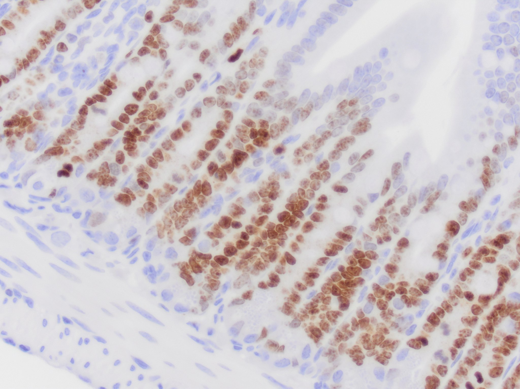
Immunohistochemistry-Paraffin: Ki67/MKI67 Antibody (1297A) [NBP2-54791] -
Immunohistochemistry-Paraffin: Ki67/MKI67 Antibody (1297A) [NBP2-54791] - Ki67/MKI67 immunoreactivity in mouse small intestine. NBP2-54791 was diluted to 0.5ug/mL in Antibody Diluent and was left on sections for 1h at room temperature. Secondary was Horse Anti-Rabbit HRP. Image from verified customer review. in A431 Human Cell Line.")
Ki67/MKI67 (1297A) in A431 Human Cell Line.
Ki67/MKI67 (1297A) was detected in immersion fixed A431 human skin carcinoma cell line using Rabbit anti-Ki67/MKI67 (1297A) Protein G Purified Recombinant Monoclonal Antibody conjugated to Alexa Fluor® 488 (Catalog # NBP2-54791AF488) (green) at 2 µg/mL overnight at 4C. Cells were counterstained with DAPI (blue). Cells were imaged using a 100X objective and digitally deconvolved. in U-2 OS Human Cell Line by Flow Cytometry.")
Detection of Ki67/MKI67 (1297A) in U-2 OS Human Cell Line by Flow Cytometry.
An intracellular stain was performed on U-2 OS human osteosarcoma cell line with Rabbit anti-Ki67/MKI67 (1297A) Protein-G purified Monoclonal Antibody conjugated to Alexa Fluor® 488 (Catalog # NBP2-54791AF488, blue histogram) or matched control antibody (Catalog # NBP2-24982, orange histogram). [NBP2-54791]")
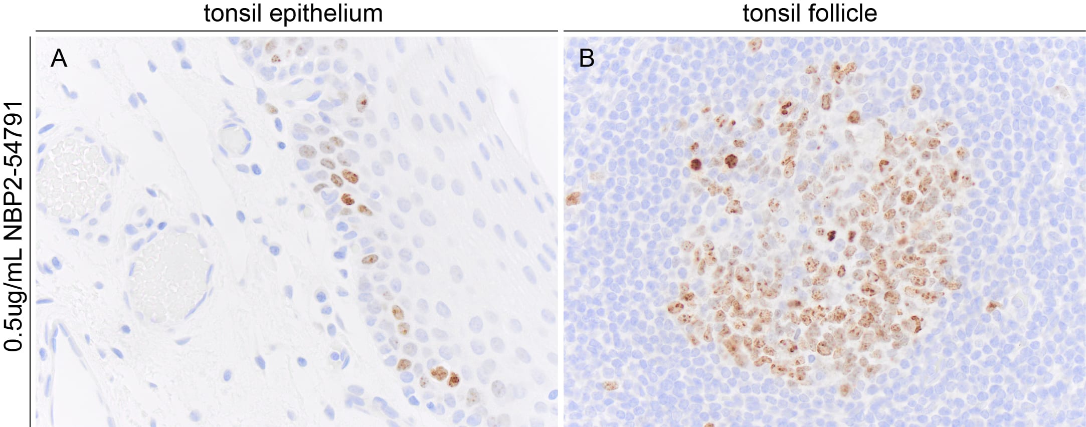
Immunohistochemistry-Paraffin: Rabbit Monoclonal Ki67/MKI67 Antibody (1297A) [NBP2-54791]
Immunohistochemistry-Paraffin: Rabbit Monoclonal Ki67/MKI67 Antibody (1297A) [NBP2-54791] - Ki67 immunoreactivity in FFPE sections of human tonsil. NBP2-54791 was used at 500ng per mL and was left on tissue sections for 1hr at room temperature. Image from a verified customer review. in A431 Human Cell Line.")
Ki67/MKI67 Antibody(1297A) in A431 Human Cell Line.
Ki67/MKI67 (1297A) was detected in immersion fixed A431 human skin carcinoma cell line using Rabbit anti-Ki67/MKI67 (1297A) Protein G Purified Monoclonal Antibody conjugated to FITC (Catalog # NBP2-54791F) (green) at 5 µg/mL overnight at 4C. Cells were counterstained with DAPI (blue). Cells were imaged using a 100X objective and digitally deconvolved.Applications for Ki67/MKI67 Antibody (1297A) - BSA Free
Application
Recommended Usage
Flow (Intracellular)
1 ug/mL
Flow Cytometry
1 ug/mL
Immunocytochemistry/ Immunofluorescence
1-10 ug/mL
Immunohistochemistry
3-15 ug/mL
Immunohistochemistry-Paraffin
3-15 ug/mL
Reviewed Applications
Read 2 reviews rated 5 using NBP2-54791 in the following applications:
Flow Cytometry Panel Builder
Bio-Techne Knows Flow Cytometry
Save time and reduce costly mistakes by quickly finding compatible reagents using the Panel Builder Tool.
Advanced Features
- Spectra Viewer - Custom analysis of spectra from multiple fluorochromes
- Spillover Popups - Visualize the spectra of individual fluorochromes
- Antigen Density Selector - Match fluorochrome brightness with antigen density

Formulation, Preparation, and Storage
Purification
Protein A or G purified
Formulation
PBS
Format
BSA Free
Preservative
0.02% Sodium Azide
Concentration
1.0 mg/ml
Shipping
The product is shipped with polar packs. Upon receipt, store it immediately at the temperature recommended below.
Stability & Storage
Store at 4C short term. Aliquot and store at -20C long term. Avoid freeze-thaw cycles.
Background: Ki67/MKI67
Detection of Ki67 by immunostaining is commonly used as a proliferation marker in solid tumors, as well as certain hematological malignancies (3-5). The Ki67 index, which reports on positive Ki67 stained tumor cell nuclei, has been extensively studied as a prognostic biomarker in cancers such as breast cancer and cervical cancer.
References
1. Gerdes J, Schwab U, Lemke H, Stein H. (1983) Production of a mouse monoclonal antibody reactive with a human nuclear antigen associated with cell proliferation. Int J Cancer. 31:13-20. PMID: 6339421
2. Starborg M, Gell K, Brundell E and Hoog C. (1996) The murine Ki-67 cell proliferation antigen accumulates in the nucleolar and heterochromatic regions of interphase cells and at the periphery of the mitotic chromosomes in a process essential for cell cycle progression. J Cell Sci. 109:143-153. 1996
3. Karamitopoulou E, Perentes E, Tolnay M, Probst A. (1998) Prognostic significance of MIB-1, p53, and bcl-2 immunoreactivity in meningiomas. Hum Pathol. 29(2):140-5. PMID: 9490273
4. Geyer FC, Rodrigues DN, Weigelt B and Reis-Filho JS. (2012) Molecular classification of estrogen receptor-positive/luminal breast cancers. Adv Anat Pathol. 19(1):39-53. PMID: 22156833
5. Ikenberg H, Bergeron C, Schmidt D, Griesser H, Alameda F, Angeloni C, Bogers J, Dachez R, Denton K, Hariri J, Keller T, von Knebel Doeberitz M, Neumann HH, Puig-Tintore LM, Sideri M, Rehm S, Ridder R; PALMS Study Group. (2013) Screening for cervical cancer precursors with p16/Ki-67 dual-stained cytology: results of the PALMS study. J Natl Cancer Inst. 105(20):1550-7. PMID: 24096620
Long Name
Antigen Identified by Monoclonal Antibody Ki67
Alternate Names
Ki-67, KIA, MIB-1, MKI67, PPP1R105, TSG126, antiKi67, Ki67 flow cytometry, Ki-67 flow cytometry, Ki67 ihc, Ki-67 ihc, Ki67 mouse, Ki-67 mouse, Ki67 western blot, Ki-67 western blot
Gene Symbol
MKI67
Additional Ki67/MKI67 Products
Product Documents for Ki67/MKI67 Antibody (1297A) - BSA Free
Certificate of Analysis
To download a Certificate of Analysis, please enter a lot or batch number in the search box below.
Product Specific Notices for Ki67/MKI67 Antibody (1297A) - BSA Free
This product is for research use only and is not approved for use in humans or in clinical diagnosis. Primary Antibodies are guaranteed for 1 year from date of receipt.
Related Research Areas
Citations for Ki67/MKI67 Antibody (1297A) - BSA Free
 Powered by Bioz
Customer Reviews for Ki67/MKI67 Antibody (1297A) - BSA Free (2)
5 out of 5
2 Customer Ratings
Have you used Ki67/MKI67 Antibody (1297A) - BSA Free?
Submit a review and receive an Amazon gift card!
$25/€18/£15/$25CAN/¥2500 Yen for a review with an image
$10/€7/£6/$10CAN/¥1110 Yen for a review without an image
Submit a review
Customer Images
Showing
1
-
2 of
2 reviews
Showing All
Filter By:
-
Application: Immunohistochemistry-ParaffinSample Tested: TonsilSpecies: HumanVerified Customer | Posted 04/28/2025Ki67 immunoreactivity in FFPE sections of human tonsil. NBP254791 was used at 500ng per mL and was left on tissue sections for 1hr at room temperature.

-
Application: Immunohistochemistry-ParaffinSample Tested: Small intestineSpecies: MouseVerified Customer | Posted 05/12/2023Ki67 immunoreactivity in mouse small intestine. NBP2-54791 was diluted to 0.5ug/mL in Antibody Diluent and was left on sections for 1h at room temperature. Secondary was Horse Anti-Rabbit HRP.Requires at least heat-induced epitope retrieval. I used Target Retrieval Solution. Slides were placed in retrieval solution that was preheated to 95 degrees Celcius and left for 20m.
There are no reviews that match your criteria.
Protocols
Find general support by application which include: protocols, troubleshooting, illustrated assays, videos and webinars.
- 7-Amino Actinomycin D (7-AAD) Cell Viability Flow Cytometry Protocol
- Antigen Retrieval Protocol (PIER)
- Antigen Retrieval for Frozen Sections Protocol
- Appropriate Fixation of IHC/ICC Samples
- Cellular Response to Hypoxia Protocols
- Chromogenic IHC Staining of Formalin-Fixed Paraffin-Embedded (FFPE) Tissue Protocol
- Chromogenic Immunohistochemistry Staining of Frozen Tissue
- ClariTSA™ Fluorophore Kits
- Detection & Visualization of Antibody Binding
- Extracellular Membrane Flow Cytometry Protocol
- Flow Cytometry Protocol for Cell Surface Markers
- Flow Cytometry Protocol for Staining Membrane Associated Proteins
- Flow Cytometry Staining Protocols
- Flow Cytometry Troubleshooting Guide
- Fluorescent IHC Staining of Frozen Tissue Protocol
- Graphic Protocol for Heat-induced Epitope Retrieval
- Graphic Protocol for the Preparation and Fluorescent IHC Staining of Frozen Tissue Sections
- Graphic Protocol for the Preparation and Fluorescent IHC Staining of Paraffin-embedded Tissue Sections
- Graphic Protocol for the Preparation of Gelatin-coated Slides for Histological Tissue Sections
- ICC Cell Smear Protocol for Suspension Cells
- ICC Immunocytochemistry Protocol Videos
- ICC for Adherent Cells
- IHC Sample Preparation (Frozen sections vs Paraffin)
- Immunocytochemistry (ICC) Protocol
- Immunocytochemistry Troubleshooting
- Immunofluorescence of Organoids Embedded in Cultrex Basement Membrane Extract
- Immunofluorescent IHC Staining of Formalin-Fixed Paraffin-Embedded (FFPE) Tissue Protocol
- Immunohistochemistry (IHC) and Immunocytochemistry (ICC) Protocols
- Immunohistochemistry Frozen Troubleshooting
- Immunohistochemistry Paraffin Troubleshooting
- Intracellular Flow Cytometry Protocol Using Alcohol (Methanol)
- Intracellular Flow Cytometry Protocol Using Detergents
- Intracellular Nuclear Staining Flow Cytometry Protocol Using Detergents
- Intracellular Staining Flow Cytometry Protocol Using Alcohol Permeabilization
- Intracellular Staining Flow Cytometry Protocol Using Detergents to Permeabilize Cells
- Preparing Samples for IHC/ICC Experiments
- Preventing Non-Specific Staining (Non-Specific Binding)
- Primary Antibody Selection & Optimization
- Propidium Iodide Cell Viability Flow Cytometry Protocol
- Protocol for Heat-Induced Epitope Retrieval (HIER)
- Protocol for Liperfluo
- Protocol for Making a 4% Formaldehyde Solution in PBS
- Protocol for VisUCyte™ HRP Polymer Detection Reagent
- Protocol for the Characterization of Human Th22 Cells
- Protocol for the Characterization of Human Th9 Cells
- Protocol for the Fluorescent ICC Staining of Cell Smears - Graphic
- Protocol for the Fluorescent ICC Staining of Cultured Cells on Coverslips - Graphic
- Protocol for the Preparation & Fixation of Cells on Coverslips
- Protocol for the Preparation and Chromogenic IHC Staining of Frozen Tissue Sections
- Protocol for the Preparation and Chromogenic IHC Staining of Frozen Tissue Sections - Graphic
- Protocol for the Preparation and Chromogenic IHC Staining of Paraffin-embedded Tissue Sections
- Protocol for the Preparation and Chromogenic IHC Staining of Paraffin-embedded Tissue Sections - Graphic
- Protocol for the Preparation and Fluorescent ICC Staining of Cells on Coverslips
- Protocol for the Preparation and Fluorescent ICC Staining of Non-adherent Cells
- Protocol for the Preparation and Fluorescent ICC Staining of Stem Cells on Coverslips
- Protocol for the Preparation and Fluorescent IHC Staining of Frozen Tissue Sections
- Protocol for the Preparation and Fluorescent IHC Staining of Paraffin-embedded Tissue Sections
- Protocol for the Preparation of Gelatin-coated Slides for Histological Tissue Sections
- Protocol for the Preparation of a Cell Smear for Non-adherent Cell ICC - Graphic
- Protocol: Annexin V and PI Staining by Flow Cytometry
- Protocol: Annexin V and PI Staining for Apoptosis by Flow Cytometry
- TUNEL and Active Caspase-3 Detection by IHC/ICC Protocol
- The Importance of IHC/ICC Controls
- Troubleshooting Guide: Fluorokine Flow Cytometry Kits
- Troubleshooting Guide: Immunohistochemistry
- View all Protocols, Troubleshooting, Illustrated assays and Webinars
FAQs for Ki67/MKI67 Antibody (1297A) - BSA Free
Showing
1
-
2 of
2 FAQs
Showing All
-
Q: Whats the concentration of this Ki67/MKI67 Antibody?
A: The concentration is lot specific available upon request.
-
Q: Whats the concentration of this Ki67/MKI67 Antibody?
A: Optimal concentrations and conditions for each application should be determined by the user.
-
Q: Whats the concentration of this Ki67/MKI67 Antibody?
A: The concentration is lot specific available upon request.
-
Q: Whats the concentration of this Ki67/MKI67 Antibody?
A: Optimal concentrations and conditions for each application should be determined by the user.
Loading...