AHR Antibody - BSA Free
Novus Biologicals | Catalog # NB100-2289
![Knockdown Validated: AHR Antibody - BSA Free [NB100-2289]](https://resources.rndsystems.com/images/products/AHR-Antibody-Knockdown-Validated-NB100-2289-img0014.jpg "Western Blot: AHR Antibody - BSA Free [NB100-2289]")
Key Product Details
Validated by
Knockout/Knockdown
Species Reactivity
Validated:
Human, Mouse, Rat, Guinea Pig
Cited:
Human, Mouse, Rat
Applications
Validated:
Immunohistochemistry, Immunohistochemistry-Paraffin, Western Blot, ELISA, Flow Cytometry, Immunocytochemistry/ Immunofluorescence, Simple Western, Immunoprecipitation, Knockdown Validated
Cited:
Western Blot, Flow Cytometry, Chemotaxis
Label
Unconjugated
Antibody Source
Polyclonal Rabbit IgG
Format
BSA Free
Loading...
Product Specifications
Immunogen
Bacterially expressed human Aryl hydrocarbon Receptor (C-terminus). [UniProt# P35869]
Reactivity Notes
Rat reactivity reported in scientific literature (PMID: 23887904). Porcine reactivity reported from a verified customer review.
Localization
First cytoplasmic, upon binding with ligand and interaction with a HSP90, it translocates to the nucleus.
Clonality
Polyclonal
Host
Rabbit
Isotype
IgG
Theoretical MW
96 kDa.
Disclaimer note: The observed molecular weight of the protein may vary from the listed predicted molecular weight due to post translational modifications, post translation cleavages, relative charges, and other experimental factors.
Disclaimer note: The observed molecular weight of the protein may vary from the listed predicted molecular weight due to post translational modifications, post translation cleavages, relative charges, and other experimental factors.
Scientific Data Images for AHR Antibody - BSA Free
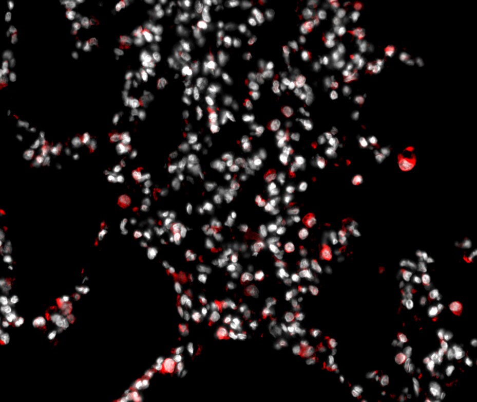
![Immunohistochemistry: AHR Antibody - BSA Free [NB100-2289]](https://resources.rndsystems.com/images/products/AHR-Antibody-Immunohistochemistry-NB100-2289-img0005.jpg "Immunohistochemistry: AHR Antibody - BSA Free [NB100-2289]")
Immunohistochemistry: AHR Antibody - BSA Free [NB100-2289]
Immunohistochemistry: AHR Antibody [NB100-2289] - Staining of Aryl hydrocarbon Receptor in mouse prostate.![Immunocytochemistry/ Immunofluorescence: AHR Antibody - BSA Free [NB100-2289]](https://resources.rndsystems.com/images/products/AHR-Antibody-Immunocytochemistry-Immunofluorescence-NB100-2289-img0012.jpg "Immunocytochemistry/ Immunofluorescence: AHR Antibody - BSA Free [NB100-2289]")
Immunocytochemistry/ Immunofluorescence: AHR Antibody - BSA Free [NB100-2289]
Immunocytochemistry/Immunofluorescence: AHR Antibody [NB100-2289] - HeLa cells were fixed for 10 minutes using 10% formalin and then permeabilized for 5 minutes using 1X PBS + 0.5% Triton X-100. The cells were incubated with anti-AHR at 5 ug/mL overnight at 4C and detected with an anti-rabbit Dylight 488 (Green) at a 1:500 dilution. Nuclei were counterstained with DAPI (Blue). Cells were imaged using a 40X objective.![Western Blot: AHR AntibodyBSA Free [NB100-2289]](https://resources.rndsystems.com/images/products/AHR-Antibody-Western-Blot-NB100-2289-img0008.jpg "Western Blot: AHR AntibodyBSA Free [NB100-2289]")
Western Blot: AHR AntibodyBSA Free [NB100-2289]
Western Blot: AHR Antibody [NB100-2289] - Detection of AhR in mouse liver cytosol using NB 100-2289.![Immunocytochemistry/ Immunofluorescence: AHR Antibody - BSA Free [NB100-2289]](https://resources.rndsystems.com/images/products/AHR-Antibody-Immunocytochemistry-Immunofluorescence-NB100-2289-img0015.jpg "Immunocytochemistry/ Immunofluorescence: AHR Antibody - BSA Free [NB100-2289]")
Immunocytochemistry/ Immunofluorescence: AHR Antibody - BSA Free [NB100-2289]
Immunocytochemistry/Immunofluorescence: AHR Antibody [NB100-2289] - HepG2 cells were fixed for 10 minutes using 4% paraformaldehyde for 10 minutes and permeabilized in 0.05% Triton X-100 in PBS for 5 minutes. The cells were incubated with anti-AHR at 1 ug/ml overnight at 4C and detected with an anti-rabbit Dylight 488 (Green) at a 1:1000 dilution. Nuclei were counterstained with DAPI (Blue). Cells were imaged using a 40X objective.![Flow Cytometry: AHR Antibody - BSA Free [NB100-2289]](https://resources.rndsystems.com/images/products/AHR-Antibody-Flow-Cytometry-NB100-2289-img0017.jpg "Flow Cytometry: AHR Antibody - BSA Free [NB100-2289]")
Flow Cytometry: AHR Antibody - BSA Free [NB100-2289]
Flow Cytometry: AHR Antibody [NB100-2289] - An intracellular stain was performed on U937 cells with NB100-2289 (blue) and a matched isotype control NBP2-24891 (orange). Cells were fixed with 4% PFA and then permeabilized with 0.1% saponin. Cells were incubated in an antibody dilution of 1.0 ug/mL for 30 minutes at room temperature, followed by Rabbit IgG (H+L) Cross-Adsorbed Secondary Antibody, Dylight 550 (SA5-10033, Thermo Fisher).![Immunocytochemistry/ Immunofluorescence: AHR Antibody - BSA Free [NB100-2289]](https://resources.rndsystems.com/images/products/AHR-Antibody-Immunocytochemistry-Immunofluorescence-NB100-2289-img0010.jpg "Immunocytochemistry/ Immunofluorescence: AHR Antibody - BSA Free [NB100-2289]")
Immunocytochemistry/ Immunofluorescence: AHR Antibody - BSA Free [NB100-2289]
Immunocytochemistry/Immunofluorescence: AHR Antibody [NB100-2289] - Aryl hydrocarbon Receptor antibody was tested in HeLa cells with DyLight 488 (green). Nuclei and alpha-tubulin were counterstained with DAPI (blue) and Dylight 550 (red).![Flow Cytometry: AHR Antibody - BSA Free [NB100-2289]](https://resources.rndsystems.com/images/products/AHR-Antibody-Flow-Cytometry-NB100-2289-img0013.jpg "Flow Cytometry: AHR Antibody - BSA Free [NB100-2289]")
Flow Cytometry: AHR Antibody - BSA Free [NB100-2289]
Flow Cytometry: AHR Antibody [NB100-2289] - An intracellular stain was performed on U-87 cells with AHR Antibody NB100-2289B (blue) and a matched isotype control (orange). Both antibodies were conjugated to Biotin. Cells were fixed with 4% PFA and then permeabilized with 0.1% saponin. Cells were incubated in an antibody dilution of 2.5 ug/mL for 30 minutes at room temperature, followed by Streptavidin - R-Phycoerythrin Protein (2012-1000, Novus Biologicals).![Simple Western: AHR AntibodyBSA Free [NB100-2289]](https://resources.rndsystems.com/images/products/AHR-Antibody-Simple-Western-NB100-2289-img0011.jpg "Simple Western: AHR AntibodyBSA Free [NB100-2289]")
Simple Western: AHR AntibodyBSA Free [NB100-2289]
Simple Western: AHR Antibody [NB100-2289] - Image shows a specific band for Aryl hydrocarbon receptor in 0.5 mg/mL of HepG2 lysate. This experiment was performed under reducing conditions using the 12-230 kDa separation system.
Western Blot: AHR Antibody - BSA Free [NB100-2289] -
Western Blot: AHR Antibody - BSA Free [NB100-2289] - Activation of AHR/Nrf2 pathway by PFT-alpha. (A) qPCR to detect mRNA level of CYP1A1, NQO1, HO1 & TRXR1 upon 20 h PFT-alpha treatment (20 µM) in MCF7 p53KO cells (upper part) & T47D cells (lower part). Relative expression level of CYP1A1 is shown in log2 scale; relative expression level of NQO1, HO1 & TRXR1 are shown as fold change, both normalized with DMSO treatment. All values represent the mean ± SD of two times independent experiments performed in three replicates. (B) Knock-down efficiency of AHR siRNA as detected by qPCR (upper part) & western blot (lower part). Relative expression level is shown as fold change normalized to scramble siRNA. All values represent the mean ± SD of two independent experiments performed in three replicates. (C) DCF-DA staining of ROS levels upon doxorubicin treatment (1 µM, 8 h) with or without PFT-alpha (20 µM, 12 h pre-treatment) in MCF7 p53KO cells transfected with scramble siRNA & siAHR. For quantification of ROS levels (lower panel), the values are reported as percentage relative to DMSO treatment group. (D) qPCR to detect mRNA level of Nrf2 targets NQO1, HO1 & TRXR1 upon 20 h PFT-alpha treatment (20 µM) in MCF7 p53KO cells transfected with scramble siRNA & siAHR. Image collected & cropped by CiteAb from the following publication (https://pubmed.ncbi.nlm.nih.gov/31974452), licensed under a CC-BY license. Not internally tested by Novus Biologicals.
AHR in A431 Human Cell Line.
AHR was detected in immersion fixed A431 human skin carcinoma cell line using Rabbit anti-AHR Affinity Purified Polyclonal Antibody conjugated to FITC (Catalog # NB100-2289F) (green) at 10 µg/mL overnight at 4C. Cells were counterstained with DAPI (blue). Cells were imaged using a 100X objective and digitally deconvolved.Applications for AHR Antibody - BSA Free
Application
Recommended Usage
ELISA
1:100 - 1:2000
Flow Cytometry
2.5 ug/ml
Immunocytochemistry/ Immunofluorescence
1:500-1:1000
Immunohistochemistry
1:100
Immunohistochemistry-Paraffin
1:100
Immunoprecipitation
1:10-1:500
Simple Western
1:200
Western Blot
1:500-1:2000
Application Notes
In Western blot a band is seen ~90 to 105 kDa representing AHR (molecular weight varies by species and by strain). Prior to immunostaining paraffin tissues, antigen retrieval with sodium citrate buffer (pH 6.0) is recommended.
In Simple Western only 10 - 15 uL of the recommended dilution is used per data point.
See Simple Western Antibody Database for Simple Western validation: Tested in HepG2 lysate 0.5 mg/mL, separated by Size, antibody dilution of 1:200, apparent MW was 74 kDa. Separated by Size-Wes, Sally Sue/Peggy Sue.
The observed molecular weight of the protein may vary from the listed predicted molecular weight due to post translational modifications, post translation cleavages, relative charges, and other experimental factors.
Reviewed Applications
Read 2 reviews rated 4.5 using NB100-2289 in the following applications:
Flow Cytometry Panel Builder
Bio-Techne Knows Flow Cytometry
Save time and reduce costly mistakes by quickly finding compatible reagents using the Panel Builder Tool.
Advanced Features
- Spectra Viewer - Custom analysis of spectra from multiple fluorochromes
- Spillover Popups - Visualize the spectra of individual fluorochromes
- Antigen Density Selector - Match fluorochrome brightness with antigen density

Formulation, Preparation, and Storage
Purification
Immunogen affinity purified
Formulation
PBS
Format
BSA Free
Preservative
0.02% Sodium Azide
Concentration
1.0 mg/ml
Shipping
The product is shipped with polar packs. Upon receipt, store it immediately at the temperature recommended below.
Stability & Storage
Store at 4C. Do not freeze.
Background: AHR
Long Name
Aryl Hydrocarbon Receptor
Alternate Names
BHLHE76
Gene Symbol
AHR
Additional AHR Products
Product Documents for AHR Antibody - BSA Free
Certificate of Analysis
To download a Certificate of Analysis, please enter a lot or batch number in the search box below.
Product Specific Notices for AHR Antibody - BSA Free
This product is for research use only and is not approved for use in humans or in clinical diagnosis. Primary Antibodies are guaranteed for 1 year from date of receipt.
Citations for AHR Antibody - BSA Free
 Powered by Bioz
Customer Reviews for AHR Antibody - BSA Free (2)
4.5 out of 5
2 Customer Ratings
Have you used AHR Antibody - BSA Free?
Submit a review and receive an Amazon gift card!
$25/€18/£15/$25CAN/¥2500 Yen for a review with an image
$10/€7/£6/$10CAN/¥1110 Yen for a review without an image
Submit a review
Customer Images
Showing
1
-
2 的
2 reviews
Showing All
Filter By:
-
Application: Immunohistochemistry-ParaffinSample Tested: lungSpecies: PigVerified Customer | Posted 10/26/2020FFPE pig lung was deparaffinized and antigen retrieval was performed. Block in 1%BSA/PBS and incubate with antibody at a dilution of 1:100 for 1 hour at room temperature. 20X image.

-
Application: ELISASample Tested: Guinea Pig Liver ExtractSpecies: OtherVerified Customer | Posted 06/04/2009
There are no reviews that match your criteria.
Protocols
View specific protocols for AHR Antibody - BSA Free (NB100-2289):
Immunocytochemistry Protocol
Culture cells to appropriate density on suitable glass coverslips in 35 mm culture dishes or 6-well plates.
1. Remove culture medium and add 10% formalin to the dish. Fix at room temperature for 5-10 minutes.
2. Remove the formalin and add 0.5% Triton-X 100 in TBS to permeabilize the cells. Incubate for 5-10 minutes.
3. Remove the permeabilization buffer and add wash buffer (i.e. PBS or PBS with 0.1% Tween-20). Be sure to not let the specimen dry out. Gently wash three times for 10 minutes.
4. Alternatively, cells can be fixed with -20C methanol for 10 min at room temperature. Remove the methanol and rehydrate in PBS for 10 min before proceeding.
5. To block nonspecific antibody binding incubate in 10% normal goat serum for 1 hour at room temperature.
6. Add primary antibody at appropriate dilution and incubate at room temperature for 1 hour or at 4 degrees C overnight.
7. Remove primary antibody and replace with wash buffer. Gently wash three times for 10 minutes.
8. Add secondary antibody at the appropriate dilution. Incubate for 1 hour at room temperature.
9. Remove antibody and replace with wash buffer. Gently wash three times for 10 minutes.
10. Nuclei can be staining with 4',6' diamino phenylindole (DAPI) at 0.1 ug/ml, or coverslips can be directly mounted in media containing DAPI.
11. Cells can now be viewed with a fluorescence microscope.
*The above information is only intended as a guide. The researcher should determine what protocol best meets their needs. Please follow proper laboratory procedures for the disposal of formalin.
Immunohistochemistry-Paraffin Embedded Sections
Antigen Unmasking:
Bring slides to a boil in 10 mM sodium citrate buffer (pH 6.0) then maintain at a sub-boiling temperature for 10 minutes. Cool slides on bench-top for 30 minutes.
Staining:
1. Wash sections in deionized water three times for 5 minutes each.
2. Wash sections in wash buffer for 5 minutes.
3. Block each section with 100-400 ul blocking solution for 1 hour at room temperature.
4. Remove blocking solution and add 100-400 ul diluted primary antibody. Incubate overnight at 4 degrees C.
5. Remove antibody solution and wash sections in wash buffer three times for 5 minutes each.
6. Add 100-400 ul biotinylated diluted secondary antibody. Incubate 30 minutes at room temperature.
7. Remove secondary antibody solution and wash sections three times with wash buffer for 5 minutes each.
8. Add 100-400 ul Streptavidin-HRP reagent to each section and incubate for 30 minutes at room temperature.
9. Wash sections three times in wash buffer for 5 minutes each.
10. Add 100-400 ul DAB substrate to each section and monitor staining closely.
11. As soon as the sections develop, immerse slides in deionized water.
12. Counterstain sections in hematoxylin.
13. Wash sections in deionized water two times for 5 minutes each.
14. Dehydrate sections.
15. Mount coverslips.
Western Blot Protocol
1. Perform SDS-PAGE on samples to be analyzed, loading 40 ug of total protein per lane.
2. Transfer proteins to membrane according to the instructions provided by the manufacturer of the membrane and transfer apparatus.
3. Stain according to standard Ponceau S procedure (or similar product) to assess transfer success, and mark molecular weight standards where appropriate.
4. Rinse the blot.
5. Block the membrane using standard blocking buffer for at least 1 hour.
6. Wash the membrane in wash buffer three times for 10 minutes each.
7. Dilute primary antibody in blocking buffer and incubate 1 hour at room temperature.
8. Wash the membrane in wash buffer three times for 10 minutes each.
9. Apply the diluted HRP conjugated secondary antibody in blocking buffer (as per manufacturers instructions) and incubate 1 hour at room temperature.
10. Wash the blot in wash buffer three times for 10 minutes each (this step can be repeated as required to reduce background).
11. Apply the detection reagent of choice in accordance with the manufacturers instructions.
Note: Tween-20 can be added to the blocking or antibody dilution buffer at a final concentration of 0.05-0.2%.
Find general support by application which include: protocols, troubleshooting, illustrated assays, videos and webinars.
- 7-Amino Actinomycin D (7-AAD) Cell Viability Flow Cytometry Protocol
- Antigen Retrieval Protocol (PIER)
- Antigen Retrieval for Frozen Sections Protocol
- Appropriate Fixation of IHC/ICC Samples
- Cellular Response to Hypoxia Protocols
- Chromogenic IHC Staining of Formalin-Fixed Paraffin-Embedded (FFPE) Tissue Protocol
- Chromogenic Immunohistochemistry Staining of Frozen Tissue
- ClariTSA™ Fluorophore Kits
- Detection & Visualization of Antibody Binding
- ELISA Sample Preparation & Collection Guide
- ELISA Troubleshooting Guide
- Extracellular Membrane Flow Cytometry Protocol
- Flow Cytometry Protocol for Cell Surface Markers
- Flow Cytometry Protocol for Staining Membrane Associated Proteins
- Flow Cytometry Staining Protocols
- Flow Cytometry Troubleshooting Guide
- Fluorescent IHC Staining of Frozen Tissue Protocol
- Graphic Protocol for Heat-induced Epitope Retrieval
- Graphic Protocol for the Preparation and Fluorescent IHC Staining of Frozen Tissue Sections
- Graphic Protocol for the Preparation and Fluorescent IHC Staining of Paraffin-embedded Tissue Sections
- Graphic Protocol for the Preparation of Gelatin-coated Slides for Histological Tissue Sections
- How to Run an R&D Systems DuoSet ELISA
- How to Run an R&D Systems Quantikine ELISA
- How to Run an R&D Systems Quantikine™ QuicKit™ ELISA
- ICC Cell Smear Protocol for Suspension Cells
- ICC Immunocytochemistry Protocol Videos
- ICC for Adherent Cells
- IHC Sample Preparation (Frozen sections vs Paraffin)
- Immunocytochemistry (ICC) Protocol
- Immunocytochemistry Troubleshooting
- Immunofluorescence of Organoids Embedded in Cultrex Basement Membrane Extract
- Immunofluorescent IHC Staining of Formalin-Fixed Paraffin-Embedded (FFPE) Tissue Protocol
- Immunohistochemistry (IHC) and Immunocytochemistry (ICC) Protocols
- Immunohistochemistry Frozen Troubleshooting
- Immunohistochemistry Paraffin Troubleshooting
- Immunoprecipitation Protocol
- Intracellular Flow Cytometry Protocol Using Alcohol (Methanol)
- Intracellular Flow Cytometry Protocol Using Detergents
- Intracellular Nuclear Staining Flow Cytometry Protocol Using Detergents
- Intracellular Staining Flow Cytometry Protocol Using Alcohol Permeabilization
- Intracellular Staining Flow Cytometry Protocol Using Detergents to Permeabilize Cells
- Preparing Samples for IHC/ICC Experiments
- Preventing Non-Specific Staining (Non-Specific Binding)
- Primary Antibody Selection & Optimization
- Propidium Iodide Cell Viability Flow Cytometry Protocol
- Protocol for Heat-Induced Epitope Retrieval (HIER)
- Protocol for Liperfluo
- Protocol for Making a 4% Formaldehyde Solution in PBS
- Protocol for VisUCyte™ HRP Polymer Detection Reagent
- Protocol for the Characterization of Human Th22 Cells
- Protocol for the Characterization of Human Th9 Cells
- Protocol for the Fluorescent ICC Staining of Cell Smears - Graphic
- Protocol for the Fluorescent ICC Staining of Cultured Cells on Coverslips - Graphic
- Protocol for the Preparation & Fixation of Cells on Coverslips
- Protocol for the Preparation and Chromogenic IHC Staining of Frozen Tissue Sections
- Protocol for the Preparation and Chromogenic IHC Staining of Frozen Tissue Sections - Graphic
- Protocol for the Preparation and Chromogenic IHC Staining of Paraffin-embedded Tissue Sections
- Protocol for the Preparation and Chromogenic IHC Staining of Paraffin-embedded Tissue Sections - Graphic
- Protocol for the Preparation and Fluorescent ICC Staining of Cells on Coverslips
- Protocol for the Preparation and Fluorescent ICC Staining of Non-adherent Cells
- Protocol for the Preparation and Fluorescent ICC Staining of Stem Cells on Coverslips
- Protocol for the Preparation and Fluorescent IHC Staining of Frozen Tissue Sections
- Protocol for the Preparation and Fluorescent IHC Staining of Paraffin-embedded Tissue Sections
- Protocol for the Preparation of Gelatin-coated Slides for Histological Tissue Sections
- Protocol for the Preparation of a Cell Smear for Non-adherent Cell ICC - Graphic
- Protocol: Annexin V and PI Staining by Flow Cytometry
- Protocol: Annexin V and PI Staining for Apoptosis by Flow Cytometry
- Quantikine HS ELISA Kit Assay Principle, Alkaline Phosphatase
- Quantikine HS ELISA Kit Principle, Streptavidin-HRP Polymer
- R&D Systems Quality Control Western Blot Protocol
- Sandwich ELISA (Colorimetric) – Biotin/Streptavidin Detection Protocol
- Sandwich ELISA (Colorimetric) – Direct Detection Protocol
- TUNEL and Active Caspase-3 Detection by IHC/ICC Protocol
- The Importance of IHC/ICC Controls
- Troubleshooting Guide: ELISA
- Troubleshooting Guide: Fluorokine Flow Cytometry Kits
- Troubleshooting Guide: Immunohistochemistry
- Troubleshooting Guide: Western Blot Figures
- Western Blot Conditions
- Western Blot Protocol
- Western Blot Protocol for Cell Lysates
- Western Blot Troubleshooting
- Western Blot Troubleshooting Guide
- View all Protocols, Troubleshooting, Illustrated assays and Webinars
Loading...
Associated Pathways
