
Aquaporin 1/AQP1 Antibody (1/A5F6)
Novus Biologicals | Catalog # NB600-749

![Immunohistochemistry: Aquaporin 1/AQP1 Antibody (1/A5F6) [NB600-749]](https://resources.rndsystems.com/images/products/Aquaporin-1-AQP1-Antibody-1-A5F6-Immunohistochemistry-NB600-749-img0002.jpg "Immunohistochemistry: Aquaporin 1/AQP1 Antibody (1/A5F6) [NB600-749]")
Scientific images may be resized, cropped, or adjusted for brightness or contrast for presentation purposes. These updates are limited to presentation and do not affect the underlying scientific interpretation of the data. Where available, additional source data may be provided upon request.
Key Product Details
Validated by
Species Reactivity
Validated:
Cited:
Applications
Validated:
Cited:
Label
Antibody Source
Product Specifications
Immunogen
Reactivity Notes
Please note that this antibody is reactive to Mouse and derived from the same host, Mouse. Additional Mouse on Mouse blocking steps may be required for IHC and ICC experiments. Please contact Technical Support for more information.
Localization
Specificity
Clonality
Host
Isotype
Scientific Data Images for Aquaporin 1/AQP1 Antibody (1/A5F6)
![Immunohistochemistry: Aquaporin 1/AQP1 Antibody (1/A5F6) [NB600-749]](https://resources.rndsystems.com/images/products/Aquaporin-1-AQP1-Antibody-1-A5F6-Immunohistochemistry-NB600-749-img0001.jpg "Immunohistochemistry: Aquaporin 1/AQP1 Antibody (1/A5F6) [NB600-749]")
Immunohistochemistry: Aquaporin 1/AQP1 Antibody (1/A5F6) [NB600-749]
Aquaporin-1-AQP1-Antibody-1-A5F6-Immunohistochemistry-NB600-749-img0001.jpg![Immunohistochemistry: Aquaporin 1/AQP1 Antibody (1/A5F6) [NB600-749]](https://resources.rndsystems.com/images/products/Aquaporin-1-AQP1-Antibody-1-A5F6-Immunohistochemistry-NB600-749-img0003.jpg "Immunohistochemistry: Aquaporin 1/AQP1 Antibody (1/A5F6) [NB600-749]")
Immunocytochemistry/ Immunofluorescence: Aquaporin 1/AQP1 Antibody (1/A5F6) [NB600-749] -
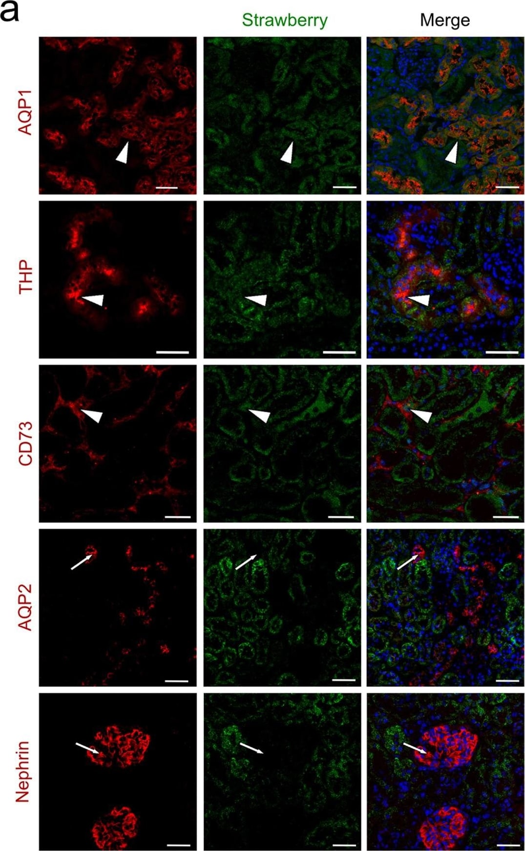
Immunocytochemistry/ Immunofluorescence: Aquaporin 1/AQP1 Antibody (1/A5F6) [NB600-749] - Intraparenchymal delivery of lentiviral vectors preferentially infects epithelial cells of both kidneys & urinary bladder.(a) Confocal images of Strawberry expression in renal sections of ELS infected kidneys 60 days post injection. Sections were stained with the indicated antibodies & merged images with DAPI (blue) are presented. Preferential transduction was observed in proximal (AQP1) & distal (THP) renal tubular epithelial cells & renal fibroblasts (CD73) (white arrowheads). Strawberry was not localised to collecting ducts (AQP2) or podocytes (Nephrin) (white arrows). Scale bars, 50 μm. (b) Confocal images of Strawberry expression in the liver, spleen, lung, urinary bladder & left & right kidney of ELS infected animals 60 days post left intrarenal injection (top panels) & uninjected control animals (bottom panels). Sections were stained with an antibody against Strawberry (green) & merged images with DAPI (blue) are presented. Scale bars, 50 μm. Image collected & cropped by CiteAb from the following publication (https://pubmed.ncbi.nlm.nih.gov/26046460), licensed under a CC-BY license. Not internally tested by Novus Biologicals.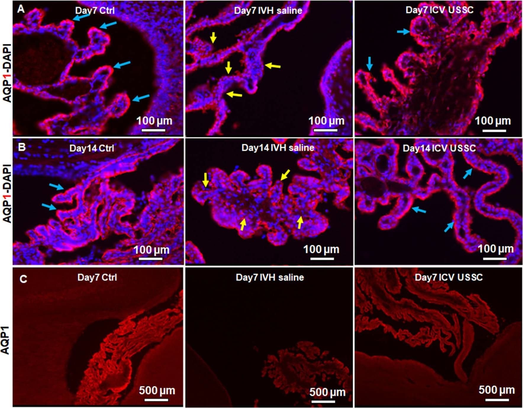
Immunocytochemistry/ Immunofluorescence: Aquaporin 1/AQP1 Antibody (1/A5F6) [NB600-749] -
Immunocytochemistry/ Immunofluorescence: Aquaporin 1/AQP1 Antibody (1/A5F6) [NB600-749] - USSC administration recovered aquaporin1 (AQP1) expression in the choroid plexus after PHH. (A) Representative immunofluorescence image of cryosections labeled with AQP1 specific antibody (arrows showing choroid epithelium immunoreactivity) on postnatal days 7. Strong AQP1 immunoreactivity in the control (blue arrows), reduced signal (yellow arrows) in IVH followed by recovered expression (blue arrows), & images from left to right respectively at postnatal day 7. CPE (Choroid plexus epithelial cells). Images were taken at 20× objective; blue = DAPI stain, 20 μm sections. Scale bar 100 μm. (B) Strong AQP1 immunoreactivity in the control (blue arrows), reduced signal (yellow arrows) in IVH followed by recovered expression (blue arrows), & the images from left to right respectively at day 14. Images were taken at 20× objective; blue = DAPI stain, 20 μm sections. Scale bar 100 μm. (C) Representative immunofluorescence image of cryosections labeled with aquaporin1. As seen in the high power image (panel-B above), the strong AQP1 immunoreactivity is evident over the entire choroid plexus in the control, reduced signal in IVH followed by recovered strong AQP1 expression observed in images from left to right respectively at day 14. Images were taken at 4× objective; 20 μm sections. The scale bar is 500 μm. Fluorescence images were taken using “Nikon Eclipse 90i microscope” (Nikon Instruments, Japan). Image collected & cropped by CiteAb from the following publication (https://pubmed.ncbi.nlm.nih.gov/33897371), licensed under a CC-BY license. Not internally tested by Novus Biologicals.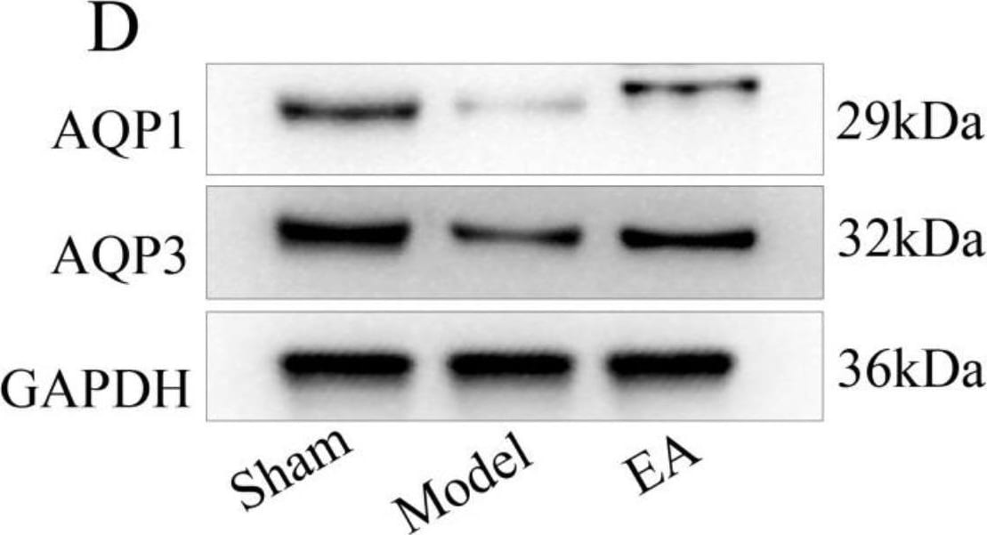
Western Blot: Aquaporin 1/AQP1 Antibody (1/A5F6) [NB600-749] -
AQP1and AQP3 protein expression in disc degeneration. (A) Representative microscopic photographs of immunofluorescence staining showing AQP1 and AQP3 immunoreaction-positive products. Nuclei counterstained with DAPI showed blue, with AQP1 and AQP3 positive products appearing green. Scale bar: 20 μm. The average optical density (AOD) was measured. (B,C) Bar graphs showing the AQP1 and AQP3 immunoreaction-positive products. (D) Western blot analysis of AQP1 and AQP3 protein levels. GAPDH was analyzed as a housekeeping gene. (E, F) Bar graphs showing the protein levels of AQP1 and AQP3. (G, H) Bar graphs showing the mRNA levels of AQP1 and AQP3. Data are expressed as mean +/- SD; **P < 0.01 compared to the sham compressed group; ##P < 0.01 and #P < 0.05 compared to the model group Image collected and cropped by CiteAb from the following open publication (https://pubmed.ncbi.nlm.nih.gov/40128722), licensed under a CC-BY license. Not internally tested by Novus Biologicals.Applications for Aquaporin 1/AQP1 Antibody (1/A5F6)
ELISA
Immunocytochemistry/ Immunofluorescence
Immunohistochemistry
Immunohistochemistry-Frozen
Immunohistochemistry-Paraffin
Western Blot
Reviewed Applications
Read 1 review rated 4 using NB600-749 in the following applications:
Formulation, Preparation, and Storage
Purification
Formulation
Preservative
Concentration
Shipping
Stability & Storage
Background: Aquaporin 1/AQP1
Alternate Names
Gene Symbol
UniProt
Additional Aquaporin 1/AQP1 Products
Product Documents for Aquaporin 1/AQP1 Antibody (1/A5F6)
Certificate of Analysis
To download a Certificate of Analysis, please enter a lot or batch number in the search box below.
Product Specific Notices for Aquaporin 1/AQP1 Antibody (1/A5F6)
This product is for research use only and is not approved for use in humans or in clinical diagnosis. Primary Antibodies are guaranteed for 1 year from date of receipt.
Related Research Areas
Citations for Aquaporin 1/AQP1 Antibody (1/A5F6)
 Powered by Bioz
Customer Reviews for Aquaporin 1/AQP1 Antibody (1/A5F6) (1)
Have you used Aquaporin 1/AQP1 Antibody (1/A5F6)?
Submit a review and receive an Amazon gift card!
$25/€18/£15/$25CAN/¥2500 for a review with an image
$10/€7/£6/$10CAN/¥1110 for a review without an image
Submit a review
Customer Images
-
Application: ImmunofluorescenceSample Tested: Rat Lung TissueSpecies: RatVerified Customer | Posted 11/30/2010

There are no reviews that match your criteria.
Protocols
Find general support by application which include: protocols, troubleshooting, illustrated assays, videos and webinars.
- Antigen Retrieval Protocol (PIER)
- Antigen Retrieval for Frozen Sections Protocol
- Appropriate Fixation of IHC/ICC Samples
- Cellular Response to Hypoxia Protocols
- Chromogenic IHC Staining of Formalin-Fixed Paraffin-Embedded (FFPE) Tissue Protocol
- Chromogenic Immunohistochemistry Staining of Frozen Tissue
- ClariTSA™ Fluorophore Kits
- Detection & Visualization of Antibody Binding
- ELISA Sample Preparation & Collection Guide
- ELISA Troubleshooting Guide
- Fluorescent IHC Staining of Frozen Tissue Protocol
- Graphic Protocol for Heat-induced Epitope Retrieval
- Graphic Protocol for the Preparation and Fluorescent IHC Staining of Frozen Tissue Sections
- Graphic Protocol for the Preparation and Fluorescent IHC Staining of Paraffin-embedded Tissue Sections
- Graphic Protocol for the Preparation of Gelatin-coated Slides for Histological Tissue Sections
- How to Run an R&D Systems DuoSet ELISA
- How to Run an R&D Systems Quantikine ELISA
- How to Run an R&D Systems Quantikine™ QuicKit™ ELISA
- ICC Cell Smear Protocol for Suspension Cells
- ICC Immunocytochemistry Protocol Videos
- ICC for Adherent Cells
- IHC Sample Preparation (Frozen sections vs Paraffin)
- Immunocytochemistry (ICC) Protocol
- Immunocytochemistry Troubleshooting
- Immunofluorescence of Organoids Embedded in Cultrex Basement Membrane Extract
- Immunofluorescent IHC Staining of Formalin-Fixed Paraffin-Embedded (FFPE) Tissue Protocol
- Immunohistochemistry (IHC) and Immunocytochemistry (ICC) Protocols
- Immunohistochemistry Frozen Troubleshooting
- Immunohistochemistry Paraffin Troubleshooting
- Preparing Samples for IHC/ICC Experiments
- Preventing Non-Specific Staining (Non-Specific Binding)
- Primary Antibody Selection & Optimization
- Protocol for Heat-Induced Epitope Retrieval (HIER)
- Protocol for Making a 4% Formaldehyde Solution in PBS
- Protocol for VisUCyte™ HRP Polymer Detection Reagent
- Protocol for the Fluorescent ICC Staining of Cell Smears - Graphic
- Protocol for the Fluorescent ICC Staining of Cultured Cells on Coverslips - Graphic
- Protocol for the Preparation & Fixation of Cells on Coverslips
- Protocol for the Preparation and Chromogenic IHC Staining of Frozen Tissue Sections
- Protocol for the Preparation and Chromogenic IHC Staining of Frozen Tissue Sections - Graphic
- Protocol for the Preparation and Chromogenic IHC Staining of Paraffin-embedded Tissue Sections
- Protocol for the Preparation and Chromogenic IHC Staining of Paraffin-embedded Tissue Sections - Graphic
- Protocol for the Preparation and Fluorescent ICC Staining of Cells on Coverslips
- Protocol for the Preparation and Fluorescent ICC Staining of Non-adherent Cells
- Protocol for the Preparation and Fluorescent ICC Staining of Stem Cells on Coverslips
- Protocol for the Preparation and Fluorescent IHC Staining of Frozen Tissue Sections
- Protocol for the Preparation and Fluorescent IHC Staining of Paraffin-embedded Tissue Sections
- Protocol for the Preparation of Gelatin-coated Slides for Histological Tissue Sections
- Protocol for the Preparation of a Cell Smear for Non-adherent Cell ICC - Graphic
- Quantikine HS ELISA Kit Assay Principle, Alkaline Phosphatase
- Quantikine HS ELISA Kit Principle, Streptavidin-HRP Polymer
- R&D Systems Quality Control Western Blot Protocol
- Sandwich ELISA (Colorimetric) – Biotin/Streptavidin Detection Protocol
- Sandwich ELISA (Colorimetric) – Direct Detection Protocol
- TUNEL and Active Caspase-3 Detection by IHC/ICC Protocol
- The Importance of IHC/ICC Controls
- Troubleshooting Guide: ELISA
- Troubleshooting Guide: Immunohistochemistry
- Troubleshooting Guide: Western Blot Figures
- Western Blot Conditions
- Western Blot Protocol
- Western Blot Protocol for Cell Lysates
- Western Blot Troubleshooting
- Western Blot Troubleshooting Guide
- View all Protocols, Troubleshooting, Illustrated assays and Webinars
FAQs for Aquaporin 1/AQP1 Antibody (1/A5F6)
-
Q: Are any of your Aquaporin-1 antibodies suitable for live cell staining? I am looking at doing flow cytometry on live cells and further isolation of the positive cells to purify the population.
A:
A list of our Aquaporin-1 antibodies suitable for use in flow cytometry can be found using this link. Of note, NB600-741 is known to bind to an extracellular domain, so should be suitable for your use.