B 细胞培养与分析

B 细胞如何调节免疫应答?
B 细胞在适应性免疫应答中发挥核心作用,主要功能如下:
- 产生抗体
- 识别并呈递抗原至 T 细胞
- 分泌调节性细胞因子,调控其他免疫细胞活性
- 形成免疫记忆
本页面汇总了适用于 B 细胞研究的各类实验工具,包括用于 B 细胞培养和活化的细胞因子、抗体,用于 B 细胞特征鉴定的抗体,以及单指标、多指标免疫分析试剂盒,全方位满足 B 细胞实验分析需求。
B 细胞培养
B 细胞在外周血淋巴细胞中占比较低,因此多数后续实验,如功能研究、感染性疾病研究、疫苗及治疗性抗体开发等,都需要先进行体外扩增培养。
小鼠 B 细胞可通过磁珠负选法从脾细胞中纯化,而人源 B 细胞则可通过负选法或 CD19+ 正选法从外周血单个核细胞中分离。随后,B 细胞通常会在表达 CD40 配体的基质饲养细胞层上进行体外培养,培养基中补充 IL-4、IL-2、IL-21、BAFF 等特异性细胞因子。额外搭配其他细胞因子,还可诱导 B 细胞定向分化、促进抗体分泌及特异性抗体亚型(如 IgA、IgG、IgE、IgM)的生成。
用于 B 细胞培养与分化的细胞因子
R&D Systems 提供种类丰富的细胞因子,可助力您优化 B 细胞培养。我们的所有蛋白产品均经过严格质控,活性稳定、批次差异小,能高效稳定实现 B 细胞扩增与分化。产品线覆盖研究级、无动物源临床前级、GMP 级蛋白试剂,可满足基础研究至临床研发中的各类研究需求。
| BAFF/BLyS/TNFSF13B | CD40 配体 | IFN-γ* | IL-2*† | IL-4* | IL-5 |
| IL-6* | IL-10* | IL-15*† | IL-21*† | RANK配体 | TNF-α* |
* 这些蛋白均有 GMP 级可供选择。
† 这些蛋白的 GMP 级和无动物源临床前级均有液体剂型可供选择。
CellXVivo™ 人 B 细胞扩增试剂盒
CellXVivo 免疫细胞扩增分化试剂盒搭配优化细胞因子组合与成熟实验方案,可实现各类免疫细胞的体外扩增和定向分化。CellXVivo 人 B 细胞扩增试剂盒所含试剂,足以将初始 107 个人 B 细胞群扩增 3-5 倍。
| 产品名称 |
| CellXVivo人 B 细胞扩增试剂盒 |
ExCellerate™B 细胞扩增培养基
ExCellerate B 细胞培养基配方经过专项优化,既能高效促进 B 细胞增殖,又可维持培养体系条件稳定。采用无异源成分配方,适配 B 细胞离体培养。可支持靶抗原特异性 B 细胞克隆形成,也适用于制备抗体的小鼠 B 细胞杂交瘤细胞系的培养,同时兼容多种物种来源的 B 细胞。
| 产品名称 |
| ExCellerate无异源成分 B 细胞培养基 |
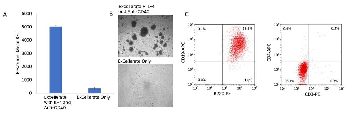
在 ExCellerate™ 人 B 细胞扩增培养基中,添加重组小鼠 IL-4 和 抗小鼠 CD40 抗体,用于小鼠 B 细胞体外扩增。采用MagCellect™小鼠 B 细胞分离试剂盒 (货号 MAGM204)从小鼠脾细胞中分离 B 细胞,分别使用添加/不添加重组小鼠 IL-4(货号 404-ML)、大鼠抗小鼠 CD40/TNFRSF5 单克隆抗体(货号 MAB440)的 ExCellerate B 细胞培养基 (货号 CCM031)进行培养扩增。(A)使用Rezasurin(刃天青;货号 AR002)监测小鼠 B 细胞的扩增情况。数据显示,添加 IL-4 和抗小鼠 CD40 抗体的 ExCellerate B细胞培养基能够有效促进小鼠 B 细胞的大量扩增。(B)小鼠 B 细胞集落的代表性明场图像。(C) 扩增所得小鼠 B 细胞纯度极高,B220+、CD19+ 双阳性占比超 98%,且 CD3、CD4 均为阴性。
B 细胞活化
B 细胞活化需要双重信号刺激:
- 第一信号由抗原与 B 细胞受体(BCR)结合所提供,随后 BCR-抗原复合物发生内吞、抗原降解,最终 B 细胞通过自身表面 MHC II 类分子完成抗原呈递。
- 第二信号是一种共刺激信号,可由抗原直接提供,也可通过 B 细胞与 T 细胞之间的相互作用产生。T 细胞通过识别 B 细胞表面的抗原-MHC II 复合物,与 B 细胞发生相互作用。这种相互作用促使 B 细胞表面 CD40 与 T 细胞表面 CD40L 等共刺激分子的结合,进而刺激 T 细胞活化,使其分泌 IL-4、IL-5、IL-6、IL-21 等细胞因子,从而驱动 B 细胞活化。B细胞活化后大量增殖并形成生发中心,最终分化为分泌抗体的浆细胞和记忆 B 细胞。
体外细胞培养体系中,可使用抗 IgD 或 IgM 抗体模拟 BCR 活化信号,并通过 CD40 配体或激动型抗 CD40 抗体联合细胞因子(如 IL-2、IL-4、IL-21、BAFF)模拟 T 细胞依赖性共刺激信号,从而促进 B 细胞活化。
B 细胞活化抗体
| CD40 | IgD | IgM |
B 细胞阻断与中和抗体
阻断抗体与中和抗体常用于研究细胞膜表面蛋白的生物学功能。我们提供多款靶向调控 B 细胞活性关键配体-受体对的阻断/中和抗体。这些抗体均经与蛋白生物活性检测相同的生物实验体系验证,确保有效阻断/中和对应细胞功能。
| APRIL | BAFF | BCMA | CD22 |
| CD40 | CD40L | TACI |
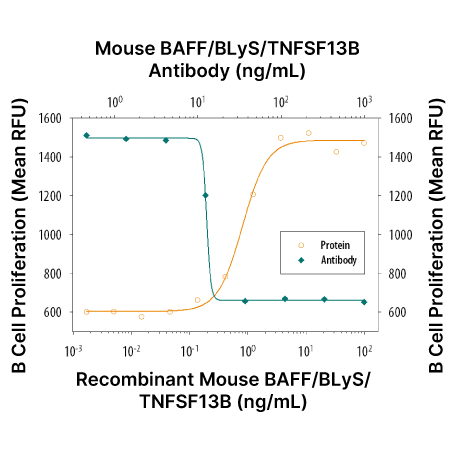
抗小鼠 BAFF 抗体中和 BAFF 诱导小鼠 B 细胞增殖实验。 在山羊 F(ab’)2 抗小鼠 IgM 存在下,重组小鼠 BAFF (货号 8876-BF)以剂量依赖性方式刺激小鼠 B 细胞增殖(橙色曲线)。在这些条件下,3 ng/mL 重组小鼠 BAFF 所诱导的增殖效应,可被梯度浓度的山羊抗小鼠 BAFF 抗原亲和纯化多克隆抗体(货号 AF2106)有效中和抑制(绿色曲线)。ND50 通常为 0.01-0.04 µg/mL。
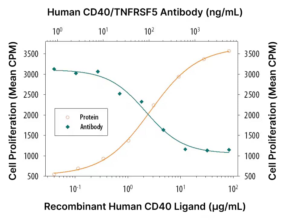
抗人 CD40 抗体中和 CD40 配体诱导的 B 细胞增殖实验。在 重组人 IL-4 (货号 204-IL)存在条件下,重组人 CD40 配体 (货号 6245-CL)以剂量依赖性方式刺激人 B 细胞富集的外周血单个核细胞增殖(橙色曲线)。梯度浓度 小鼠抗人 CD40 单克隆抗体(货号 MAB6322)可有效中和10 µg/mL CD40配体引发的细胞增殖效应(绿色曲线)。体系中加入 5 µg/mL 该抗体时,可在重组人 IL-4 协同作用下,中和约 80% 的重组人 CD40 配体诱导的细胞增殖活性。
B 细胞标志物
科研人员常根据特定细胞表面和细胞内标志物的表达差异,区分不同 B 细胞亚群。我们精选的一抗可搭配 30 余种荧光标记物,灵活实现多重检测实验,直接检测细胞表面和胞内靶标蛋白。所有抗体均经特异性与重现性验证,适配流式细胞术、免疫细胞化学/免疫荧光(ICC/IF)、免疫组化(IHC)、蛋白免疫印迹等多种实验体系。下表列出了常用于鉴别不同 B 细胞亚群的标志物。
滤泡 B 细胞
滤泡 B 细胞是小鼠和人体内数量最多的成熟 B 细胞亚群,主要分布于淋巴结和脾脏的滤泡中。该类细胞可在次级淋巴器官中再循环,活化后可分化为短寿命抗体分泌浆细胞和记忆 B 细胞。
| 人源标志物 | 表达 | 小鼠标志物 | 表达 |
| CD10/脑啡肽酶 | 阴性 | B220/CD45 R | 阳性 |
| CD19 | Positive | CD1d | 中 |
| CD20/MS4A1 | 阳性 | CD19 | 中 |
| CD21 | 阳性 | CD21 | 低 |
| CD22/Siglec-2 | 阳性 | CD23/Fcε RII | 阳性 |
| CD23/Fcε RII | 阳性 | CD43 | 阴性 |
| CD24 | 低 | CXCR5 | 阳性 |
| CD27 | 阴性 | IgD | 高 |
| CD38 | 低 | IgM | 低 |
| CXCR5 | 阳性 | L-选择素/CD62L | 阳性 |
| HLA-DR | 阳性 | MHC II 类 | 阳性 |
| TACI | 阳性 | ||
| IgD | 高 | ||
| IgM | 低 |
边缘区 B 细胞
边缘区 B 细胞是分布于脾脏边缘区白髓与红髓之间的一类固有样 B 细胞亚群,在抵御血源性病原体方面发挥着关键作用。边缘区 B 细胞表达多反应性 BCR,能够结合多种抗原;活化后分泌低亲和力抗体,能够迅速清除体内病原体和细胞凋亡碎片。
| 人源标志物 | 表达 | 小鼠标志物 | 表达 |
| CD1c/BDCA-1 | 阳性 | B220/CD45 R | 阳性 |
| CD19 | 阳性 | C1q R1/CD93 | 阴性 |
| CD20/MS4A1 | 阳性 | CD1d | 阳性 |
| CD21 | 阳性 | CD19 | 中 |
| CD23/Fcε RII | 阴性/低 | CD21 | 高 |
| CD27 | 阳性 | CD22/Siglec-2 | 阳性 |
| FCRL3/FcRH3 | 阳性 | CD23/Fcε RII | 阴性 |
| IgD | 低 | CD43 | 阴性 |
| IgM | 阳性 | IgD | 低 |
| TACI | 阳性 | IgM | 高 |
记忆 B 细胞
B细胞活化后,会在淋巴结和脾脏 B 细胞滤泡形成的生发中心内分化为记忆 B 细胞。记忆 B 细胞可随血液全身循环,其携带的 BCR 仅特异性识别最初触发其形成的抗原。再次接触同种抗原时,记忆 B 细胞会迅速启动免疫应答,合成分泌高亲和力特异性抗体,抵御二次感染。
| 人源标志物 | 表达 | 小鼠标志物 | 表达 |
| B7-1/CD80 | 阳性 | B220/CD45 R | 阳性 |
| B7-2/CD86 | 阳性 | B7-1/CD80 | 阳性 |
| C1q R1/CD93 | 阴性 | CD19 | 阳性 |
| CD19 | 阳性 | CD21 | 阳性 |
| CD20/MS4A1 | 阳性 | CD27 | 中/阳性 |
| CD21 | 阳性 | CD40 | 阳性 |
| CD27 | 中/阳性 | MHC II 类 | 阳性 |
| HLA-DR | 阳性 | ||
| TACI | 阳性 |
浆细胞
浆细胞是专职分泌抗体的终末分化细胞,机体接触抗原后,短期和长效抗体免疫应答均依赖其发挥作用。该类细胞已彻底分化成熟,不再具备增殖能力,主要功能是大量持续分泌抗体,帮助机体抵御外来病原体入侵。
| 人源标志物 | 表达 | 小鼠标志物 | 表达 |
| BCMA | 阳性 | B220/CD45 R | 低 |
| BLIMP1 | 阳性 | BCMA | 阳性 |
| CD10/脑啡肽酶 | 阴性 | BLIMP1 | 阳性 |
| CD19 | 低 | CD19 | 阴性 |
| CD20/MS4A1 | 阴性/低 | CD27 | 高 |
| CD27 | 阳性 | CD38 | 低 |
| CD38 | 高 | CXCR4 | 高 |
| CXCR4 | 阳性 | IgD | 阴性 |
| HLA-DR | 低 | MHC II 类 | 阴性/低 |
| IgD | 阴性 | 多配体聚糖-1/CD138 | 阳性 |
| 多配体聚糖-1/CD138 | 高 |
调节性 B 细胞
调节性 B 细胞是一类数量稀少的 B 细胞亚群,具备免疫抑制功能,可抑制炎症反应、维持机体免疫稳态。这些细胞可分泌 IL-10、IL-35、TGF-β 等细胞因子,从而抑制促炎性效应 T 细胞的分化、增殖与活化。
| 人源标志物 | 表达 | 小鼠标志物 | 表达 |
| CD1d | 阳性 | CD1d | 高 |
| CD5 | 阳性 | CD5 | 阳性 |
| CD19 | 阳性 | CD19 | 阳性 |
| CD21 | 阳性 | CD23/Fcε RII | 阴性/低 |
| CD24 | 阳性 | CD24 | 阳性 |
| IL-10 | 阳性 | TIM-1 | 阳性 |
| IL-35 | 阳性 | IL-10 | 阳性 |
| TGF-β | 阳性 | IL-35 | 阳性 |
| TGF-β | 阳性 |
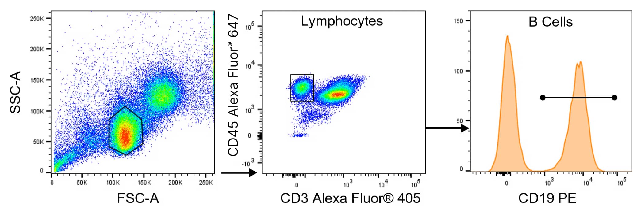
多色流式细胞术检测 B 细胞。采用 CD19 B 细胞组合抗体试剂盒(货号 FMC-P-003)对人外周血单个核细胞(PBMC)进行染色,实现 B 细胞检测。试剂盒组分:Alexa Fluor® 405 偶联小鼠抗人 CD3 单克隆抗体、Alexa Fluor® 647偶联小鼠抗人 CD45 单克隆抗体、PE偶联小鼠抗人 CD19 单克隆抗体。Alexa Fluor® 是美国俄勒冈州尤金市 Molecular Probes, Inc. 的注册商标。
B细胞细胞因子检测 —— 单指标 ELISA 试剂盒
活化后,B 细胞主要分泌 CCL17、CCL22,两者均可结合 Th2 细胞高表达的 CCR4 受体。外界刺激信号与其他免疫细胞分泌的细胞因子,会进一步调控 B 细胞的细胞因子分泌功能。依据不同信号调控,活化 B 细胞既能分泌促炎细胞因子(如 IL-1β、IL-6、TNF-α),也能分泌抗炎细胞因子(如 IL-10)。
因此,B 细胞不仅依靠产生抗体、形成免疫记忆与免疫应答,还能通过分泌细胞因子调控各类免疫细胞功能,在免疫调控中占据重要地位。
如需通过细胞因子分泌谱鉴定分析 B 细胞,我们提供多款可溶性蛋白定量免疫检测产品:包含 即用型 Quantikine™ ELISA 试剂盒、90分钟快速检测 QuicKit™ ELISA 试剂盒,以及组分齐全、可供自主开发实验的 DuoSet™ ELISA 开发试剂盒。
Quantikine ELISA 试剂盒
- 经过长期一致性与重现性验证
- 适配多种已验证的样本类型
- 试剂与稀释液体系经过优化,检测结果精准、灵敏度高
Quantikine QuicKit ELISA 试剂盒
- 90 分钟即可完成实验并获取数据
- Quantikine 高品质标准,批间一致性与重现性俱佳
- 精简操作流程,仅需一次洗涤操作
DuoSet ELISA 开发试剂盒
- 甄选经过功能验证的配对抗体,保障优异实验效果
- 靶标种类丰富,覆盖千余种指标,涵盖多个物种的新型靶标
- 适配多种实验平台,支持 384 孔板检测
用于 B 细胞鉴定的单指标 ELISA 试剂盒 —— 按分子分类产品
| CCL17 | CCL22 | GM-CSF | IFN-γ | IL-1 β | IL-2 |
| IL-4 | IL-6 | IL-10 | TGF-β 1 | TNF-α |
ELISpot 试剂盒与 ELISpot 开发模块
ELISpot 试剂盒是基于微孔板的高灵敏度检测体系,用于检测分泌细胞因子的细胞。完整的 ELISpot 试剂盒开箱即用,无需进行实验开发或优化。作为完整试剂盒的替代方案,我们还提供 ELISpot 开发模块,支持自主搭建定制化 ELISpot 实验方案。
- 灵敏度高,可检出频率低于十万分之一细胞的免疫应答
- 无需体外细胞扩增
- 仅需少量原代细胞,适合高通量批量检测
| IFN-γ | IgG | IgM | IL-2 | IL-4 | IL-5 |
| IL-6 | IL-10 | IL-13 | IL-17 | TNF-α |
了解更多ELISpot 检测相关信息。
用于细胞因子分泌分析的多重检测
Proteome Profiler™ 抗体芯片
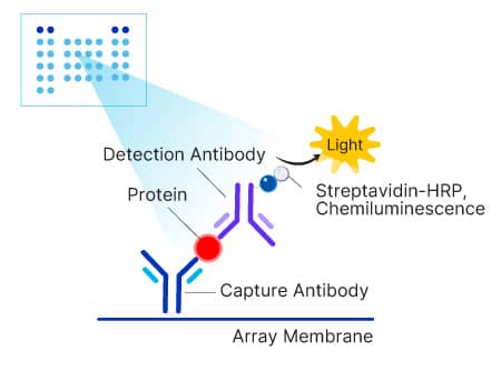
Proteome Profiler 抗体芯片基于固相膜载体构建,单份样本可同步检测多达 119 种蛋白。操作简便、性价比高,是早期探索发现阶段理想的半定量多重检测工具。该芯片利用化学发光法进行信号检测,可参照传统蛋白免疫印迹的方式,对膜上蛋白表达水平进行分析判读。
| Proteome Profiler 抗体芯片 | 检测指标数量 | 检测指标 |
| 人趋化因子抗体芯片 | 31 重 | CCL1·CCL2·CCL3/CCL4·CCL5·CCL7·CCL14·CCL15·CCL17·CCL18·CCL19·CCL20·CCL21·CCL22·CCL26·CCL28·趋化素·CX3CL1·CXCL1·CXCL4·CXCL5·CXCL7·CXCL8·CXCL9·CXCL10·CXCL11·CXCL12·CXCL16·CXCL17·IL-16·中期因子·XCL1 |
| 人细胞因子抗体芯片 | 36 重 | C5a·CCL1·CCL2·CCL5·CXCL1·CXCL10·CXCL11·CXCL12·CD40 配体·G-CSF·GM-CSF·ICAM-1·IFN-γ·IL-1α·IL-1β·IL-1ra·IL-2·IL-4·IL-5·IL-6·IL-8·IL-10·IL-12p70·IL-13·IL-16·IL-17·IL-17E·IL-18·IL-21·IL-27·IL-32α·MIF·MIP-1α/MIP-1β·丝氨酸蛋白酶抑制剂 E1·TNF-α·TREM-1 |
| 人 XL 细胞因子抗体芯片 | 105 重 | 脂联素·血管生成素·血管生成素-1·血管生成素-2·载脂蛋白 A1·BAFF·BDNF·C5a·CCL2·CCL3/CCL4/MIP-1α/β·CCL5·CCL7·CCL17·CCL19·CCL20·CD14·CD30·CD31·CD40 配体·几丁质酶 3 样蛋白·补体因子 D·C 反应蛋白·Cripto-1·CXCL1·CXCL4·CXCL5·CXCL9·CXCL10·CXCL11·CXCL12·胱抑素 C·Dkk-1·DPPIV·EGF·内皮糖蛋白·EMMPRIN·Fas 配体·碱性 FGF·FGF-7/KGF·FGF-19·Flt-3 配体·G-CSF·GDF-15·GM-CSF·生长激素·HGF·ICAM-1·IFN-γ·IGFBP-2·IGFBP-3·IL-1α·IL-1β·IL-1ra·IL-2·IL-3·IL-4·IL-5·IL-6·IL-8·IL-10·IL-11·IL-12 p70·IL-13·IL-15·IL-16·IL-17A·IL-18 BPa·IL-19·IL-22·IL-23·IL-24·IL-27·IL-31·IL-32α/β/γ·IL-33·IL-34·激肽释放酶 3·瘦素·LIF·脂质运载蛋白-2·M-CSF·MIF·MMP-9·髓过氧化物酶·骨桥蛋白·PDGF-AA·PDGF-AB/BB·正五聚蛋白 3·RAGE·RBP4·松弛素-2·抵抗素·丝氨酸蛋白酶抑制剂 E1·SHBG·ST2·TFF3·TfR·TGF-α·血小板反应蛋白-1·TIM-1·TNF-α·uPAR·VCAM-1·VEGF·维生素 D 结合蛋白 |
| 小鼠趋化因子抗体芯片 | 25 重 | C5a·CCL2·CCL3/CCL4·CCL5·CCL6/C10·CCL8·CCL9/10/MIP-1γ·CCL11·CCL12·CCL21·CCL22·CCL27·CCL28·趋化素·CX3CL1·CXCL1·CXCL2·CXCL9·CXCL10·CXCL11·CXCL12·CXCL13·CXCL16·IL-16·LIX |
| 小鼠细胞因子抗体芯片 | 40 重 | C5a·CCL1·CCL2·CCL3·CCL4·CCL5·CCL11·CCL12·CCL17·CXCL1·CXCL2·CXCL9·CXCL10·CXCL11·CXCL12·CXCL13·G-CSF·GM-CSF·ICAM-1·IFN-γ·IL-1α·IL-1β·IL-1ra·IL-2·IL-3·IL-4·IL-5·IL-6·IL-7·IL-10·IL-12 p70·IL-13 · IL-16·IL-17·IL-23·IL-27·M-CSF·TIMP-1·TNF-α·TREM-1 |
| 小鼠 XL 细胞因子抗体芯片 | 111 重 | 脂联素·双调蛋白·血管生成素-1·血管生成素-2·类血管生成素-3·BAFF·C1q R1/CD93·C5a·CCL2·CCL3/CCL4/MIP-1α/β·CCL5·CCL6/C10·CCL11·CCL12·CCL17·CCL19·CCL20·CCL21·CCL22·CD14·CD40·CD160·趋化素·几丁质酶 3 样蛋白·凝血因子 III/组织因子·补体因子 D·C 反应蛋白·CX3CL1·CXCL1·CXCL2·CXCL9·CXCL10·CXCL11·CXCL13·CXCL16·胱抑素 C·Dkk-1·DPPIV·EGF·内皮糖蛋白·内皮抑素·胎球蛋白 A·酸性 FGF·FGF-21·Flt-3 配体·Gas6·G-CSF·GDF-15·GM-CSF·HGF·ICAM-1·IFN-γ·IGFBP-1·IGFBP-2·IGFBP-3·IGFBP-5·IGFBP-6·IL-1α·IL-1β·IL-1ra·IL-2·IL-3·IL-4·IL-5·IL-6·IL-7·IL-10·IL-11·IL-12 p40·IL-13·IL-15·IL-17A·IL-22·IL-23·IL-27·IL-28·IL-33·LDL R·瘦素·LIF·脂质运载蛋白-2·LIX·M-CSF·MMP-2·MMP-3·MMP-9·髓过氧化物酶·骨桥蛋白·骨保护素·PD-ECGF·PDGF-BB·正五聚蛋白 2·正五聚蛋白 3·骨膜蛋白·Pref-1·增殖蛋白·前蛋白转化酶 9·RAGE·RBP 4·Reg3G·抵抗素·E-选择素/CD62E·P-选择素/CD62P·丝氨酸蛋白酶抑制剂 E1·丝氨酸蛋白酶抑制剂 F1·血小板生成素·TIM-1·TNF-α·VCAM-1·VEGF·WISP-1 |
了解更多Proteome Profiler 抗体芯片相关信息。
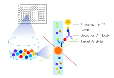
Luminex® 检测组合
Luminex 检测组合是一种基于微球的多重检测技术,单份样本可同步检测多达 50 种指标,大幅节省样本用量、实验时间和成本。可选用靶标超 490 种,涵盖人、小鼠、大鼠、猪、非人灵长类等物种。Luminex 检测组合均经过严格质量质控,实验数据稳定可靠、重现性优异。
Luminex 检测提供 3 种灵活选用规格:
- 探索级检测:可灵活搭配组合检测指标,支持个性化定制
- 固定式高性能组合:成品现货,下单即可发货。
- 可调配高性能组合:可挑选部分指标使用,也可选用整套指标
| Luminex 高性能组合 - 固定式 | 检测指标 |
| 人细胞因子组合 15 重 | IFN-α2·IFN-γ·IL-1α·IL-1β·IL-1ra·IL-2·IL-3·IL-4·IL-6·IL-7·IL-9·IL-10·IL-15·IL-33·VEGF |
| 人免疫治疗组合 25 重 | CD40 配体·GM-CSF·颗粒酶 B·IFN-α2·IFN-γ·IL-1α·IL-1β·IL-1ra·IL-2·IL-4·IL-6·IL-8·IL-9·IL-10·IL-12 p70·IL-13·IL-15·IL-17A·IL-33·IP-10·MCP-1·MIP-1α·MIP-1β·PD-L1·TNF-α |
| 人 XL 细胞因子组合 46 重 | CD40 配体·EGF·嗜酸细胞活化趋化因子·碱性 FGF·Flt-3 配体·G-CSF·GM-CSF·颗粒酶 B·GRO α·GRO β·IFN-α2·IFN-β·IFN-γ·IL-1α·IL-1β·IL-1ra·IL-2·IL-3·IL-4·IL-5·IL-6·IL-7·IL-8·IL-9·IL-10·IL-12 p70·IL-13·IL-15·IL-17A·IL-17E·IL-33·IP-10·MCP-1·MIP-1α·MIP-1β·MIP-3α·MIP-3β·PDGF-AA·PDGF-AB/BB·PD-L1·RANTES·TGF-α·TNF-α·TNF-β·TRAIL·VEGF |
| 小鼠 XL 细胞因子组合 45 重 | BAFF·CCL2·CCL3·CCL4·CCL5·CCL11·CCL19·几丁质酶 3 样蛋白 1·CXCL1·CXCL10·EGF·碱性 FGF·Flt-3配体·G-CSF·GDF-15·GM-CSF·ICAM-1·IFN-γ·IL-1α·IL-1β·IL-2·IL-3·IL-4·IL-5·IL-6·IL-7·IL-9·IL-10·IL-11·IL-12 p70·IL-13·IL-16·IL-17A·IL-18·IL-21·IL-27·IL-31·LDL R·LIF·LIX·M-CSF·TIMP-1·TNF-α·VEGF |
| Luminex 高性能组合 - 可调配 | 检测指标 |
| 人细胞因子组合 A | CCL2·CCL3·CCL4·CCL5·CXCL5·碱性 FGF·G-CSF·GM-CSF·IFN-γ·IL-1α·IL-1β·IL-1ra·IL-2·IL-4·IL-5·IL-6·IL-8·IL-10·IL-17A·血小板生成素·TNF-α·VEGF |
| 人高灵敏度细胞因子组合A | GM-CSF · IFN-γ · IL-1β · IL-2 · IL-4 · IL-5 · IL-6 · IL-8 · IL-10 · IL-12 · TNF-α · VEGF |
| 人高灵敏度细胞因子组合B | GM-CSF · IFN-γ · IL-1β · IL-2 · IL-5 · IL-6 · IL-7 · IL-13 · IL-15 · IL-17A · IL-17F · IL-22 · IL-23 ·IL-31 · IL-33 · IL-36β · TNF-α |
| 人 XL 细胞因子组合 | CCL2·CCL3·CCL4·CCL5·CCL11·CCL19·CCL20·CD40 配体·CXCL1·CXCL2·CXCL10·EGF·bFGF·Flt-3 配体·G-CSF·GM-CSF·颗粒酶 B·IFN-α2·IFN-β·IFN-γ·IL-1α·IL-1β·IL-1ra·IL-2·IL-3·IL-4·IL-5·IL-6·IL-7·IL-8·IL-9·IL-10·IL-12 p70·IL-13·IL-15·IL-17A·IL-17E·IL-33·PD-L1·PDGF-AA·PDGF-AB/BB·TGF-α·TNF-α·TNF-β·TRAIL·VEGF |
| 小鼠 XL 细胞因子组合 | BAFF·CCL2·CCL3·CCL4·CCL5·CCL11·CCL19·几丁质酶 3 样蛋白 1·CXCL1·CXCL10·EGF·碱性 FGF·Flt-3 配体·G-CSF·GDF-15·GM-CSF·ICAM-1·IFN-γ·IL-1α·IL-1β·IL-1ra·IL-2·IL-3·IL-4·IL-5·IL-6·IL-7·IL-9·IL-10·IL-11·IL-12 p70·IL-13·IL-16·IL-17A·IL-18·IL-21·IL-27·IL-31·LDL R·LIF·LIX·M-CSF·TIMP-1·TNF-α·VEGF |
了解更多 Luminex 检测相关信息。
Luminex® 是 Luminex Corporation 的注册商标。
B 细胞培养与分析其他产品
细胞分离试剂盒
包含正选、负选试剂盒,可分离免疫细胞用于体外培养实验。
无动物源重组蛋白
助力实验从临床前研究平稳过渡至临床制剂生产,培养体系组分明确可控,消除动物源试剂带来的监管审批及伦理相关隐忧。我们的无动物源蛋白采用与无动物源 GMP 级蛋白相同的表达体系和生产工艺制备。
GMP 级蛋白
严格遵照生产规范制备,可作为细胞治疗生产流程中的辅助材料使用。产品经过全套严苛质量检测,附带完整溯源资料,原料来源和生产流程全程可查。同时提供液态剂型、工艺级大包装和封闭工艺适配型 GMP 级细胞因子。了解更多。
细胞增殖与活力检测
用于研究细胞增殖、活力和细胞毒性的试剂,包括荧光报告染料、MTT 检测试剂盒等。
精选内容
B 细胞发育海报
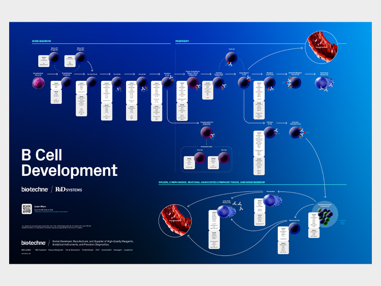
包含人、小鼠 B 细胞各发育阶段专属标志物,轻松完成不同分化阶段细胞鉴定分型。
人免疫细胞标志物海报
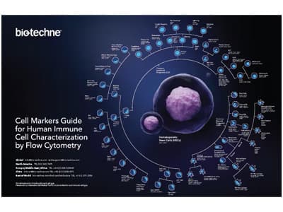
汇总流式细胞术区分各类免疫细胞所用标志物,便于快速查阅参考。
Simple Reader™ 紧凑型酶标仪

Simple Reader™ 酶标仪外形小巧、经济高效,检测结果精准,助力节约实验成本与场地。
人细胞因子与趋化因子家族周期表海报
梳理各类细胞因子家族分类知识,既是实用科研工具书,也可装点实验室。
批量采购,控本增效
单品大批量采购或多品类合并下单,均可享受优惠集采价格。